



Dehydroevodiamine
Based on 3 publication(s) in Google Scholar
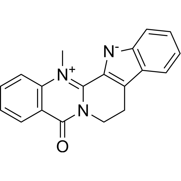
Dehydroevodiamine is a blood-brain barrier-permeable, orally effective AChE and BACE1 inhibitor (IC50 = 40.96 μM). Dehydroevodiamine is isolated and extracted from Evodia rutaecarpa. Dehydroevodiamine attenuates neurotoxicity through multiple targets by inhibiting BACE1 to block Aβ production, inhibiting AChE activity, scavenging ROS, and suppressing calcium influx. Dehydroevodiamine can be used for research on Alzheimer's disease, cerebral ischemia, vascular dementia, and other cognitive disorder-related diseases.
Nur für Forschungszwecke. Wir verkaufen nicht an Patienten.
- Reinheit : 98.55%
- CAS. Nr.: 67909-49-3
- Formel: C19H15N3O
- Molecular Weight:301.34
-
Speicherung:
4°C, protect from light
* In solvent : -80°C, 6 months; -20°C, 1 month (protect from light)

To place orders, for customer services and technical support, please contact: MedChemExpress USA
Tel: 609-228-6898 E-mail: [email protected] [email protected]


-
 Biologische Aktivität
-
Chemical Information
-
Lösungsmittel & Löslichkeit
- Protokoll
- Reinheit & Dokumentation
- Verweise
-
 Help & FAQs


-

HY-N2106R
Dehydroevodiamine (Standard)Reference Standard -
HY-32735
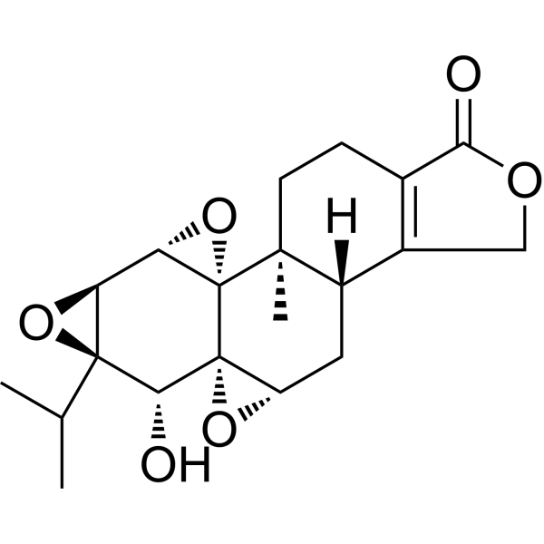
TriptolideImmunosuppressive Agent -
HY-N1445
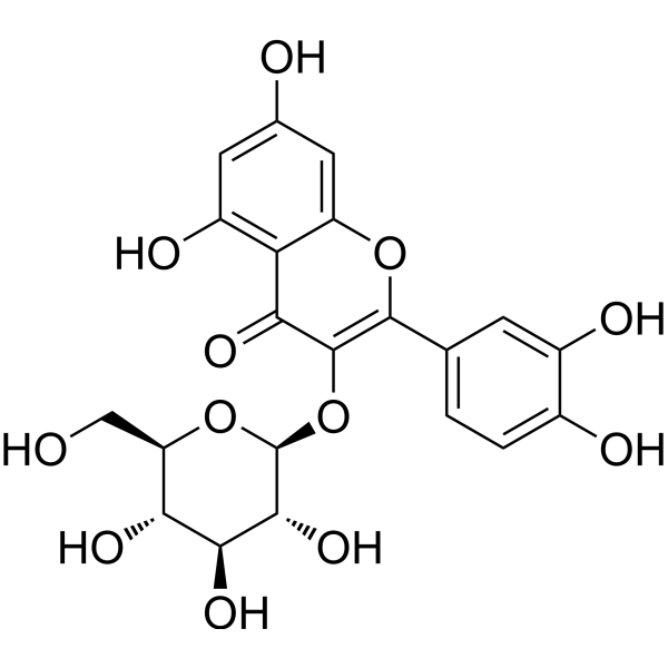
IsoquercitrinNO Synthase Inhibitor -
HY-N6966
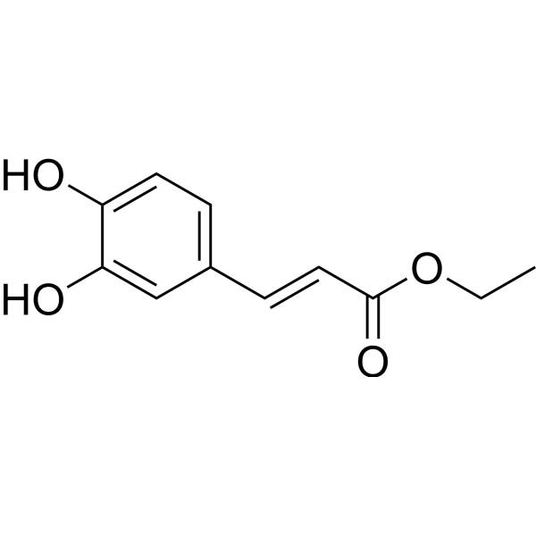
Ethyl CaffeateCOX Inhibitor -
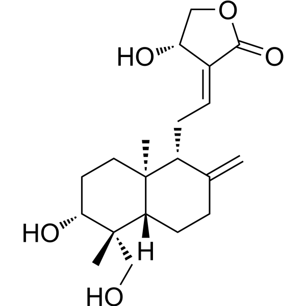
HY-N0191
AndrographolideNF-κB Inhibitor -
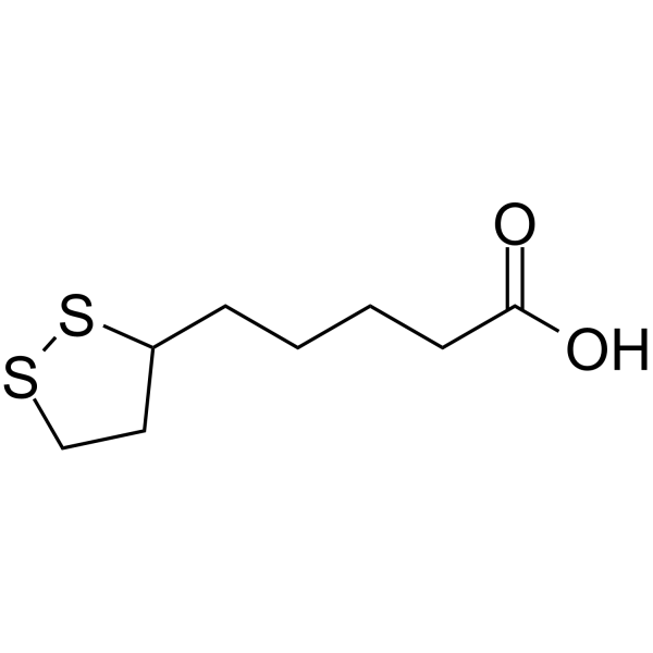
HY-N0492
α-Lipoic AcidAntioxidant -
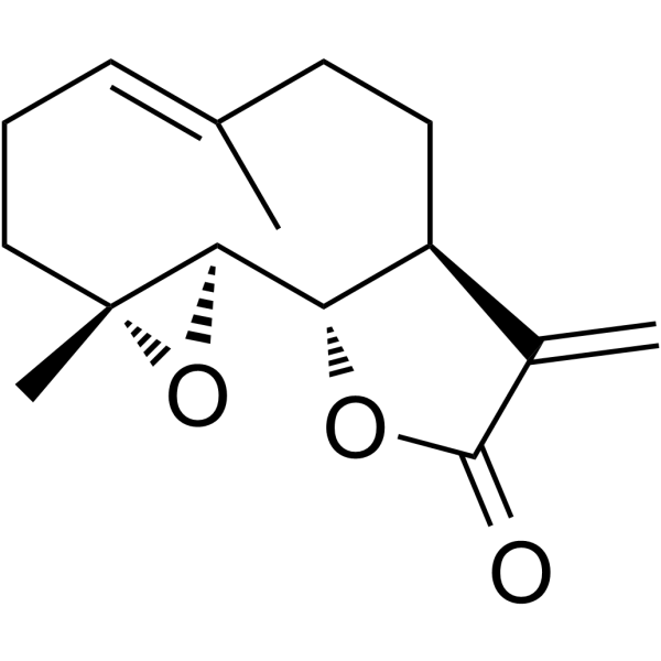
HY-N0141
ParthenolideNF-κB Inhibitor -
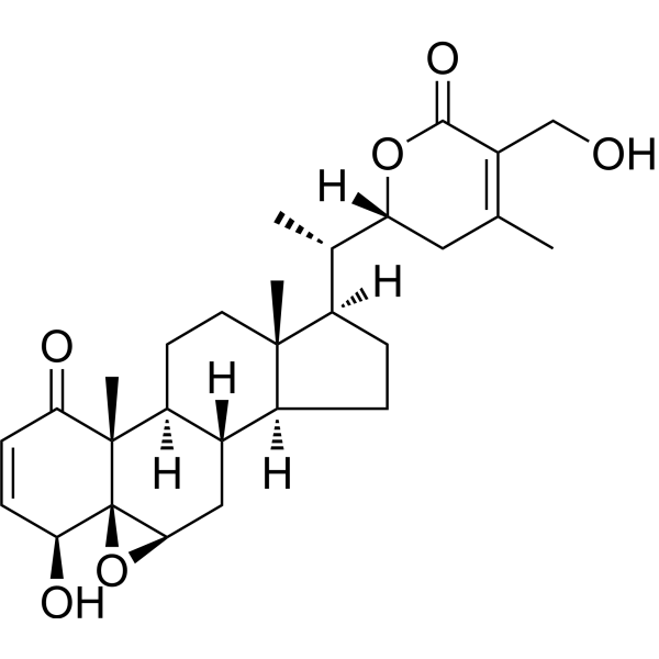
HY-N2065
Withaferin ANF-κB Inhibitor -
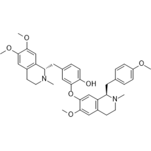
HY-N0441
NeferineAutophagy Modulator -
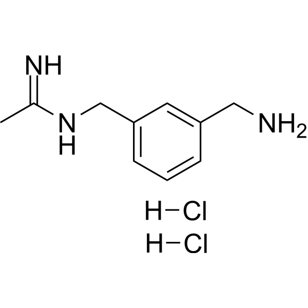
HY-18731
1400W DihydrochlorideNO Synthase Inhibitor -
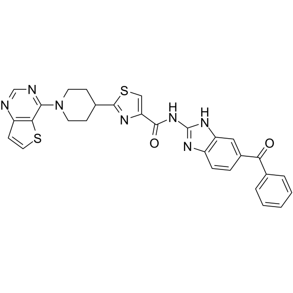
HY-10496
SC75741NF-κB Inhibitor -
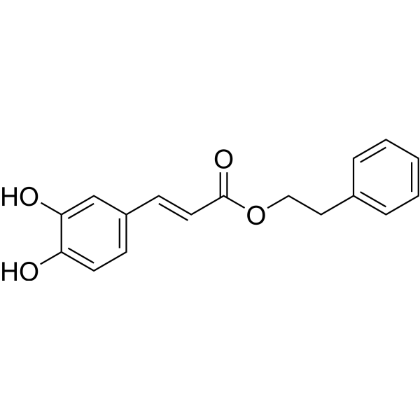
HY-N0274
Caffeic acid phenethyl esterApoptosis Inducer
Publications Citing Use of MedChemExpress (MCE) Dehydroevodiamine
MoreAlle Endogenous Metabolite Isoform-spezifische Produkte anzeigen
More

Biologische Aktivität
Beschreibung
IC50 & Target
[1]|
BACE1 40.96 μM (IC50) |
AChE |
In Vitro
Dehydroevodiamine (0.5-4 μM; 4 h) attenuates Aβ1-42 (HY-P1362)-induced neurotoxicity and reduces ROS production in primary cortical neurons in a dose-dependent manner[3].
Dehydroevodiamine () (10 μM; 5 min) reduces the KCl-dependent increase in intracellular calcium levels in primary cortical neurons[3].
Dehydroevodiamine (25-100 µM; 1 h) acts as a competitive inhibitor of BACE1 in mouse brain homogenates, with an IC50 of 40.96 µM[2].
MedChemExpress (MCE) has not independently confirmed the accuracy of these methods. They are for reference only.
-
Cell Line:Primary cortical neurons from embryonic day 18 Sprague-Dawley rat embryos
-
Concentration:0.5, 1, 2, 4 μM
-
Incubation Time:4 h
-
Result:Reduced cell viability by approximately 60% following exposure to 25 μM Aβ1-42 peptide.
Enhanced cell viability in a dose-dependent manner with values of 55.89% at 2 μM and 57.84% at 4 μM compared to Aβ1-42-exposed neurons.
In Vivo
Dehydroevodiamine (10 mg/kg; p.o.; single administration) ameliorates spatial memory impairment in the Scopolamine (HY-N0296)-induced amnesia model in Wistar rats[3].
Dehydroevodiamine (10 mg/kg; p.o.; once daily; 21 days) ameliorates learning and spatial memory impairment in an Aβ1-42 intracerebroventricular infusion-induced Wistar rat model[3].
Dehydroevodiamine (10-20 mg/kg (p.o.) or 6.25 mg/kg (i.p.); single dose or once daily for 7 consecutive days) reverses memory acquisition and spatial working memory deficits in Scopolamine-induced Sprague-Dawley rat models[4].
Dehydroevodiamine (6.25 mg/kg; i.p.; single administration 30 min before ischemia or once daily for 7 consecutive days after ischemia) reduces cerebral infarct size and attenuates neuronal damage in the hippocampus and cognitive deficits in a middle cerebral artery occlusion (MCA)-induced focal cerebral ischemia model in Sprague-Dawley rats[4].
Dehydroevodiamine (6.25 mg/kg; i.p.; once daily; for 7 consecutive days) significantly reduces neuronal death in the hippocampus and dentate gyrus and ameliorates memory impairment in a Sprague-Dawley rat model of electrolytic lesion of the entorhinal cortex (EC)[4].
Dehydroevodiamine (0.03-0.3 mg/kg; i.v.; single administration; observation for 1-10 min) selectively increases cerebral cortical blood flow without affecting other cardiopulmonary functions in anesthetized adult cats[1].
MedChemExpress (MCE) has not independently confirmed the accuracy of these methods. They are for reference only.
-
Animal Model:Wistar rats (7-week-old)[3]
-
Dosage:10 mg/kg
-
Administration:p.o.; once daily; 21 days
-
Result:Reduced escape latency to 4.4 sec.
Increased passive avoidance latency to 204.85 sec.
-
Animal Model:Sprague-Dawley (Male, 200-250 g, middle cerebral artery occlusion)[4]
-
Dosage:6.25 mg/kg
-
Administration:i.p.; once daily; for 7 days
-
Result:Decreased the number of damaged neurons in the CA1 region of the hippocampus to 4.3% compared with 42.3% in the nontreated group.
Recovered the latency time to the sham control level in the passive avoidance test.
-
Animal Model:Sprague-Dawley (Male, 200-250 g, unilateral electrolytic lesion of the entorhinal cortex)[4]
-
Dosage:6.25 mg/kg
-
Administration:i.p.; once daily; for 7 days
-
Result:Increased the latency time significantly.
Reduced neuronal damage to 9.23% in CA1, 4.2% in CA3, and 13.2% in the dentate gyrus.
-
Animal Model:Adult cats (either sex, 2.3-3.5 kg, anesthetized)[1]
-
Dosage:0.03, 0.1, and 0.3 mg/kg
-
Administration:i.v.; single dose; 1-10 min
-
Result:Selectively increased cerebral cortical blood flow without affecting other cardiopulmonary functions.
-
Animal Model:Tg2576 (transgenic, over-expressing human APP695 with the Swedish double mutation K670N, M671L, 7 months old at start, mixed gender)[2]
-
Dosage:0.5 mg/kg
-
Administration:i.p.; once daily; 4 months
-
Result:Ameliorated memory impairment and reduced cortical soluble Aβ and amyloid plaques.


Chemical Information
-
CAS. Nr. 67909-49-3
-
Appearance Solid
-
Molecular Weight 301.34
-
Formel C19H15N3O
-
Color Light yellow to yellow
-
SMILES
O=C1N2C(C([N-]C3=C4C=CC=C3)=C4CC2)=[N+](C)C5=C1C=CC=C5
-
Structure Classification
-
Initial Source
-
Versand
Room temperature in continental US; may vary elsewhere.
-
Speicherung
4°C, protect from light
* In solvent : -80°C, 6 months; -20°C, 1 month (protect from light)


Publications (3)
-
Journal Impact Factor
-
Most Recent
-
BMC Complement Med Ther
Hepatotoxicity prediction for traditional Chinese medicine: a two-step in silico framework integrating network and machine learning approaches. [Abstract]2026 Apr 2;26(1):177. PMID: 41923057 -
Chem Biol Drug Des
CD163 and TYROBP Are Two Therapeutic Targets for Hyperglycemia-Induced Mesangial Cell Stress. [Abstract]2025 Dec;106(6):e70213. PMID: 41329505 -
J Anim Physiol Anim Nutr
Dihydromyricetin Suppresses Lipopolysaccharide-Induced Intestinal Injury Through Reducing Reactive Oxygen Species Generation and NOD-Like Receptor Pyrin Domain Containing 3 Inflammasome Activation. [Abstract]2025 Mar;109(2):610-622. PMID: 39618421


Lösungsmittel & Löslichkeit
In Vitro:
DMSO : 10 mg/mL (33.19 mM; ultrasonic and warming and heat to 60°C; Hygroscopic DMSO has a significant impact on the solubility of product, please use newly opened DMSO)
Please refer to the solubility information to select the appropriate solvent. Once prepared, please aliquot and store the solution to prevent product inactivation from repeated freeze-thaw cycles.
Storage method and period of stock solution: -80°C, 6 months; -20°C, 1 month (protect from light). When stored at -80°C, please use it within 6 months. When stored at -20°C, please use it within 1 month.
Please refer to the solubility information to select the appropriate solvent. Once prepared, please aliquot and store the solution to prevent product inactivation from repeated freeze-thaw cycles.
Storage method and period of stock solution: -80°C, 6 months; -20°C, 1 month (protect from light). When stored at -80°C, please use it within 6 months. When stored at -20°C, please use it within 1 month.
Konzentration (Stammlösung) × Volumen (Stammlösung) = Konzentration (Ziellösung) × Volumen (Ziellösung)
In Vivo:
Select the appropriate dissolution method based on your experimental animal and administration route.
- For the following dissolution methods, please ensure to first prepare a clear stock solution using an In Vitro approach and then sequentially add co-solvents:
- To ensure reliable experimental results, the clarified stock solution can be appropriately stored based on storage conditions. As for the working solution for In Vivo experiments, it is recommended to prepare freshly and use it on the same day.
- The percentages shown for the solvents indicate their volumetric ratio in the final prepared solution. If precipitation or phase separation occurs during preparation, heat and/or sonication can be used to aid dissolution.
Add each solvent one by one: 10% DMSO 90% (20% SBE-β-CD in Saline)
Solubility: ≥ 1 mg/mL (3.32 mM); Clear solution
This protocol yields a clear solution of ≥ 1 mg/mL (saturation unknown).
Taking 1 mL working solution as an example, add 100 μL DMSO stock solution (10.0 mg/mL) to 900 μL 20% SBE-β-CD in Saline, and mix evenly.
Preparation of 20% SBE-β-CD in Saline (4°C, storage for one week): 2 g SBE-β-CD powder is dissolved in 10 mL Saline, completely dissolve until clear.
For the following dissolution methods, please prepare the working solution directly:
It is recommended to prepare fresh solutions and use them promptly within a short period of time.
The percentages shown for the solvents indicate their volumetric ratio in the final prepared solution. If precipitation or phase separation occurs during preparation, heat and/or sonication can be used to aid dissolution.
Add each solvent one by one: 0.5% CMC-Na/saline water
Solubility: 10 mg/mL (33.19 mM); Suspended solution; Need ultrasonic
In Vivo Dissolution Calculator
Please enter the basic information of animal experiments:
-
-
-
-
Recommended: Prepare an additional quantity of animals to account for potential losses during experiments.
Please enter your animal formula composition:
-
%DMSO +
Recommended: Keep the proportion of DMSO in working solution below 2% if your animal is weak.
-
%+
-
+%Tween-80 + +
-
%Saline +
The co-solvents required include: DMSO, . All of co-solvents are available by MedChemExpress (MCE). , Tween 80. All of co-solvents are available by MedChemExpress (MCE).
Working solution concentration: 0.22 mg/mL
Method for preparing stock solution: mg drug dissolved in μL DMSO. Stock solution concentration: mg/mL. * In solvent : -80°C, 6 months; -20°C, 1 month (protect from light)
1. Take μL DMSO stock solution;
2. Add μL .
μL , mix evenly;
3. Then add μL Tween 80, mix evenly;
4. Then add μL
Please ensure that the stock solution in the first step is dissolved to a clear state, and add co-solvents in sequence. You can use ultrasonic heating (ultrasonic cleaner, recommended frequency 20-40 kHz), vortexing, etc. to assist dissolution.


Reinheit & Dokumentation
-
Data Sheet (300 KB)
-
SDS (394 KB)
- English - EN (394 KB)
- Français - FR (394 KB)
- Deutsch - DE (394 KB)
- Norwegian - NO (394 KB)
- Español - ES (394 KB)
- Swedish - SV (394 KB)
- Italian - IT (394 KB)
- Korean - KR (394 KB)
- Portuguese - PT (394 KB)
-
Handling Instructions (2659 KB)
Verweise
[1]. Haji A, et al. Increased feline cerebral blood flow induced by dehydroevodiamine hydrochloride from Evodia rutaecarpa. Journal of natural products. 1994 Mar;57(3):387-9. [Content Brief]
[3]. Shin KY, et al. Dehydroevodiamine·HCl enhances cognitive function in memory-impaired rat models. The Korean journal of physiology & pharmacology : official journal of the Korean Physiological Society and the Korean Society of Pharmacology. 2017 Jan;21(1):55-64. [Content Brief]


Complete Stock Solution Preparation Table
Please refer to the solubility information to select the appropriate solvent. Once prepared, please aliquot and store the solution to prevent product inactivation from repeated freeze-thaw cycles.
Storage method and period of stock solution: -80°C, 6 months; -20°C, 1 month (protect from light). When stored at -80°C, please use it within 6 months. When stored at -20°C, please use it within 1 month.
| Optional Solvent | Concentration Solvent Mass | 1 mg | 5 mg | 10 mg | 25 mg |
|---|---|---|---|---|---|
| DMSO | 1 mM | 3.3185 mL | 16.5926 mL | 33.1851 mL | 82.9628 mL |
| 5 mM | 0.6637 mL | 3.3185 mL | 6.6370 mL | 16.5926 mL | |
| 10 mM | 0.3319 mL | 1.6593 mL | 3.3185 mL | 8.2963 mL | |
| 15 mM | 0.2212 mL | 1.1062 mL | 2.2123 mL | 5.5309 mL | |
| 20 mM | 0.1659 mL | 0.8296 mL | 1.6593 mL | 4.1481 mL | |
| 25 mM | 0.1327 mL | 0.6637 mL | 1.3274 mL | 3.3185 mL | |
| 30 mM | 0.1106 mL | 0.5531 mL | 1.1062 mL | 2.7654 mL |


Dehydroevodiamine Related Classifications
HY-N2106 Related Classifications
 Powered by Bioz