



BI-4916
Based on 7 publication(s) in Google Scholar
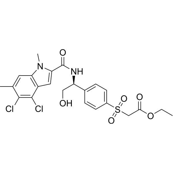
BI-4916 is a proagent of BI-4924. BI-4924 is a NADH/NAD+-competitive PHGDH inhibitor. BI-4916 inhibits cancer cell migration. BI-4916 can be used for cancer, inflammation and infection study.
For research use only. We do not sell to patients.
The BI-4916 was designed by Boehringer Ingelheim and could be obtained free of charge through the Boehringer Ingelheim open innovation portal opnMe.com, associated with its negative control.
- Purity : 98.67%
- CAS No.: 2244451-48-5
- Formula: C23H24Cl2N2O6S
- Molecular Weight:527.42
-
Storage:Powder -20°C, 3 years , 4°C, 2 years ; In solvent -80°C, 2 years , -20°C, 1 year

To place orders, for customer services and technical support, please contact: MedChemExpress USA
Tel: 609-228-6898 E-mail: [email protected] [email protected]


-
 Biological Activity
-
Chemical Information
-
Solvent & Solubility
- Protocols
- Purity & Documentation
- References
-
 Help & FAQs


-
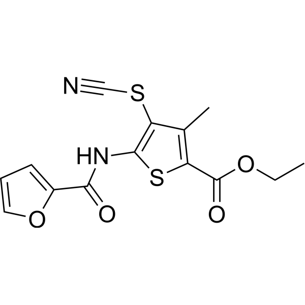
HY-100012
CBR-5884PHGDH Inhibitor -
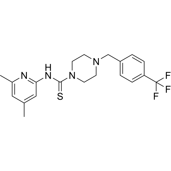
HY-101966
NCT-503PHGDH Inhibitor -
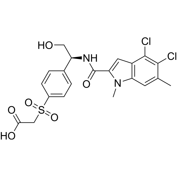
HY-126254
BI-4924PHGDH Inhibitor -
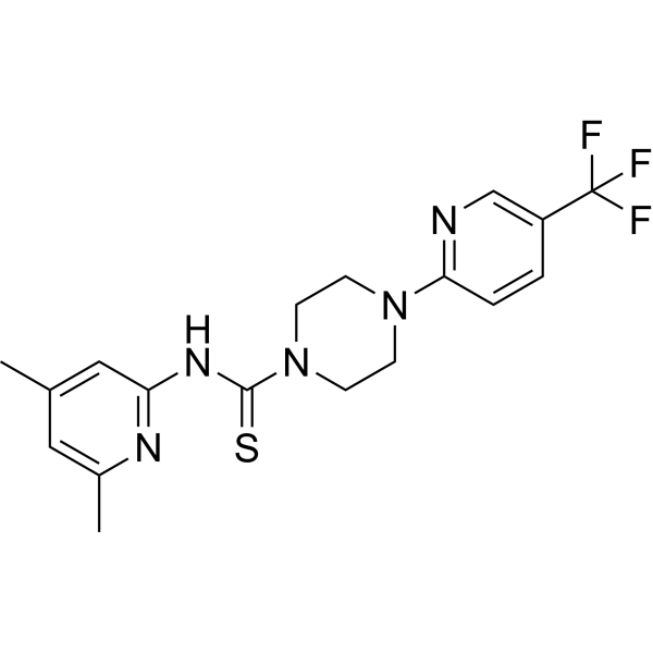
HY-117240
NCT-502PHGDH Inhibitor -
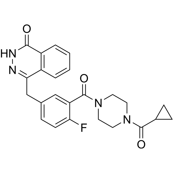
HY-10162
OlaparibPARP Inhibitor -
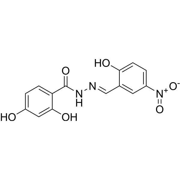
HY-123269
PKUMDL-WQ-2101PHGDH inhibitor -

HY-13259
MG-132Proteasome Inhibitor -
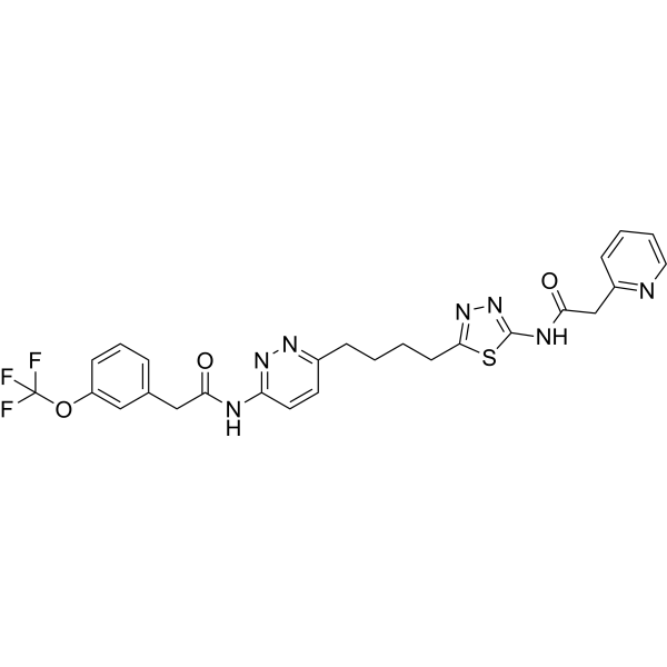
HY-12248
TelaglenastatGLS1 Inhibitor -
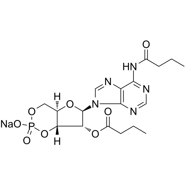
HY-B0764
Bucladesine sodiumcAMP Analog -
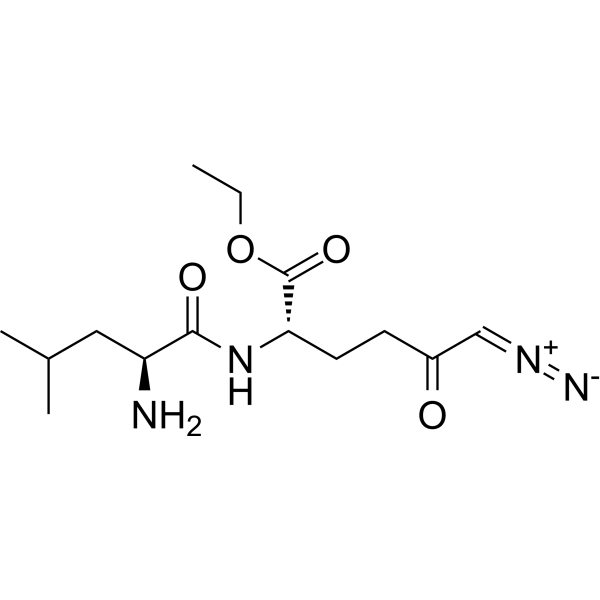
HY-122218
JHU-083Glutaminase Antagonist -

HY-101966R
NCT-503 (Standard)Reference Standard -
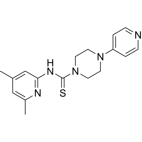
HY-121733
PHGDH-inactiveNegative Control
Publications Citing Use of MedChemExpress (MCE) BI-4916
More- Nat Metab. 2024 Aug;6(8):1529-1548. [Abstract]
- Nat Commun. 2022 May 16;13(1):2699. [Abstract]
- Mol Metab. 2025 Dec:102:102275. [Abstract]
- Am J Physiol Cell Physiol. 2025 Nov 1;329(5):C1560-C1576. [Abstract]
- iScience. 2024 Feb 8;27(3):109173. [Abstract]
- University of Maryland. 2024.
- bioRxiv. 2023 Jan 23.


Biological Activity
Description
Cellular Effect
|
Cell Line

|
Type | Value | Description | References |
|---|---|---|---|---|
| MDA-MB-468 | IC50 |
29 nM
Compound: BI-4916
|
Inhibition of PHGDH in human MDA-MB-468 cells assessed as reduction in [13C]-serine incubated for 1 hr using [13C]glucose as substrate by LC-MS/MS analysis
Inhibition of PHGDH in human MDA-MB-468 cells assessed as reduction in [13C]-serine incubated for 1 hr using [13C]glucose as substrate by LC-MS/MS analysis
|
[PMID: 31365252] |
| MDA-MB-468 | IC50 |
2 μM
Compound: BI-4916
|
Inhibition of PHGDH in human MDA-MB-468 cells assessed as reduction in [13C]-serine incubated for 72 hrs using [13C]glucose as substrate by LC-MS/MS analysis
Inhibition of PHGDH in human MDA-MB-468 cells assessed as reduction in [13C]-serine incubated for 72 hrs using [13C]glucose as substrate by LC-MS/MS analysis
|
[PMID: 31365252] |
In Vitro
BI-4916 (15 μM, 24 h) reduces breast cancer cell migration[2].
BI-4916 (15 μM, 24 h) disrupts cGAS-STING signaling, impairing the epithelial response against viral and bacterial infection and fueling experimental enteritis[3].
MedChemExpress (MCE) has not independently confirmed the accuracy of these methods. They are for reference only.
-
Cell Line:MDA-MB-468 cells
-
Concentration:15 μM
-
Incubation Time:24 h
-
Result:Had no adverse effect on tricarboxylic acid (TCA) cycle activity and proliferation rate.
Reduced Methotrexate (MTX) (HY-14519) mediated cell migration.
Was effective to reduce the migratory capacity of MTX resistant, pro-migratory MDA-MB-468 cells.
-
Cell Line:wild type Mode-K cells (SV40 large T-antigen-immortalised murine small intestinal epithelial cell line)
-
Concentration:15 μM
-
Incubation Time:24 h
-
Result:Resulted in the decreased phosphorylation of STING and its downstream target TBK1 dual perturbed with serine/glycine (S/G) starvation.
- Scratch/Wound-Healing Migration Assay
- Research Protocol for Infectious Diseases
- Cell migration
- Chemotaxis Gradient Chamber Assay 1
- Under-Agarose Cell Migration Assay
- Neural Crest/Neuronal Cell Migration Explant Assay
- Transwell/Boyden Chamber Migration Assay
- 3D Collagen/Hydrogel Matrix Migration Assay
- Research Protocol for Inflammation-related Diseases
- Cell-Exclusion Zone Migration Assay
- Chemotaxis Gradient Chamber Assay 2


Chemical Information
-
CAS No. 2244451-48-5
-
Appearance Solid
-
Molecular Weight 527.42
-
Formula C23H24Cl2N2O6S
-
Color White to yellow
-
SMILES
O=C(OCC)CS(=O)(C1=CC=C([C@H](NC(C(N2C)=CC3=C2C=C(C)C(Cl)=C3Cl)=O)CO)C=C1)=O
-
Shipping
Room temperature in continental US; may vary elsewhere.
-
Storage
Powder -20°C 3 years 4°C 2 years In solvent -80°C 2 years -20°C 1 year


Publications (7)
-
Journal Impact Factor
-
Most Recent
-
Nat Metab
The unique catalytic properties of PSAT1 mediate metabolic adaptation to glutamine blockade. [Abstract]2024 Aug;6(8):1529-1548. PMID: 39192144 -
Nat Commun
Mitochondria preserve an autarkic one-carbon cycle to confer growth-independent cancer cell migration and metastasis. [Abstract]2022 May 16;13(1):2699. PMID: 35577770 -
Mol Metab
Canagliflozin synergises with serine restriction mediating anti-leukaemic effects in T-cell acute lymphoblastic leukaemia. [Abstract]2025 Dec:102:102275. PMID: 41120088 -
Am J Physiol Cell Physiol
Glycolytic metabolism and biomass production from glucose in human skeletal muscle growth. [Abstract]2025 Nov 1;329(5):C1560-C1576. PMID: 41071646 -
iScience
Serine metabolism is crucial for cGAS-STING signaling and viral defense control in the gut. [Abstract]2024 Feb 8;27(3):109173. PMID: 38496294 -
-


Solvent & Solubility
In Vitro:
DMSO : 250 mg/mL (474.01 mM; Need ultrasonic; Hygroscopic DMSO has a significant impact on the solubility of product, please use newly opened DMSO)
Please refer to the solubility information to select the appropriate solvent. Once prepared, please aliquot and store the solution to prevent product inactivation from repeated freeze-thaw cycles.
Storage method and period of stock solution: -80°C, 2 years; -20°C, 1 year. When stored at -80°C, please use it within 2 years. When stored at -20°C, please use it within 1 year.
Please refer to the solubility information to select the appropriate solvent. Once prepared, please aliquot and store the solution to prevent product inactivation from repeated freeze-thaw cycles.
Storage method and period of stock solution: -80°C, 2 years; -20°C, 1 year. When stored at -80°C, please use it within 2 years. When stored at -20°C, please use it within 1 year.
Concentration (start) × Volume (start) = Concentration (final) × Volume (final)
In Vivo:
Select the appropriate dissolution method based on your experimental animal and administration route.
- For the following dissolution methods, please ensure to first prepare a clear stock solution using an In Vitro approach and then sequentially add co-solvents:
- To ensure reliable experimental results, the clarified stock solution can be appropriately stored based on storage conditions. As for the working solution for In Vivo experiments, it is recommended to prepare freshly and use it on the same day.
- The percentages shown for the solvents indicate their volumetric ratio in the final prepared solution. If precipitation or phase separation occurs during preparation, heat and/or sonication can be used to aid dissolution.
In Vivo Dissolution Calculator
Please enter the basic information of animal experiments:
-
-
-
-
Recommended: Prepare an additional quantity of animals to account for potential losses during experiments.
Please enter your animal formula composition:
-
%DMSO +
Recommended: Keep the proportion of DMSO in working solution below 2% if your animal is weak.
-
%+
-
+%Tween-80 + +
-
%Saline +
The co-solvents required include: DMSO, . All of co-solvents are available by MedChemExpress (MCE). , Tween 80. All of co-solvents are available by MedChemExpress (MCE).
Working solution concentration: 0.22 mg/mL
Method for preparing stock solution: mg drug dissolved in μL DMSO. Stock solution concentration: mg/mL.
1. Take μL DMSO stock solution;
2. Add μL .
μL , mix evenly;
3. Then add μL Tween 80, mix evenly;
4. Then add μL
Please ensure that the stock solution in the first step is dissolved to a clear state, and add co-solvents in sequence. You can use ultrasonic heating (ultrasonic cleaner, recommended frequency 20-40 kHz), vortexing, etc. to assist dissolution.


Purity & Documentation
-
Data Sheet (275 KB)
-
SDS (251 KB)
- English - EN (251 KB)
- Français - FR (251 KB)
- Deutsch - DE (251 KB)
- Norwegian - NO (251 KB)
- Español - ES (251 KB)
- Swedish - SV (251 KB)
- Italian - IT (251 KB)
- Korean - KR (251 KB)
- Portuguese - PT (251 KB)
-
Handling Instructions (2659 KB)
References
[1]. Weinstabl H, et al. Intracellular Trapping of the Selective Phosphoglycerate Dehydrogenase (PHGDH) Inhibitor BI-4924 Disrupts Serine Biosynthesis. J Med Chem. 2019 Jul 31. [Content Brief]
[2]. Kiweler N, et al. Mitochondria preserve an autarkic one-carbon cycle to confer growth-independent cancer cell migration and metastasis. Nat Commun. 2022 May 16;13(1):2699. [Content Brief]
[3]. Becker B, et al. Serine metabolism is crucial for cGAS-STING signaling and viral defense control in the gut. iScience. 2024 Feb 8;27(3):109173. [Content Brief]


Complete Stock Solution Preparation Table
Please refer to the solubility information to select the appropriate solvent. Once prepared, please aliquot and store the solution to prevent product inactivation from repeated freeze-thaw cycles.
Storage method and period of stock solution: -80°C, 2 years; -20°C, 1 year. When stored at -80°C, please use it within 2 years. When stored at -20°C, please use it within 1 year.
| Optional Solvent | Concentration Solvent Mass | 1 mg | 5 mg | 10 mg | 25 mg |
|---|---|---|---|---|---|
| DMSO | 1 mM | 1.8960 mL | 9.4801 mL | 18.9602 mL | 47.4006 mL |
| 5 mM | 0.3792 mL | 1.8960 mL | 3.7920 mL | 9.4801 mL | |
| 10 mM | 0.1896 mL | 0.9480 mL | 1.8960 mL | 4.7401 mL | |
| 15 mM | 0.1264 mL | 0.6320 mL | 1.2640 mL | 3.1600 mL | |
| 20 mM | 0.0948 mL | 0.4740 mL | 0.9480 mL | 2.3700 mL | |
| 25 mM | 0.0758 mL | 0.3792 mL | 0.7584 mL | 1.8960 mL | |
| 30 mM | 0.0632 mL | 0.3160 mL | 0.6320 mL | 1.5800 mL | |
| 40 mM | 0.0474 mL | 0.2370 mL | 0.4740 mL | 1.1850 mL | |
| 50 mM | 0.0379 mL | 0.1896 mL | 0.3792 mL | 0.9480 mL | |
| 60 mM | 0.0316 mL | 0.1580 mL | 0.3160 mL | 0.7900 mL | |
| 80 mM | 0.0237 mL | 0.1185 mL | 0.2370 mL | 0.5925 mL | |
| 100 mM | 0.0190 mL | 0.0948 mL | 0.1896 mL | 0.4740 mL |


BI-4916 Related Classifications
HY-126253 Related Classifications
 Powered by Bioz