



Other Analysis Dyes
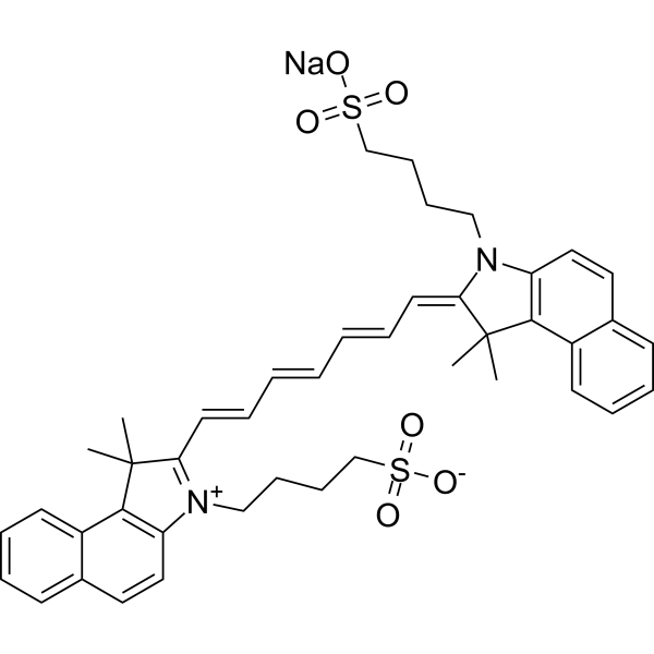
- Formel: C43H47N2NaO6S2
- Molecular Weight: 774.96
Indocyanine green (Foxgreen) is a low toxicic fluorescent agent that has been widely used in medical diagnostics, such as determining cardiac output, hepatic function, and liver blood flow, and for ophthalmic angiography (Ex/Em = 785/813 nm).
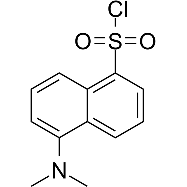
- Formel: C12H12ClNO2S
- Molecular Weight: 269.75
Dansyl chloride is a reagent that produces stable blue or blue-green fluorescent sulfonamide adducts in the reaction of aliphatic and aromatic amines with primary amino groups, and is widely used for modified amino acids, protein sequencing and amino acid analysis.
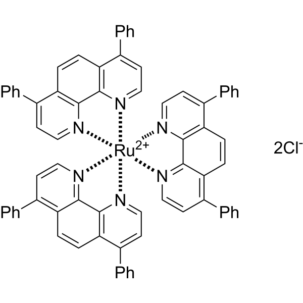
- Formel: C72H48Cl2N6Ru
- Molecular Weight: 1169.17
Tris(4,7-diphenyl-1,10-phenanthroline)ruthenium(II) dichloride ([Ru(dpp)3]2+) is an electrochemiluminescent (ECL) probe and oxygen-sensitive sensor. Tris(4,7-diphenyl-1,10-phenanthroline)ruthenium(II) dichloride can be used to modify electrode surfaces for the detection of sulfates (S2O82-) and oxalates, based on electrochemical reactions that generate excited-state species, releasing photons through irreversible redox reactions. Tris(4,7-diphenyl-1,10-phenanthroline)ruthenium(II) dichloride utilizes the oxygen quenching of fluorescence mechanism, where fluorescence intensity reflects the metabolic rate of living microorganisms or oxygen levels within cells/tumors, allowing for real-time detection. Tris(4,7-diphenyl-1,10-phenanthroline)ruthenium(II) dichloride's main applications include microbial detection, antibacterial activity studies, and tumor microenvironment research.
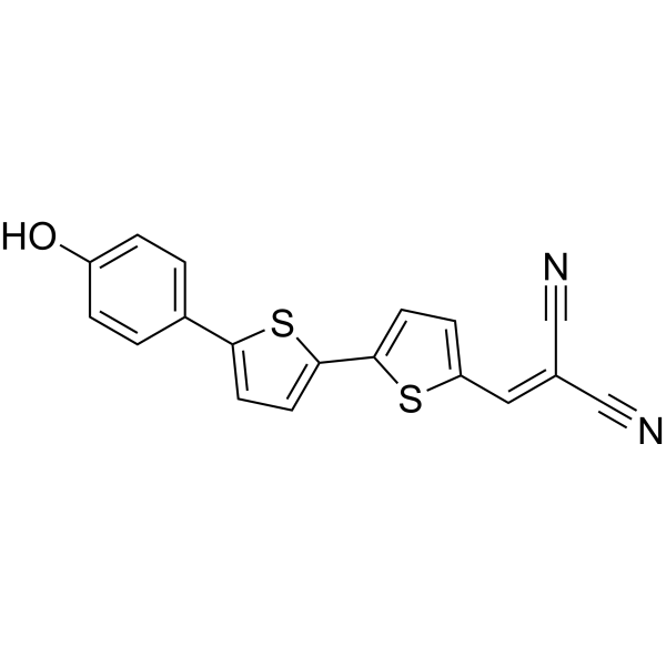
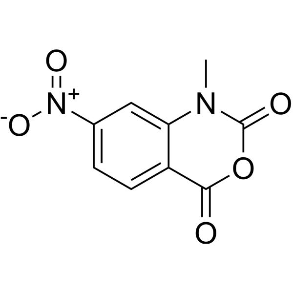
- Formel: C9H6N2O5
- Molecular Weight: 222.15
1-Methyl-7-nitroisatoic anhydride (1M7) is a reagent that detects local nucleotide flexibility, for probing 2'-hydroxyl reactivity, can be used for RNA structure analysis.

- Formel: C20H17N7O4S
- Molecular Weight: 451.46
LumiPK is an environment-sensitive probe that specifically targets the liver isoform of pyruvate kinase (PKL). LumiPK exhibits high affinity for PKL with a Kd of 37 nM. LumiPK integrates the 4-sulfonamido-7-aminobenzoxadiazole (SBD) fluorophore into a phthalazine scaffold, and its fluorescence intensity changes with the polarity of the chemical environment, thereby directly reflecting protein occupancy. LumiPK binds only to the tetrameric form of PKL, with the binding site being an allosteric pocket formed at the subunit interface, and key water-mediated interactions enhance its potency; in polar environments such as buffer, its fluorescence is significantly quenched, whereas upon binding to the nonpolar allosteric pocket of PKL, its fluorescence is enhanced. In fluorescence detection, its excitation/emission wavelengths are Ex/Em = 450/550 nm.

- Formel: C45H35NNa2O10S2
- Molecular Weight: 859.87
di-SFA is a fluorescent probe used to measure transmembrane potential (Vm) across the plasma membrane. di-SFA contains a 3-hydroxychromone fluorophore that undergoes excited-state intramolecular proton transfer (ESIPT), producing dual emission from the normal (N*) and tautomeric (T*) states. di-SFA inserts deeply into the hydrophobic region of lipid bilayers. The sulfonate groups give di-SFA a double negative charge in aqueous media. Ratiometric measurements are performed in lipid vesicles using Ex/Em = 450/527 nm and Ex/Em = 450/605 nm.

- Formel: C86H98ClF6N13O15S2
- Molecular Weight: 1767.35
T-sCPY-s500R-H is a NADPH-based FRET ratiometric biosensor probe. T-sCPY-s500R-H forms a 1:1 FRET pair upon binding to the eDHFR-HaloTag fusion protein through conjugation of the donor s500R (green fluorescence) and the acceptor sCPY (red fluorescence) with the HaloTag ligand and the eDHFR ligand (TMP), respectively; NADPH binding to eDHFR induces a conformational change that converts sCPY from a colorless spirolactam form to a red fluorescent form, enabling quantification of NADPH levels through the ratio of red/green fluorescence intensity (Ex = 488 or 490 nm; green channel Em = 500-580 nm; red channel Em = 600-700 nm). T-sCPY-s500R-H is applicable to research in fields such as cell metabolism studies, oxidative stress and NADPH homeostasis analysis, tumor metabolism, and NADPH-modulating drug screening.
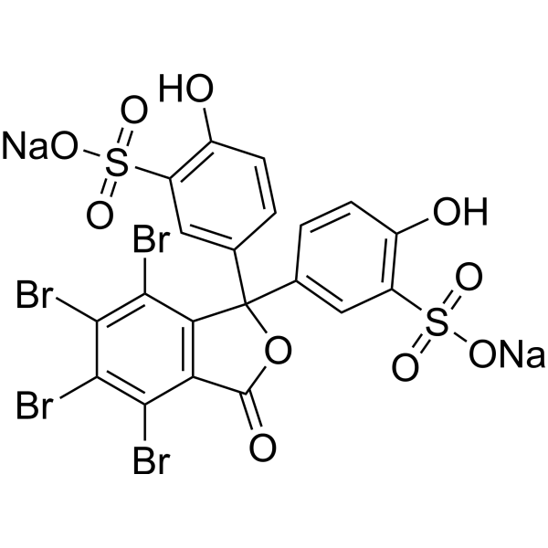
- Formel: C20H8Br4Na2O10S2
- Molecular Weight: 838.00
Sulfobromophthalein (Bromosulfophthalein) disodium salt is an organic anion dye used in the study of a variety of membrane carriers expressed in animal tissues and involved in transport of agents and metabolites.
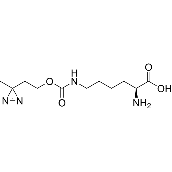
- Formel: C11H20N4O4
- Molecular Weight: 272.30
DiAzKs (H-L-Photo-lysine) is a diazirine-containing lysine amino acid and is a photo-cross-linker. DiAzKs can site-selective incorporated into proteins and is used to crosslink protein-protein interactions in vitro and in living cells. DiAzKs acts as a UV light-activated photo-crosslinking probe.
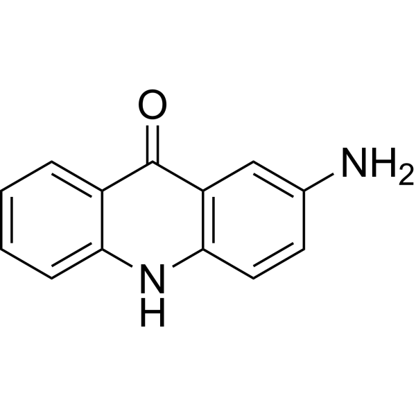
- Formel: C13H10N2O
- Molecular Weight: 210.24
2-Aminoacridone is a widely used fluorophore (λexc=428 nm, λem=525 nm).
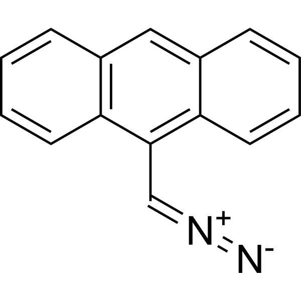
- Formel: C15H10N2
- Molecular Weight: 218.25
9-Anthryldiazomethane is a fluorescent labeling reagent, which can be used for detecting fatty acids and derivatives.


- Formel: C85H77N2NaO6S
- Molecular Weight: 1277.59
TPE-SQ5 is a ratiometric Fluorescent probe for hypochlorite (ClO-) detection with a limit of detection of 5.6 nM. TPE-SQ5 exhibits high selectivity and high sensitivity towards ClO-. TPE-SQ5 can be oxidized by ClO- to form an epoxide, which quenches its deep red fluorescence; subsequently, the epoxide decomposes into a hemicyanine and a sulfonic acid-substituted oxindole, producing blue-green fluorescence to achieve a ratiometric response. TPE-SQ5 can form nanoparticles with Pluronic F-127 via the nanoprecipitation method, and these nanoparticles possess cell permeability. TPE-SQ5 has unique Ex/Em spectra in different solvents and nanoparticle forms, with emission peaks at 643 nm (380 nm excitation, 95% H2O/THF), 631 nm (564 nm excitation, DMSO), 652 nm (536 nm excitation, THF) and 630 nm (524 nm excitation, nanoparticles); the Ex/Em settings used for cell imaging are 488/550-650 nm (red channel) and 405/450-550 nm (green channel).

- Formel: C48H47BrN2O6
- Molecular Weight: 827.80
TPh-PyBz-β-gal is a fluorescent probe used for the detection and imaging of β-galactosidase (β-gal) in living cells. The detection mechanism of TPh-PyBz-β-gal relies on β-gal, which cleaves the glycosidic bond linking the AIE-active fluorophore and the D-galactose residue unit of the probe. The emission wavelength of TPh-PyBz-β-gal after activation is 606 nm. TPh-PyBz-β-gal has low cytotoxicity and can be applied to β-gal imaging in living cells.

- Formel: C26H20ClN7O3
- Molecular Weight: 513.94
CGTDP is a fluorescent probe used for selective labeling of active-site arginine residues, detection of local polarity, and monitoring of conformational changes in enzyme active sites. CGTDP contains a neutral red moiety serving as a polarity-sensitive fluorophore, and a phenylglyoxal unit acting as an arginine-specific labeling group; it undergoes a selective reaction via its α-dicarbonyl moiety with the guanidyl group of arginine residues in enzyme active sites to form a covalent linkage, thereby enabling the reporting of the local microenvironment at the labeled site. The excitation/emission wavelengths are Ex/Em = 398/607 nm and Ex/Em = 507/607 nm; these wavelengths blue-shift in organic solvents, such as Ex/Em = 375/569 nm and Ex/Em = 469/569 nm in THF, and Ex/Em = 375/587 nm and Ex/Em = 470/587 nm in acetonitrile. When labeled onto creatine kinase, its excitation/emission wavelengths are Ex/Em = 493/590 nm.

- Formel: C34H49Br2F2N3
- Molecular Weight: 697.58
Di-4-AN (F) EPPTEA is a voltage-sensitive dye used for membrane potential imaging and optical recording of electrical activity in excitable cells and tissues. Its detection mechanism is based on electrochromism (also known as the molecular Stark effect). The single-photon excitation/emission wavelengths measured in ethanol solution are Ex/Em = 444/610 nm, and it can be excited via 1064 nm two-photon excitation in optical recording applications. Di-4-AN (F) EPPTEA can be used to record backpropagating action potentials in individual dendritic spines of brain cortical slices, map subcellular action potential characteristics of single cardiomyocytes, and perform real-time recording of multicellular action potentials in cerebellar slices.

- Formel: C28H25NO3
- Molecular Weight: 423.50
P6-Aggrate is a fluorescent probe for aggregated proteome detection. P6-Aggrate specifically recognizes amorphous aggregated proteomes through non-covalent reversible binding, and its fluorescence enhances after heat-induced protein aggregation. P6-Aggrate reflects the polarity and compactness heterogeneity within aggregated proteomes via emission wavelength shift: short-wavelength emission (blue shift) corresponds to large aggregates with high compactness, while long-wavelength emission (red shift) corresponds to small spots with low compactness (Ex/Em = 488/520-580 nm). P6-Aggrate enables reversible monitoring of the dynamic processes of formation and clearance of stress-induced proteome aggregation such as that induced by MG132 (HY-13259) in living cells. P6-Aggrate can be used in studies related to protein homeostasis imbalance, neurodegenerative diseases and protein aggregation.

- Formel: C48H43NO3S
- Molecular Weight: 713.92
TPE-Cy is a fluorescent probe with both aggregation-induced emission (AIE) property and pH-dependent luminescent behavior. TPE-Cy exhibits significant luminescent differences under different acidic and alkaline conditions: it shows moderate to strong red emission with linear variation with pH in acidic environments and within the pH range of 5-7, weak or even no emission within the pH range of 7-10, and switches to blue emission under alkaline conditions. TPE-Cy possesses excellent cell permeability and biocompatibility, enabling accurate ratiometric pH sensing and imaging monitoring in living cells via confocal microscopy, ratiometric analysis and flow cytometry.

- Formel: C17H18N2O
- Molecular Weight: 266.34
ALDH2 probe 5 is a ALDH2 isoform-selective fluorescent probe with an emission wavelength centered at 494 nm. ALDH2 probe 5 is a turn-on fluorescent substrate for ALDH2; in its unreacted aldehyde form, twisted intramolecular charge transfer (TICT) occurs, resulting in fluorescence quenching. After ALDH2 probe 5 is catalytically oxidized to carboxylate by ALDH2, TICT is inhibited and fluorescence is significantly enhanced. ALDH2 probe 5 enables highly sensitive quantitative imaging of ALDH2 activity in recombinant enzymes, live cells, blood, tissue homogenates, and the brains of living mice. ALDH2 probe 5 can be used for studies related to neuroinflammation.



