



FAK PROTAC B5
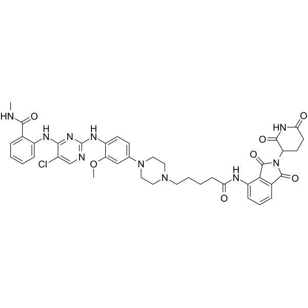
FAK PROTAC B5 is a FAK PROTAC degrader with an IC50 of 14.9 nM. FAK PROTAC B5 induces FAK degradation via the ubiquitin-proteasome pathway by simultaneously binding to FAK and the CRBN E3 ligase. FAK PROTAC B5 inhibits the proliferation, migration and invasion of cancer cells. FAK PROTAC B5 can be used for the research of non-small cell lung cancer.
(Pink: EGFR Target protein ligand; Blue: Cereblon ligand (HY-41547); Black: linker).
For research use only. We do not sell to patients.
- CAS No.: 2471525-44-5
- Formula: C41H43ClN10O7
- Molecular Weight:823.30
-
Storage:
Please store the product under the recommended conditions in the Certificate of Analysis.

To place orders, for customer services and technical support, please contact: MedChemExpress USA
Tel: 609-228-6898 E-mail: [email protected] [email protected]


-
 Biological Activity
-
Chemical Information
- Protocols
- Purity & Documentation
- References
-
 Help & FAQs


All PROTACs Isoforms
More

Biological Activity
Description
IC50 & Target
IC50: 14.9 nM (FAK)[1]
Cellular Effect
|
Cell Line

|
Type | Value | Description | References |
|---|---|---|---|---|
| A549 | IC50 |
0.14 μM
Compound: B5
|
Antiproliferative activity against human A549 cells assessed as inhibition of cell growth incubated for 72 hrs by MTT assay
Antiproliferative activity against human A549 cells assessed as inhibition of cell growth incubated for 72 hrs by MTT assay
|
[PMID: 35486993] |
In Vitro
FAK PROTAC B5 (compound B5) potently inhibits the activity of purified FAK kinase with an IC50 of 14.9 nM; it also potently inhibits the proliferation of A549 cells, with an IC50 of 0.14 μM after 72 h of treatment[1].
FAK PROTAC B5 (2.00 μM; 2 h) exhibits moderate membrane permeability in Caco-2 cell monolayers, with a Papp value of 1.4 × 10⁻⁶ cm/s in the A-to-B direction[1].
FAK PROTAC B5 (0.01-1 μM; 12 h) degrades 86.4% of FAK protein in A549 cells; this degradation process requires binding to FAK, interaction with the CRBN E3 ligase, and proceeds via the proteasomal pathway[1].
FAK PROTAC B5 (0.20-1.80 μM; 72 h) inhibits migration and invasion of A549 cells in a concentration-dependent manner[1].
MedChemExpress (MCE) has not independently confirmed the accuracy of these methods. They are for reference only.
-
Cell Line:A549 non-small cell lung cancer cells
-
Concentration:0.01, 0.1, 1 μM
-
Incubation Time:12 h
-
Result:Induced 86.4% degradation of FAK protein after 12 h of treatment.
-
Cell Line:A549 non-small cell lung cancer cells
-
Concentration:10 nM (FAK PROTAC B5)
5 μM TAE-226, 5 μM Pomalidomide, 5 μM MG-132 (pretreatment) -
Incubation Time:12 h (FAK PROTAC B5 treatment); 2 h (pretreatment)
-
Result:Almost completely prevented FAK degradation by FAK PROTAC B5 when cells were pretreated with TAE-226 (HY-13203), Pomalidomide (HY-10984), or MG-132 (HY-13259).
-
Cell Line:A549 non-small cell lung cancer cells
-
Concentration:0.20 μM, 0.60 μM, 1.80 μM
-
Incubation Time:72 h
-
Result:Inhibited A549 cell migration in a concentration-dependent manner.
Reduced 72 h wound recovery to 42.2% at 0.20 μM.
Reduced 72 h wound recovery to 33.3% at 0.60 μM.
Reduced 72 h wound recovery to 21.7% at 1.80 μM.
-
Cell Line:A549 non-small cell lung cancer cells
-
Concentration:0.20 μM, 0.60 μM, 1.80 μM
-
Incubation Time:72 h
-
Result:Inhibited A549 cell invasion in a concentration-dependent manner.
Reduced the number of invasive cells.
Exhibited activity superior to TAE-226 (HY-13203).


Chemical Information
-
CAS No. 2471525-44-5
-
Molecular Weight 823.30
-
Formula C41H43ClN10O7
-
SMILES
O=C(NC1=CC=CC(C(N2C(CC3)C(NC3=O)=O)=O)=C1C2=O)CCCCN4CCN(C5=CC=C(NC6=NC=C(Cl)C(NC7=CC=CC=C7C(NC)=O)=N6)C(OC)=C5)CC4
-
Shipping
Room temperature in continental US; may vary elsewhere.
-
Storage
Please store the product under the recommended conditions in the Certificate of Analysis.


Purity & Documentation
References

Calculators
Concentration (start) × Volume (start) = Concentration (final) × Volume (final)


FAK PROTAC B5 Related Classifications
HY-143458 Related Classifications
 Powered by Bioz