



ACT-660602

Based on 1 Customer Validation
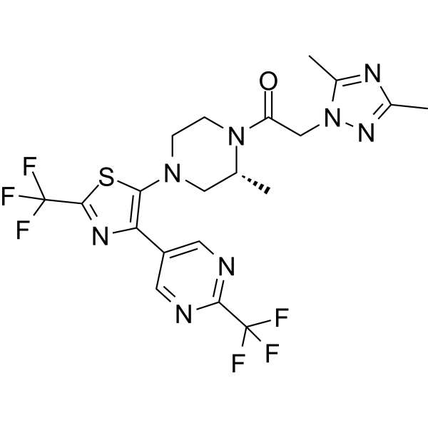
ACT-660602 is an orally active antagonist of chemokine receptor (CXCR3) with an IC50 value of 204 nM. ACT-660602 inhibits T-cell migration and shows efficacy in acute lung ingury model. ACT-660602 can be used for autoimmune diseases research.
For research use only. We do not sell to patients.
- Purity : 99.02%
- CAS No.: 1646267-59-5
- Formula: C20H20F6N8OS
- Molecular Weight:534.48
-
Storage:
4°C, sealed storage, away from moisture and light
* In solvent : -80°C, 6 months; -20°C, 1 month (sealed storage, away from moisture and light)

To place orders, for customer services and technical support, please contact: MedChemExpress USA
Tel: 609-228-6898 E-mail: [email protected] [email protected]


-
 Biological Activity
-
Chemical Information
-
Solvent & Solubility
- Protocols
- Purity & Documentation
- References
-
 Help & FAQs


-
HY-149055
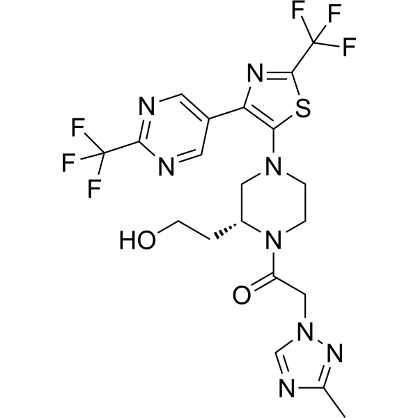
ACT-777991CXCR3 Antagonist -
HY-16711
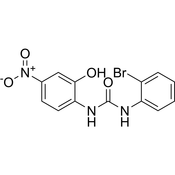
SB225002CXCR2 Antagonist -
HY-15319
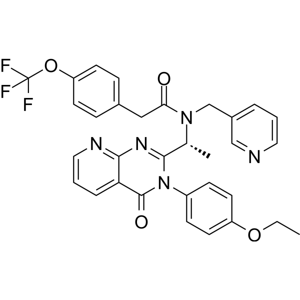
AMG 487CXCR3 Antagonist -
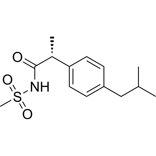
HY-15251
ReparixinCXCR1/2 Inhibitor -
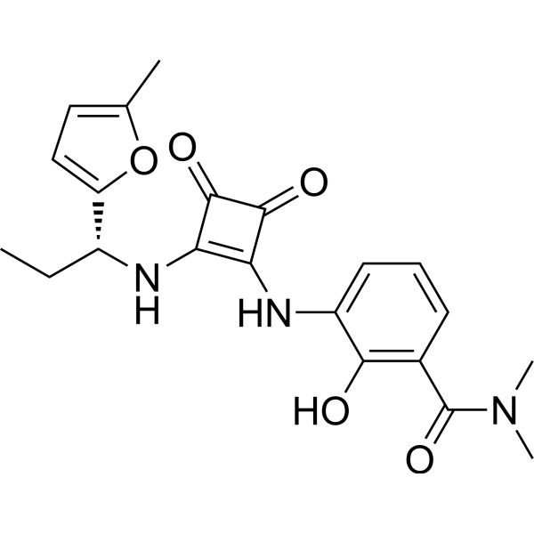
HY-10198
NavarixinCXCR1/2 Antagonist -
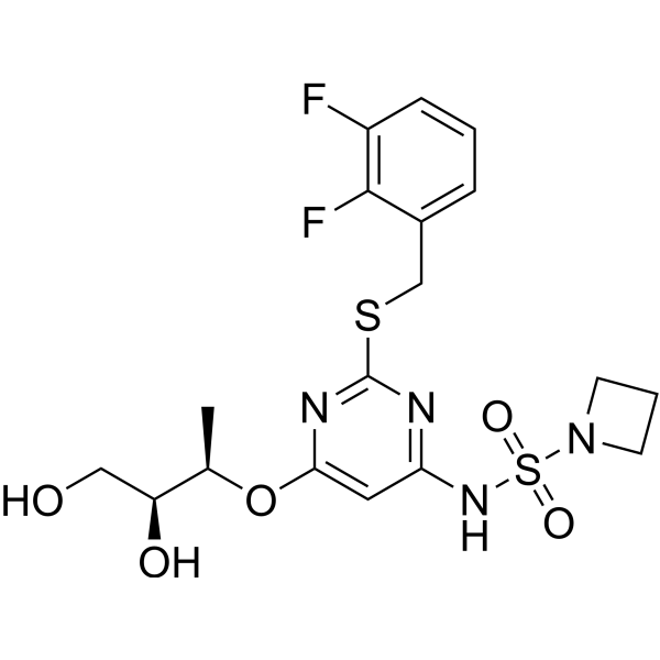
HY-19855
AZD-5069CXCR2 Antagonist -
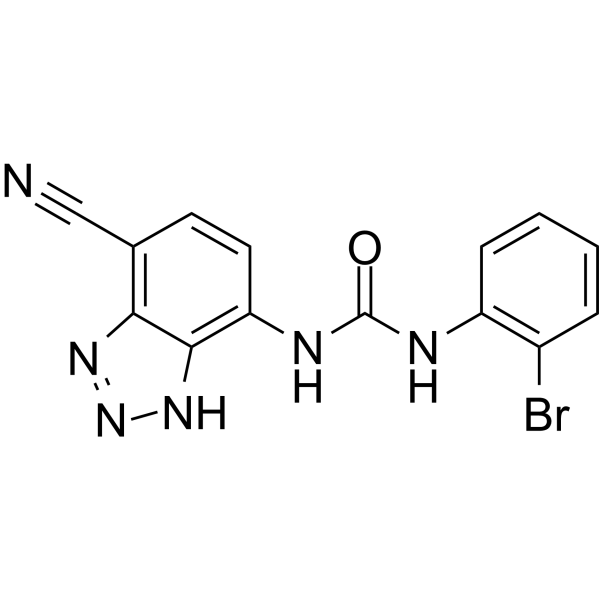
HY-50688
SB-265610CXCR2 Antagonist -
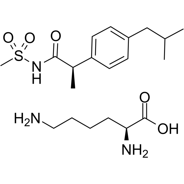
HY-15252
Reparixin L-lysine saltCXCR Inhibitor -
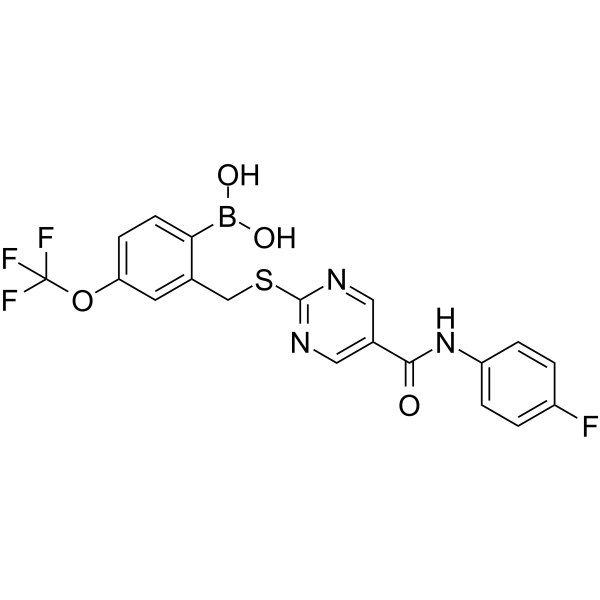
HY-119339
SX-682CXCR1/2 Inhibitor -
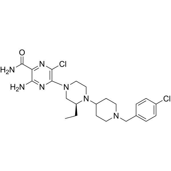
HY-10017
SCH 546738CXCR3 Antagonist -
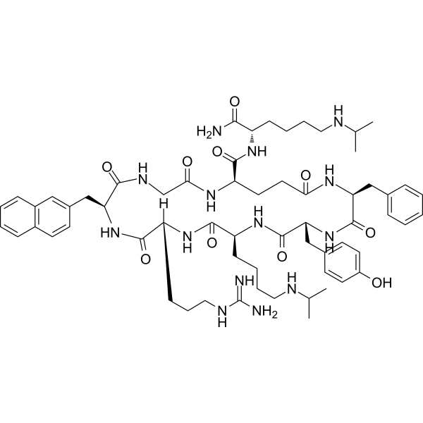
HY-12488
LY2510924CXCR Inhibitor -
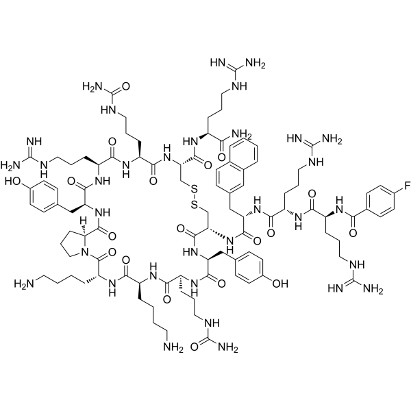
HY-P0171
MotixafortideCXCR4 Antagonist


Biological Activity
Description
IC50 & Target
|
CXCR3 204 nM (IC50) |
Cellular Effect
|
Cell Line

|
Type | Value | Description | References |
|---|---|---|---|---|
| T-cell | IC50 |
37 nM
Compound: 9j; ACT-660602
|
Inhibition of cell migration in human T cell in presence of CXCL11 as agonist by imaging method
Inhibition of cell migration in human T cell in presence of CXCL11 as agonist by imaging method
|
[PMID: 35947786] |
In Vitro
ACT-660602 shows selectivity to CXCR3 over hERG, with IC50s of 18 μM (hERG)[1].
ACT-660602 (112 nM; 6 h) inhibits cell migration and improves the metabolic stability[1].
ACT-660602 (5, 20, 100 or 500 nM) displays an non-competitive binding mode to CXCL10 and CXCL11 in different concentration, with more stable IC50s[1].
MedChemExpress (MCE) has not independently confirmed the accuracy of these methods. They are for reference only.
-
Cell Line:CD3/CD28-activated primary human T cells
-
Concentration:112 nM
-
Incubation Time:45 min
-
Result:Inhibited cell migration.
In Vivo
ACT-660602 (30 mg/kg; p.o.; once daily) displays anti-inflammatory activity and exerts efficacy in the mouse model of acute lung ingury[1].
Range for Pharmacokinetics of ACT-660602[1]
| Animal | Route | Dose (range) (mg/kg) | Cmax (range) (ng/mL) | Tmax (range) (h) | AUC (range) (ng•h/mL) | F (%) | CL (range) (mL/min/kg) | Vss (range) (L/kg) | T1/2 (range) (h) |
| Dog | p.o. | 2 | 1380 | 1 | 20000 | 8 | 1.3 | 1.7 | 14.5 |
| i.v. | 0.5 | 1300-1450 | 0.5-2.0 | 10400-32000 | / | 0.6-3.0 | 1.6-1.7 | 6.3- | |
| Rat | p.o. | 2 | 1520 | 0.5 | 14000 | 80 | 1.9 | 1.1 | 7.1 |
| i.v. | 0.5 | 1250-1860 | 0.5-1.0 | 11600-15641 | / | 1.9-1.9 | 0.9-1.3 | 5.7-8.8 |
MedChemExpress (MCE) has not independently confirmed the accuracy of these methods. They are for reference only.
-
Animal Model:LPS-induced lung inflammation model (72 h post LPS challenge)[1]
-
Dosage:30 mg/kg
-
Administration:Oral gavage; once daily
-
Result:Significantly reduced recruitment of the CXCR3+ CD8+ T cell in the bronchoalveolar lavage compartment.
- Scratch/Wound-Healing Migration Assay
- Cell migration
- Zigmond Chamber Chemotaxis Assay
- Under-Agarose Cell Migration Assay
- Neural Crest/Neuronal Cell Migration Explant Assay
- Transwell/Boyden Chamber Migration Assay
- 3D Collagen/Hydrogel Matrix Migration Assay
- Cell-Exclusion Zone Migration Assay
- Dunn/Insall Chamber Chemotaxis Assay


Chemical Information
-
CAS No. 1646267-59-5
-
Appearance Solid
-
Molecular Weight 534.48
-
Formula C20H20F6N8OS
-
Color White to off-white
-
SMILES
FC(C1=NC=C(C2=C(N3C[C@@H](C)N(C(CN4N=C(C)N=C4C)=O)CC3)SC(C(F)(F)F)=N2)C=N1)(F)F
-
Shipping
Room temperature in continental US; may vary elsewhere.
-
Storage
4°C, sealed storage, away from moisture and light
* In solvent : -80°C, 6 months; -20°C, 1 month (sealed storage, away from moisture and light)


Solvent & Solubility
In Vitro:
DMSO : 110 mg/mL (205.81 mM; Need ultrasonic; Hygroscopic DMSO has a significant impact on the solubility of product, please use newly opened DMSO)
Please refer to the solubility information to select the appropriate solvent. Once prepared, please aliquot and store the solution to prevent product inactivation from repeated freeze-thaw cycles.
Storage method and period of stock solution: -80°C, 6 months; -20°C, 1 month (sealed storage, away from moisture and light). When stored at -80°C, please use it within 6 months. When stored at -20°C, please use it within 1 month.
Please refer to the solubility information to select the appropriate solvent. Once prepared, please aliquot and store the solution to prevent product inactivation from repeated freeze-thaw cycles.
Storage method and period of stock solution: -80°C, 6 months; -20°C, 1 month (sealed storage, away from moisture and light). When stored at -80°C, please use it within 6 months. When stored at -20°C, please use it within 1 month.
Concentration (start) × Volume (start) = Concentration (final) × Volume (final)


Purity & Documentation
-
Data Sheet (277 KB)
-
SDS (252 KB)
- English - EN (252 KB)
- Français - FR (252 KB)
- Deutsch - DE (252 KB)
- Norwegian - NO (252 KB)
- Español - ES (252 KB)
- Swedish - SV (252 KB)
- Italian - IT (252 KB)
- Korean - KR (252 KB)
- Portuguese - PT (252 KB)
-
Handling Instructions (2659 KB)
References


Complete Stock Solution Preparation Table
Please refer to the solubility information to select the appropriate solvent. Once prepared, please aliquot and store the solution to prevent product inactivation from repeated freeze-thaw cycles.
Storage method and period of stock solution: -80°C, 6 months; -20°C, 1 month (sealed storage, away from moisture and light). When stored at -80°C, please use it within 6 months. When stored at -20°C, please use it within 1 month.
| Optional Solvent | Concentration Solvent Mass | 1 mg | 5 mg | 10 mg | 25 mg |
|---|---|---|---|---|---|
| DMSO | 1 mM | 1.8710 mL | 9.3549 mL | 18.7098 mL | 46.7744 mL |
| 5 mM | 0.3742 mL | 1.8710 mL | 3.7420 mL | 9.3549 mL | |
| 10 mM | 0.1871 mL | 0.9355 mL | 1.8710 mL | 4.6774 mL | |
| 15 mM | 0.1247 mL | 0.6237 mL | 1.2473 mL | 3.1183 mL | |
| 20 mM | 0.0935 mL | 0.4677 mL | 0.9355 mL | 2.3387 mL | |
| 25 mM | 0.0748 mL | 0.3742 mL | 0.7484 mL | 1.8710 mL | |
| 30 mM | 0.0624 mL | 0.3118 mL | 0.6237 mL | 1.5591 mL | |
| 40 mM | 0.0468 mL | 0.2339 mL | 0.4677 mL | 1.1694 mL | |
| 50 mM | 0.0374 mL | 0.1871 mL | 0.3742 mL | 0.9355 mL | |
| 60 mM | 0.0312 mL | 0.1559 mL | 0.3118 mL | 0.7796 mL | |
| 80 mM | 0.0234 mL | 0.1169 mL | 0.2339 mL | 0.5847 mL | |
| 100 mM | 0.0187 mL | 0.0935 mL | 0.1871 mL | 0.4677 mL |


ACT-660602 Related Classifications
HY-151096 Related Classifications
 Powered by Bioz