



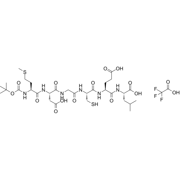
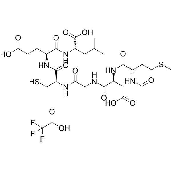
Box5 TFA
Based on 14 publication(s) in Google Scholar
Box5 TFA is a potent Wnt5a antagonist. Box5 TFA inhibits Wnt5a signaling and inhibits Wnt5a-mediated Ca2+ release. Box5 TFA inhibits cell migration. Box5 TFA has the potential for the research of melanoma.
Nos produits utilisent uniquement pour la recherche. Nous ne vendons pas aux patients.
- Pureté : 99.50%
- Formule: C30H50N6O13S2·xC2HF3O2
- Masse moléculaire:766.88 (free base)
-
Stockage:
Sealed storage, away from moisture and light, under nitrogen.
Powder -80°C, 2 years , -20°C, 1 year* In solvent : -80°C, 6 months; -20°C, 1 month (sealed storage, away from moisture and light, under nitrogen)

To place orders, for customer services and technical support, please contact: MedChemExpress USA
Tel: 609-228-6898 E-mail: [email protected] [email protected]


-
 Activité biologique
-
Chemical Information
-
Solvant et solubilité
- Protocole
- Pureté et documentation
- Références
-
 Help & FAQs


Publications Citing Use of MedChemExpress (MCE) Box5 TFA
More- Adv Sci (Weinh). 2025 Aug;12(32):e02774. [Abstract]
- J Immunother Cancer. 2025 Mar 22;13(3):e010555. [Abstract]
- J Neuroinflammation. 2024 Mar 26;21(1):75. [Abstract]
- Nano Today. 2024 Apr, 55, 102201.
- J Hazard Mater. 2026 Feb 1:503:141085. [Abstract]
- J Transl Med. 2025 Mar 6;23(1):282. [Abstract]
- J Ethnopharmacol. 2026 May 23:363:121445. [Abstract]
- J Mol Cell Biol. 2025 Dec 5:mjaf050. [Abstract]
- J Mol Cell Biol. 2025 Jul 28;17(2):mjaf002. [Abstract]
- Sci Rep. 2026 May 11;16(1):21377. [Abstract]
- J Cell Mol Med. 2026 Jun;30(11):e71170. [Abstract]
- Front Biosci (Landmark Ed). 2026 Jun 24;31(6):49794.
- Oral Dis. 2024 Nov;30(8):5140-5153. [Abstract]
- Odontology. 2026 Jan 21. [Abstract]
 Customer Validation & Images

-
WB
-
Cell Imaging/Staining
-
Cell Proliferation/Viability Assay
-
WB
-
Histological Imaging/Staining


Activité biologique
Description
IC50 & Target
|
Wnt5A |
In Vitro
Box5 TFA (100 μM) decreases the expression of rWnt5a (0.1 μg/mL) stimulated p-MARCKS in A2058 cells[1].
MedChemExpress (MCE) has not independently confirmed the accuracy of these methods. They are for reference only.
- Scratch/Wound-Healing Migration Assay
- Cell migration
- Zigmond Chamber Chemotaxis Assay
- Under-Agarose Cell Migration Assay
- Neural Crest/Neuronal Cell Migration Explant Assay
- Transwell/Boyden Chamber Migration Assay
- 3D Collagen/Hydrogel Matrix Migration Assay
- Ca2+ Staining Technique
- Cell-Exclusion Zone Migration Assay
- Dunn/Insall Chamber Chemotaxis Assay


Chemical Information
-
Appearance Solid
-
Masse moléculaire 766.88 (free base)
-
Formule C30H50N6O13S2·xC2HF3O2
-
Color White to off-white
-
Sequence
t-Boc-Met-Asp-Gly-Cys-Glu-Leu
-
Sequence Shortening
t-Boc-MDGCEL
-
Livraison
Room temperature in continental US; may vary elsewhere.
-
Stockage
Sealed storage, away from moisture and light, under nitrogen
Powder -80°C 2 years -20°C 1 year * In solvent : -80°C, 6 months; -20°C, 1 month (sealed storage, away from moisture and light, under nitrogen)


Publications (14)
-
Journal Impact Factor
-
Most Recent
-
Adv Sci (Weinh)
Single-Cell RNA Sequencing Identifies MMP11+ Cancer-Associated Fibroblasts as Drivers of Angiogenesis and Bladder Cancer Progression. [Abstract]2025 Aug;12(32):e02774. PMID: 40552583 -
J Immunother Cancer
Inhibition of stromal MAOA leading activation of WNT5A enhance prostate cancer immunotherapy by involving the transition of cancer-associated fibroblasts. [Abstract]2025 Mar 22;13(3):e010555. PMID: 40121032 -
J Neuroinflammation
Wnt5a/β-catenin-mediated epithelial-mesenchymal transition: a key driver of subretinal fibrosis in neovascular age-related macular degeneration. [Abstract]2024 Mar 26;21(1):75. PMID: 38532410
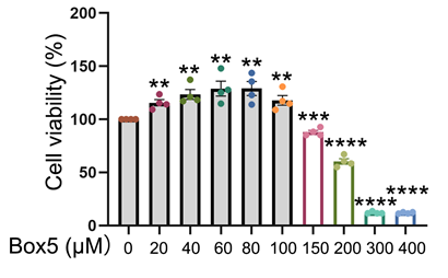
Box5 TFA purchased from MedChemExpress. Usage Cited in: J Neuroinflammation. 2024 Mar 26;21(1):75. [Abstract]
The cell viability of ARPE-19 cells examined with CCK-8 assay after treatment with the indicated doses of Box5 for 48 hours. The Vehicle Control groups for Box5 contained DMSO concentrations of 0.2% and 2%, respectively.
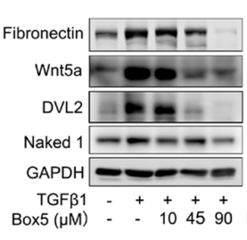
Box5 TFA purchased from MedChemExpress. Usage Cited in: J Neuroinflammation. 2024 Mar 26;21(1):75. [Abstract]
The protein levels of fibronectin, Wnt5a, Dvl2, and Naked1 in TGFβ1 (10 ng/mL)-treated ARPE-19 cells with or without different concentrations of Box5 (10, 45 and 90 μmol/L) for 48 h. Box5 suppressed the TGFβ1-mediated up-regulation of fibronectin, Wnt5a, Dvl2, and Naked1 in a dose-dependent manner.
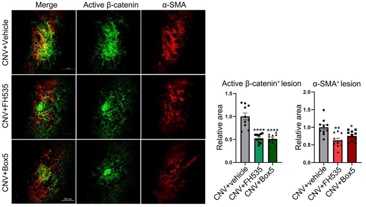
Box5 TFA purchased from MedChemExpress. Usage Cited in: J Neuroinflammation. 2024 Mar 26;21(1):75. [Abstract]
The effects of intravitreal injections of FH535 or Box5 (90 μmol/L) on β-catenin and α-SMA expression within the CNV lesions in mice at day 21 after laser induction, along with the corresponding quantitative analysis. Box5 (90 μmol/L) treatment significantly reduced the immunostaining areas of active β-catenin by 47.39% (p = 0.00002, n = 10).
-
-
J Hazard Mater
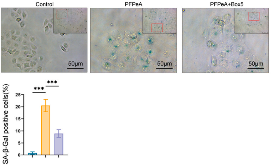
PFPeA exposure drives hepatoxicity and liver fibrosis via oxidative stress/Wnt5a-induced hepatocyte senescence. [Abstract]2026 Feb 1:503:141085. PMID: 41529635
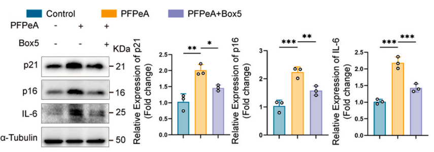
Box5 TFA purchased from MedChemExpress. Usage Cited in: J Hazard Mater. 2026 Feb 1:503:141085. [Abstract]
Protein level of p21, p16 and IL-6 after inhibiting Wnt5a with Box5 (100 μM; 24 h) in THLE-2 cells, and statistical analysis, α-Tubulin were used as control. The results showed that Box5 decreased the senescence related proteins.
Box5 TFA purchased from MedChemExpress. Usage Cited in: J Hazard Mater. 2026 Feb 1:503:141085. [Abstract]
Representative images of SA-β-Gal staining after inhibiting Wnt5a with Box5 (100 μM; 24 h) in THLE-2 cells. SA-β-Galactosidase staining confirmed that inhibition of Wnt5a decreased the senescence level of hepatocytes induced by PFPeA.
-
J Transl Med
Single-cell and spatial transcriptome profiling reveal CTHRC1+ fibroblasts promote EMT through WNT5A signaling in colorectal cancer. [Abstract]2025 Mar 6;23(1):282. PMID: 40050872 -
J Ethnopharmacol
Dendrobine ameliorates non-alcoholic fatty liver disease by inhibiting mitochondrial fission through modulation of the Wnt5a/p-CaMKII/p-Drp1 signaling axis. [Abstract]2026 May 23:363:121445. PMID: 41763618 -
J Mol Cell Biol
Canonical Wnt signaling affects calcium homeostasis in serum-treated AC16 cells through MLN-mediated SERCA2a regulation. [Abstract]2025 Dec 5:mjaf050. PMID: 41348974 -
J Mol Cell Biol
Wnt/β-catenin pathway induces cardiac dysfunction via AKAP6-mediated RyR2 phosphorylation and sarcoplasmic reticulum calcium leakage. [Abstract]2025 Jul 28;17(2):mjaf002. PMID: 40097291 -
Sci Rep
DOCK1 inhibitor Box5 (TFA) suppresses proliferation of AML cell lines and prolongs survival in AML xenograft models. [Abstract]2026 May 11;16(1):21377. PMID: 42108312 -
J Cell Mol Med
Decoding the Oncogenic Role of GNG10 in Colorectal Cancer: A Non-Canonical Wnt Pathway-Driven Mechanism. [Abstract]2026 Jun;30(11):e71170. PMID: 42252561 -
-
Oral Dis
Synovial osteoclastogenesis mediated by chondrocyte-secreted TNFα promotes TMJ condylar resorption. [Abstract]2024 Nov;30(8):5140-5153. PMID: 38720613 -
Odontology
Fusobacterium nucleatum promotes senescence of human oral mucosa fibroblasts via the Wnt5A/mTOR pathway. [Abstract]2026 Jan 21. PMID: 41559469


Solvant et solubilité
In Vitro:
DMSO : 100 mg/mL (Need ultrasonic; Hygroscopic DMSO has a significant impact on the solubility of product, please use newly opened DMSO)
In Vivo:
Select the appropriate dissolution method based on your experimental animal and administration route.
- For the following dissolution methods, please ensure to first prepare a clear stock solution using an In Vitro approach and then sequentially add co-solvents:
- To ensure reliable experimental results, the clarified stock solution can be appropriately stored based on storage conditions. As for the working solution for In Vivo experiments, it is recommended to prepare freshly and use it on the same day.
- The percentages shown for the solvents indicate their volumetric ratio in the final prepared solution. If precipitation or phase separation occurs during preparation, heat and/or sonication can be used to aid dissolution.
Add each solvent one by one: 10% DMSO 40% PEG300 5% Tween-80 45% Saline
Solubility: ≥ 2.5 mg/mL; Clear solution
This protocol yields a clear solution of ≥ 2.5 mg/mL (saturation unknown).
Taking 1 mL working solution as an example, add 100 μL DMSO stock solution (25.0 mg/mL) to 400 μL PEG300, and mix evenly; then add 50 μL Tween-80 and mix evenly; then add 450 μL Saline to adjust the volume to 1 mL.
Preparation of Saline: Dissolve 0.9 g sodium chloride in ddH₂O and dilute to 100 mL to obtain a clear Saline solution.
Add each solvent one by one: 10% DMSO 90% (20% SBE-β-CD in Saline)
Solubility: ≥ 2.5 mg/mL; Clear solution
This protocol yields a clear solution of ≥ 2.5 mg/mL (saturation unknown).
Taking 1 mL working solution as an example, add 100 μL DMSO stock solution (25.0 mg/mL) to 900 μL 20% SBE-β-CD in Saline, and mix evenly.
Preparation of 20% SBE-β-CD in Saline (4°C, storage for one week): 2 g SBE-β-CD powder is dissolved in 10 mL Saline, completely dissolve until clear.
In Vivo Dissolution Calculator
Please enter the basic information of animal experiments:
-
-
-
-
Recommended: Prepare an additional quantity of animals to account for potential losses during experiments.
Please enter your animal formula composition:
-
%DMSO +
Recommended: Keep the proportion of DMSO in working solution below 2% if your animal is weak.
-
%+
-
+%Tween-80 + +
-
%Saline +
The co-solvents required include: DMSO, . All of co-solvents are available by MedChemExpress (MCE). , Tween 80. All of co-solvents are available by MedChemExpress (MCE).
Working solution concentration: 0.22 mg/mL
Method for preparing stock solution: mg drug dissolved in μL DMSO. Stock solution concentration: mg/mL. * In solvent : -80°C, 6 months; -20°C, 1 month (sealed storage, away from moisture and light, under nitrogen)
1. Take μL DMSO stock solution;
2. Add μL .
μL , mix evenly;
3. Then add μL Tween 80, mix evenly;
4. Then add μL
Please ensure that the stock solution in the first step is dissolved to a clear state, and add co-solvents in sequence. You can use ultrasonic heating (ultrasonic cleaner, recommended frequency 20-40 kHz), vortexing, etc. to assist dissolution.


Pureté et documentation
-
Fiche technique (274 KB)
-
SDS (252 KB)
- English - EN (252 KB)
- Français - FR (252 KB)
- Deutsch - DE (252 KB)
- Norwegian - NO (252 KB)
- Español - ES (252 KB)
- Swedish - SV (252 KB)
- Italian - IT (252 KB)
- Korean - KR (252 KB)
- Portuguese - PT (252 KB)
-
Instruction de manipulation (2659 KB)
Références

Calculators
Concentration (start) × Volume (start) = Concentration (final) × Volume (final)


Box5 TFA Related Classifications
HY-123071A Related Classifications
 Powered by Bioz