



Fusaric acid

Based on 1 Customer Validation
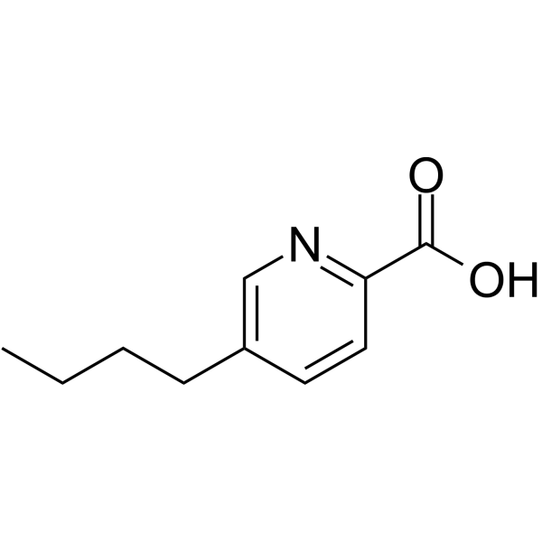
Fusaric acid is an orally active multi-pathway inhibitor with the activity of inducing oxidative stress and apoptosis. Fusaric acid can chelate divalent metal cations, damage mitochondrial membrane structure, and activate apoptosis-related proteases such as Caspase-3/7, -8, and -9. Fusaric acid also regulates Bax/Bcl-2 protein, inhibits fibrosis-related signaling pathways such as NF-κB, TGF-β1/SMADs, and PI3K/AKT/mTOR, and reduces collagen deposition. Fusaric acid is also a dopamine β-hydroxylase inhibitor, which reduces endogenous levels of norepinephrine and epinephrine in the brain, heart, spleen, and adrenal glands. Fusaric acid can play a role in myocardial fibrosis and improve cardiac hypertrophy in heart disease, and can also be used in the study of esophageal cancer and liver cancer.
Nur für Forschungszwecke. Wir verkaufen nicht an Patienten.
- Reinheit : 99.94%
- CAS. Nr.: 536-69-6
- Formel: C10H13NO2
- Molecular Weight:179.22
-
Speicherung:Powder -20°C, 3 years , 4°C, 2 years ; In solvent -80°C, 6 months , -20°C, 1 month

To place orders, for customer services and technical support, please contact: MedChemExpress USA
Tel: 609-228-6898 E-mail: [email protected] [email protected]


-
 Biologische Aktivität
-
Chemical Information
-
Lösungsmittel & Löslichkeit
- Protokoll
- Reinheit & Dokumentation
- Verweise
-
 Help & FAQs


-
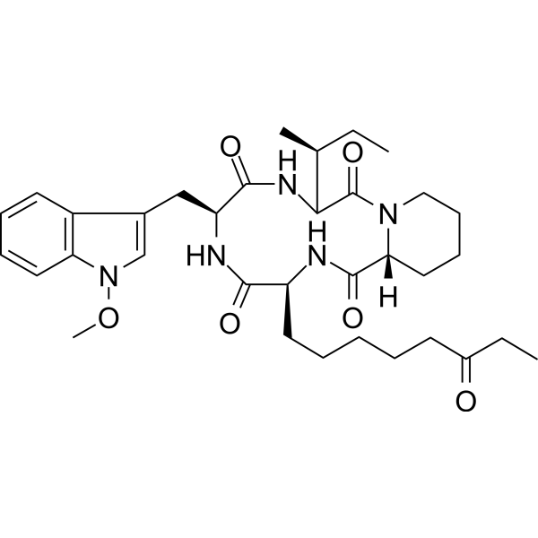
HY-N6735
ApicidinHDAC Inhibitor -
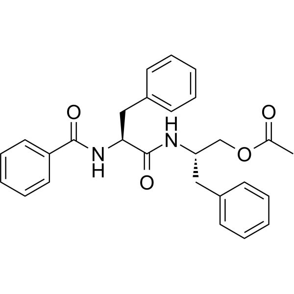
HY-N2905
Aurantiamide acetateCathepsin Inhibitor -
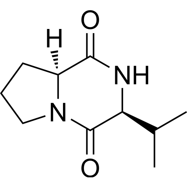
HY-P1938
Cyclo(L-Pro-L-Val)Bacterial Inhibitor/Anti-inflammatory Agent -
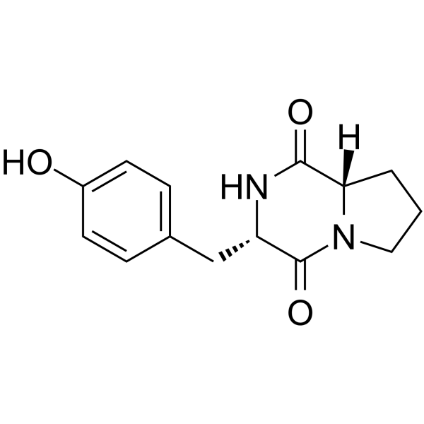
HY-P1940
MaculosinAnti-Bacterial Agent -
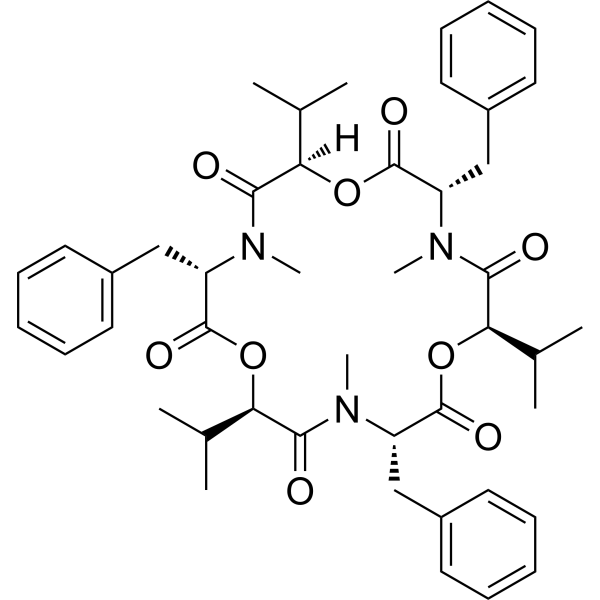
HY-N6739
BeauvericinAnticancer Fungal Mycotoxin -

HY-128483R
Fusaric acid (Standard)Dopamine β-hydroxylase Inhibitor -
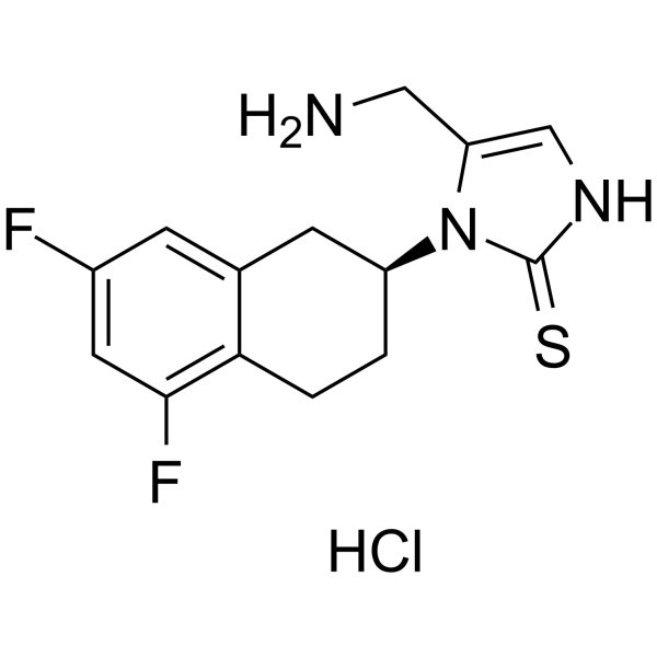
HY-13289A
Nepicastat hydrochlorideDopamine β-Hydroxylase Inhibitor -

HY-N6739R
Beauvericin (Standard)Anticancer Fungal Mycotoxin
Alle Adrenergic Receptor Isoform-spezifische Produkte anzeigen
More

Biologische Aktivität
Beschreibung
In Vitro
Fusaric acid (0-500 μg/mL; 24 h) inhibits cell viability in a dose-dependent manner in human esophageal cancer cells (SNO) with an IC50 value of 78.81 μg/mL[1].
Fusaric acid (100 μM; 24-72 h) induces programmed cell death in tobacco suspension cells, accompanied by H2O2 accumulation, decreases mitochondrial membrane potential, and activation of caspase-3-like proteases[2].
Fusaric acid (0-160 μM; 24-96 h) inhibits the cell viability of rat cardiomyocytes (H9C2) and mouse cardiac fibroblasts (mCFs), and suppresses the expression of fibrosis-related proteins (α-SMA, type I/III collagen) in WB results at 24 h, and downregulates TGF-β1/SMADs and PI3K/AKT signaling pathways[3].
MedChemExpress (MCE) has not independently confirmed the accuracy of these methods. They are for reference only.
-
Cell Line:Cardiomyocytes isolated from mice, Rat cardiomyocytes (H9C2) and Mouse cardiac fibroblasts (mCFs)
-
Concentration:1.25 μM, 2.5 μM, 5 μM, 10 μM, 20 μM, 40 μM, 80 μM, 160 μM
-
Incubation Time:6 h, 12 h, 24 h, 36 h, 48 h, 72 h, 96 h
-
Result:Only at the high concentration of 80 and 160 uM for long time (72 and 96 h), significant difference in cells, indicating a relatively low doses for time under 96 h showed no obvious cytotoxicity to cardiomyocytes isolated from mice and H9C2 cells.
In Vivo
MedChemExpress (MCE) has not independently confirmed the accuracy of these methods. They are for reference only.
-
Animal Model:Male C57BL/6 mice (18-22 g, 8 weeks old) with Isoprenaline (HY-B0468)-induced cardiac hypertrophy and fibrosis[3]
-
Dosage:50 mg/kg, 100 mg/kg
-
Administration:Oral gavage daily for 7 days before Isoprenaline treatment, then continued for another 7 days (total 15 days)
-
Result:Cardiac hypertrophy markers:
Reduced heart weight/body weight and heart weight/tibia length ratios compared to the Isoprenaline group, indicating attenuated cardiac hypertrophy.
Histological analysis:
Masson's trichrome staining showed decreased collagen deposition in heart tissues, with reduced fibrosis area compared to the Isoprenaline group. WGA staining revealed smaller cardiomyocyte cross-sectional area, indicating attenuated myocyte hypertrophy.
Echocardiography:
Improved left ventricular function, including increased fractional shortening (FS%) and reduced left ventricular end-diastolic/systolic dimensions, compared to the Isoprenaline group.
- Fibrosis/Collagen Morphometry
- Collagen: Sirius Red Staining
- Detection of Apoptosis
- TUNEL staining for apoptotic DNA fragmentation
- Annexin V plus membrane-impermeant dye apoptosis staining
- ROS/oxidative-stress fluorescent staining
- Connective Tissue: Masson's Trichrome/Collagen Trichrome Staining
- Apoptosis Solutions
- Research Protocol for Cardiovascular Diseases
- Research Protocol for Inflammation-related Diseases
- Liver Cancer Modeling
- Cardiac Morphometry


Chemical Information
-
CAS. Nr. 536-69-6
-
Appearance Solid
-
Molecular Weight 179.22
-
Formel C10H13NO2
-
Color White to off-white
-
SMILES
O=C(C1=NC=C(CCCC)C=C1)O
-
Structure Classification
-
Initial Source
Fusarium spp.
-
Versand
Room temperature in continental US; may vary elsewhere.
-
Speicherung
Powder -20°C 3 years 4°C 2 years In solvent -80°C 6 months -20°C 1 month


Lösungsmittel & Löslichkeit
In Vitro:
DMSO : 58.33 mg/mL (325.47 mM; Need ultrasonic; Hygroscopic DMSO has a significant impact on the solubility of product, please use newly opened DMSO)
H2O : 50 mg/mL (278.99 mM; Need ultrasonic)
Please refer to the solubility information to select the appropriate solvent. Once prepared, please aliquot and store the solution to prevent product inactivation from repeated freeze-thaw cycles.
Storage method and period of stock solution: -80°C, 6 months; -20°C, 1 month. When stored at -80°C, please use it within 6 months. When stored at -20°C, please use it within 1 month.
* Note: If you choose water as the stock solution, please dilute it to the working solution, then filter and sterilize it with a 0.22 μm filter before use.
Please refer to the solubility information to select the appropriate solvent. Once prepared, please aliquot and store the solution to prevent product inactivation from repeated freeze-thaw cycles.
Storage method and period of stock solution: -80°C, 6 months; -20°C, 1 month. When stored at -80°C, please use it within 6 months. When stored at -20°C, please use it within 1 month.
* Note: If you choose water as the stock solution, please dilute it to the working solution, then filter and sterilize it with a 0.22 μm filter before use.
Konzentration (Stammlösung) × Volumen (Stammlösung) = Konzentration (Ziellösung) × Volumen (Ziellösung)
In Vivo:
Select the appropriate dissolution method based on your experimental animal and administration route.
- For the following dissolution methods, please ensure to first prepare a clear stock solution using an In Vitro approach and then sequentially add co-solvents:
- To ensure reliable experimental results, the clarified stock solution can be appropriately stored based on storage conditions. As for the working solution for In Vivo experiments, it is recommended to prepare freshly and use it on the same day.
- The percentages shown for the solvents indicate their volumetric ratio in the final prepared solution. If precipitation or phase separation occurs during preparation, heat and/or sonication can be used to aid dissolution.
Add each solvent one by one: 10% DMSO 40% PEG300 5% Tween-80 45% Saline
Solubility: ≥ 2.08 mg/mL (11.61 mM); Clear solution
This protocol yields a clear solution of ≥ 2.08 mg/mL (saturation unknown).
Taking 1 mL working solution as an example, add 100 μL DMSO stock solution (20.8 mg/mL) to 400 μL PEG300, and mix evenly; then add 50 μL Tween-80 and mix evenly; then add 450 μL Saline to adjust the volume to 1 mL.
Preparation of Saline: Dissolve 0.9 g sodium chloride in ddH₂O and dilute to 100 mL to obtain a clear Saline solution.
Add each solvent one by one: 10% DMSO 90% (20% SBE-β-CD in Saline)
Solubility: ≥ 2.08 mg/mL (11.61 mM); Clear solution
This protocol yields a clear solution of ≥ 2.08 mg/mL (saturation unknown).
Taking 1 mL working solution as an example, add 100 μL DMSO stock solution (20.8 mg/mL) to 900 μL 20% SBE-β-CD in Saline, and mix evenly.
Preparation of 20% SBE-β-CD in Saline (4°C, storage for one week): 2 g SBE-β-CD powder is dissolved in 10 mL Saline, completely dissolve until clear.
In Vivo Dissolution Calculator
Please enter the basic information of animal experiments:
-
-
-
-
Recommended: Prepare an additional quantity of animals to account for potential losses during experiments.
Working solution concentration: 0.22 mg/mL
This product has good water solubility, please refer to the measured solubility data in water/PBS/Saline for details.


Reinheit & Dokumentation
-
Data Sheet (276 KB)
-
SDS (392 KB)
- English - EN (392 KB)
- Français - FR (392 KB)
- Deutsch - DE (392 KB)
- Norwegian - NO (392 KB)
- Español - ES (392 KB)
- Swedish - SV (392 KB)
- Italian - IT (392 KB)
- Korean - KR (392 KB)
- Portuguese - PT (392 KB)
-
Handling Instructions (2659 KB)
Verweise
[1]. Devnarain N, et al. Fusaric acid induces oxidative stress and apoptosis in human cancerous oesophageal SNO cells. Toxicon. 2017 Feb;126:4-11. [Content Brief]
[2]. Jiao J, et al. Hydrogen peroxide production and mitochondrial dysfunction contribute to the fusaric acid-induced programmed cell death in tobacco cells. J Plant Physiol. 2014 Aug 15;171(13):1197-203. [Content Brief]
[3]. Li X, et al. Fusaric acid (FA) protects heart failure induced by isoproterenol (ISP) in mice through fibrosis prevention via TGF-β1/SMADs and PI3K/AKT signaling pathways. Biomed Pharmacother. 2017 Sep;93:130-145. [Content Brief]
[4]. Nagatsu T, et al. Inhibition of dopamine beta-hydroxylase by fusaric acid (5-butylpicolinic acid) in vitro and in vivo. Biochem Pharmacol. 1970 Jan;19(1):35-44. [Content Brief]


Complete Stock Solution Preparation Table
Please refer to the solubility information to select the appropriate solvent. Once prepared, please aliquot and store the solution to prevent product inactivation from repeated freeze-thaw cycles.
Storage method and period of stock solution: -80°C, 6 months; -20°C, 1 month. When stored at -80°C, please use it within 6 months. When stored at -20°C, please use it within 1 month.
| Optional Solvent | Concentration Solvent Mass | 1 mg | 5 mg | 10 mg | 25 mg |
|---|---|---|---|---|---|
| H2O / DMSO | 1 mM | 5.5797 mL | 27.8987 mL | 55.7973 mL | 139.4934 mL |
| 5 mM | 1.1159 mL | 5.5797 mL | 11.1595 mL | 27.8987 mL | |
| 10 mM | 0.5580 mL | 2.7899 mL | 5.5797 mL | 13.9493 mL | |
| 15 mM | 0.3720 mL | 1.8599 mL | 3.7198 mL | 9.2996 mL | |
| 20 mM | 0.2790 mL | 1.3949 mL | 2.7899 mL | 6.9747 mL | |
| 25 mM | 0.2232 mL | 1.1159 mL | 2.2319 mL | 5.5797 mL | |
| 30 mM | 0.1860 mL | 0.9300 mL | 1.8599 mL | 4.6498 mL | |
| 40 mM | 0.1395 mL | 0.6975 mL | 1.3949 mL | 3.4873 mL | |
| 50 mM | 0.1116 mL | 0.5580 mL | 1.1159 mL | 2.7899 mL | |
| 60 mM | 0.0930 mL | 0.4650 mL | 0.9300 mL | 2.3249 mL | |
| 80 mM | 0.0697 mL | 0.3487 mL | 0.6975 mL | 1.7437 mL | |
| 100 mM | 0.0558 mL | 0.2790 mL | 0.5580 mL | 1.3949 mL |
* Note: If you choose water as the stock solution, please dilute it to the working solution, then filter and sterilize it with a 0.22 μm filter before use.


Fusaric acid Related Classifications
HY-128483 Related Classifications
 Powered by Bioz