



MLN120B
Based on 29 publication(s) in Google Scholar
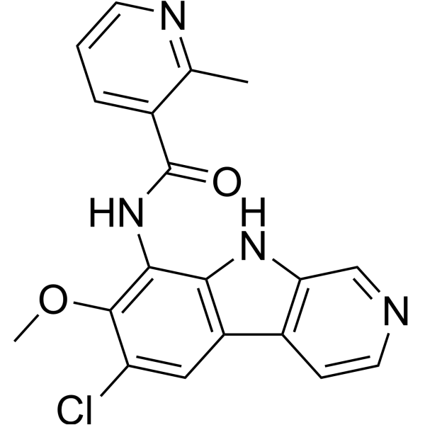
MLN120B (ML120B) is a potent, ATP competitive, and orally active inhibitor of IKKβ with an IC50 of 60 nM. MLN120B inhibits multiple myeloma cell growth in vitro and in vivo and also can be used for the research of rheumatoid arthritis.
Para uso exclusivo en investigación. No vendemos a pacientes.
- Pureza : 99.76%
- No. CAS: 783348-36-7
- Fòrmula: C19H15ClN4O2
- Peso molecular:366.80
-
Almacenamiento:Powder -20°C, 3 years , 4°C, 2 years ; In solvent -80°C, 2 years , -20°C, 1 year

To place orders, for customer services and technical support, please contact: MedChemExpress USA
Tel: 609-228-6898 E-mail: [email protected] [email protected]


-
 Actividad biológica
-
Chemical Information
-
Solvente y solubilidad
- Protocolo
- Pureza y Documentación
- Referencias
-
 Help & FAQs


-
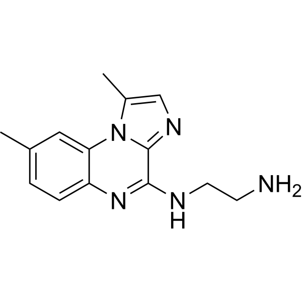
HY-10519
BMS-345541IKK-1/IKK-2 Inhibitor -
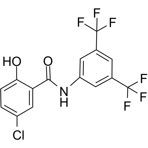
HY-10172
IMD-0354IKKβ Inhibitor -
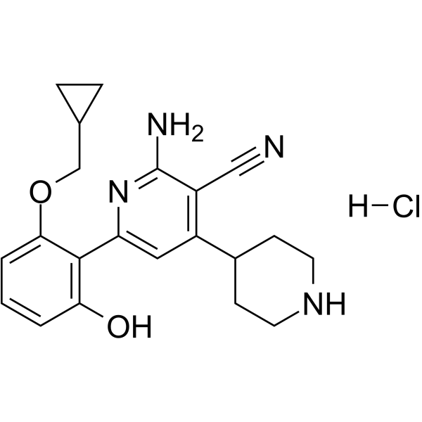
HY-13060
ACHP HydrochlorideIKKβ Inhibitor -
HY-13453
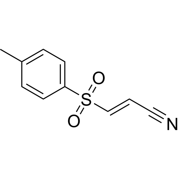
BAY 11-7082IκBα/NF-κB Inhibitor -
HY-13019
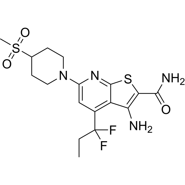
BI605906IKK Inhibitor -
HY-13687
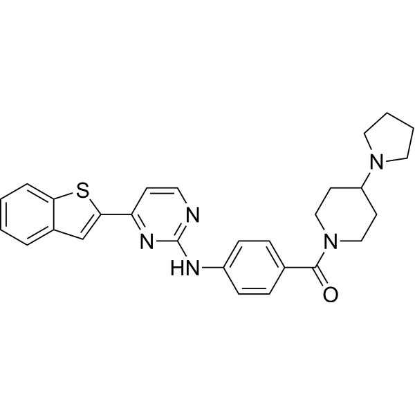
IKK 16IKK Inhibitor -
HY-10074
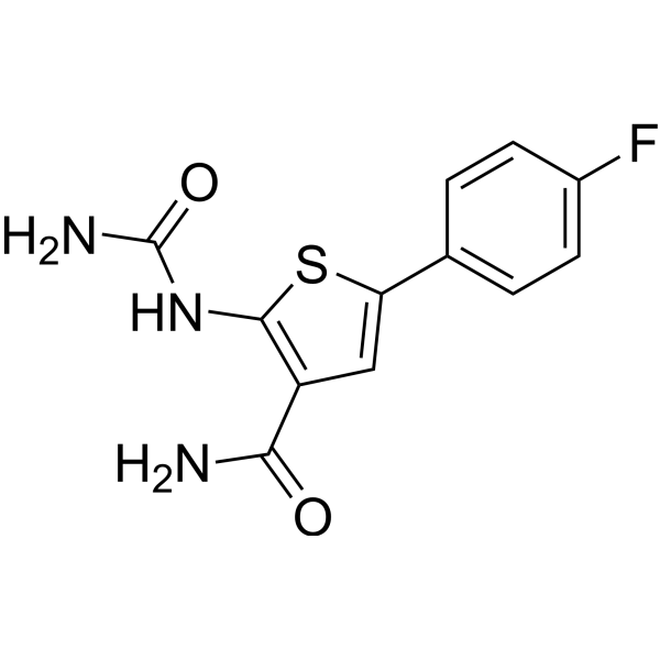
TPCA-1IKK-2 Inhibitor -
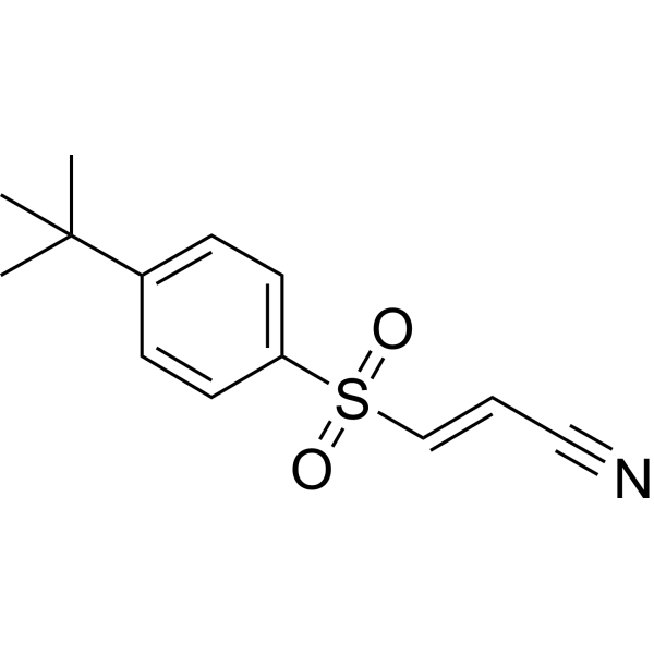
HY-10257
BAY 11-7085NF-κB Activation Inhibitor -
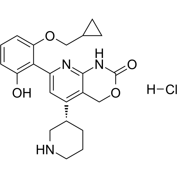
HY-50948
Bay 65-1942 hydrochlorideIKK Inhibitor -
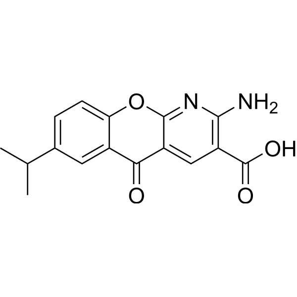
HY-B0713
AmlexanoxIKK Inhibitor -
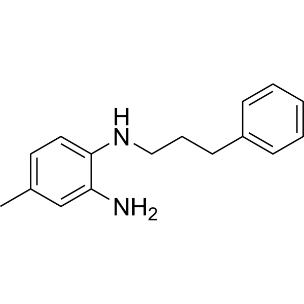
HY-13982
JSH-23NF-κB Inhibitor -

HY-10219
RapamycinmTOR Inhibitor
Publications Citing Use of MedChemExpress (MCE) MLN120B
More- Signal Transduct Target Ther. 2020 Oct 9;5(1):235. [Abstract]
- Cancer Cell. 2024 Feb 12;42(2):238-252.e9. [Abstract]
- Nature. 2022 Oct;610(7931):366-372. [Abstract]
- Blood. 2023 Nov 2.
- Blood. 2015 Nov 12;126(20):2291-301. [Abstract]
- Blood. 2015 Sep 10;126(11):1324-35. [Abstract]
- Sci Transl Med. 2021 Jan 27;13(578):eaba7308. [Abstract]
- Theranostics. 2020 Apr 6;10(11):5029-5047. [Abstract]
- Cell Death Dis. 2016 Dec 1;7(12):e2505. [Abstract]
- Acta Pharmacol Sin. 2021 May;42(5):814-823. [Abstract]
- Aging Cell. 2021 Jul;20(7):e13426. [Abstract]
- Cell Rep. 2020 Mar 31;30(13):4370-4385.e7. [Abstract]
- Cell Prolif. 2023 Sep;56(9):e13442. [Abstract]
- J Invest Dermatol. 2017 Dec;137(12):2532-2543. [Abstract]
- Biochem Pharmacol. 2018 Jun:152:45-59. [Abstract]
- Int J Mol Sci. 2026 Aug 26;27(17):7636.
- Int Immunopharmacol. 2024 Nov 15:141:113013. [Abstract]
- Sci Rep. 2021 Jul 28;11(1):15319. [Abstract]
- Int J Cancer. 2017 Mar 1;140(5):1147-1158. [Abstract]
- J Biol Chem. 2015 Apr 24;290(17):11061-74. [Abstract]
- Viruses. 2021 May 25;13(6):976. [Abstract]
- Cytokine. 2026 May:201:157135. [Abstract]
- Exp Ther Med. 2021 Feb;21(2):122. [Abstract]
- bioRxiv. 2025 February 22.
- University of Strathclyd. 2024
- bioRxiv. 2024 Mar 28.
- bioRxiv. 2023 Mar 23.
- Patent. US20220025002A1.
- SSRN. 2022 Jan 26.
 Customer Validation & Images

-
WB
-
WB


Actividad biológica
Descripciòn
IC50 & Target
[4]|
IKKβ 60 nM (IC50) |
In Vitro
MLB120B (0-20 μM; 90 minutes) inhibits phosphorylation and degradation of IκB in RPMI 8226 and INA6 cells; however, no significant inhibition is observed in MM.1S cells[1].MLB120B (1.25-20 μM; 90 minutes) completely abrogates TNF-a-induced phosphorylation and degradation of IκB in a dosedependent fashion. Phosphorylation of p65 NF-κB induced by TNF-a is also blocked by MLN120B[1].MLN120B inhibits proliferation of multiple myeloma cell lines. MM.1S, MM.1R, RPMI 8226, RPMI-LR5, RPMI-Dox40, U266, and INA6 cells. Five percent to fifty percent and 18% to 70% inhibition in proliferation is observed at doses >20 uM and [3H]thymidine uptake, respectively[1].MLN120B (1.25-40 μM; 72 hours) almost completely blocks stimulation of MM.1S, U266, and INA6 cell growth, as well as IL-6 secretion from BMSCs, induced by multiple myeloma cell adherence to BMSCs[1].MLN120B shows an inhibitory effect on LPS induced NF-κB activation in RAW267.4 cells. The IC50 values of MLN120B is 1.4 μM, 14.8 μM or 27.3 μM for NF-κB2-luc2, IL8-luc2 or TNF-AIP3-luc2 reporter transfected cells, respectively[3].
MedChemExpress (MCE) has not independently confirmed the accuracy of these methods. They are for reference only.
-
Cell Line:MM.1S cells
-
Concentration:1.25-20 μM
-
Incubation Time:90 minutes
-
Result:Inhibited p- IκB and p-P65 expression in a dose-dependent manner.
-
Cell Line:Multiple myeloma cell lines: MM.1S, MM.1R, RPMI 8226, RPMI-LR5, RPMI-Dox40, U266, and INA6 cells
-
Concentration:1.25 μM-20 μM
-
Incubation Time:72 hours
-
Result:Inhibits proliferation of multiple myeloma cell lines.
In Vivo
MedChemExpress (MCE) has not independently confirmed the accuracy of these methods. They are for reference only.
-
Animal Model:Two-month-old female Lewis rats[2]
-
Dosage:30 mg/kg, 10 mg/kg, 3 mg/kg, or 1 mg/kg
-
Administration:Oral administration; twice daily; 3 weeks
-
Result:Protected against bone and cartilage destruction in a rat model.
-
Animal Model:SCID mice implanted with human fetal bone chips and then INA6 cells are directly injected into mice[1]
-
Dosage:50 mg/kg
-
Administration:Oral administration; twice daily; 3 weeks
-
Result:Inhibited human multiple myeloma cell growth in vivo.


Chemical Information
-
No. CAS 783348-36-7
-
Appearance Solid
-
Peso molecular 366.80
-
Fòrmula C19H15ClN4O2
-
Color Off-white to light yellow
-
SMILES
O=C(C1=CC=CN=C1C)NC2=C(OC)C(Cl)=CC3=C2NC4=C3C=CN=C4
-
Synonyms
ML120B
-
Envío
Room temperature in continental US; may vary elsewhere.
-
Almacenamiento
Powder -20°C 3 years 4°C 2 years In solvent -80°C 2 years -20°C 1 year


Publications (29)
-
Journal Impact Factor
-
Most Recent
-
Signal Transduct Target Ther
2020 Oct 9;5(1):235. PMID: 33037188 -
Cancer Cell
Response to Bruton's tyrosine kinase inhibitors in aggressive lymphomas linked to chronic selective autophagy. [Abstract]2024 Feb 12;42(2):238-252.e9. PMID: 38215749 -
Nature
2022 Oct;610(7931):366-372. PMID: 36198801 -
-
Blood
ATM deficiency promotes development of murine B-cell lymphomas that resemble diffuse large B-cell lymphoma in humans. [Abstract]2015 Nov 12;126(20):2291-301. PMID: 26400962 -
Blood
NF-κB and AKT signaling prevent DNA damage in transformed pre-B cells by suppressing RAG1/2 expression and activity. [Abstract]2015 Sep 10;126(11):1324-35. PMID: 26153519 -
Sci Transl Med
A chimeric antigen receptor with antigen-independent OX40 signaling mediates potent antitumor activity. [Abstract]2021 Jan 27;13(578):eaba7308. PMID: 33504651 -
Theranostics
2020 Apr 6;10(11):5029-5047. PMID: 32308766 -
Cell Death Dis
2016 Dec 1;7(12):e2505. PMID: 27906189 -
Acta Pharmacol Sin
Activation of unfolded protein response overcomes Ibrutinib resistance in diffuse large B-cell lymphoma. [Abstract]2021 May;42(5):814-823. PMID: 32855532 -
Aging Cell
2021 Jul;20(7):e13426. PMID: 34187082 -
Cell Rep
SFTSV Infection Induces BAK/BAX-Dependent Mitochondrial DNA Release to Trigger NLRP3 Inflammasome Activation. [Abstract]2020 Mar 31;30(13):4370-4385.e7. PMID: 32234474 -
Cell Prolif
2023 Sep;56(9):e13442. PMID: 37086012 -
J Invest Dermatol
The Unknown Aspect of BAFF: Inducing IL-35 Production by a CD5+CD1dhiFcγRIIbhi Regulatory B-Cell Subset in Lupus. [Abstract]2017 Dec;137(12):2532-2543. PMID: 28844943
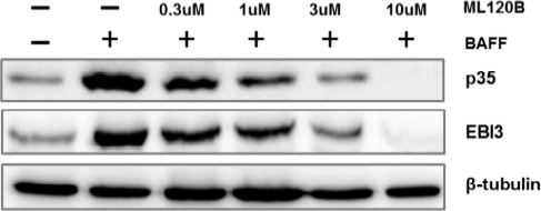
MLN120B purchased from MedChemExpress. Usage Cited in: J Invest Dermatol. 2017 Dec;137(12):2532-2543. [Abstract]
MZ B cells are pre-treated with different concentrations of MLN120B, which selectively inhibit NF-κB1 pathway, the expression of p35 and Ebi3 significantly reduced.
-
Biochem Pharmacol
Canagliflozin exerts anti-inflammatory effects by inhibiting intracellular glucose metabolism and promoting autophagy in immune cells. [Abstract]2018 Jun:152:45-59. PMID: 29551587
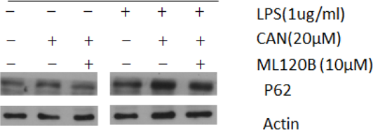
MLN120B purchased from MedChemExpress. Usage Cited in: Biochem Pharmacol. 2018 Jun:152:45-59. [Abstract]
Effects of NF-κB inhibitor (10 μM MLN120B) is measured on p62 protein in LPS-induced RAW264.7 cells.
-
-
Int Immunopharmacol
Inflammatory factor-mediated miR-155/SOCS1 signaling axis leads to Treg impairment in systemic lupus erythematosus. [Abstract]2024 Nov 15:141:113013. PMID: 39213866 -
Sci Rep
A phenotypic high-content, high-throughput screen identifies inhibitors of NLRP3 inflammasome activation. [Abstract]2021 Jul 28;11(1):15319. PMID: 34321581 -
Int J Cancer
Synergy of interleukin 10 and toll-like receptor 9 signalling in B cell proliferation: Implications for lymphoma pathogenesis. [Abstract]2017 Mar 1;140(5):1147-1158. PMID: 27668411 -
J Biol Chem
Tyrosine Kinase 2-mediated Signal Transduction in T Lymphocytes Is Blocked by Pharmacological Stabilization of Its Pseudokinase Domain. [Abstract]2015 Apr 24;290(17):11061-74. PMID: 25762719 -
Viruses
Activation of the STAT3 Signaling Pathway by the RNA-Dependent RNA Polymerase Protein of Arenavirus. [Abstract]2021 May 25;13(6):976. PMID: 34070281 -
Cytokine
The LA-NF-κB-ILC2 axis exacerbates intestinal milk protein allergy in mice by promoting ILC2 activation. [Abstract]2026 May:201:157135. PMID: 41780286 -
Exp Ther Med
Proteomic and microRNA-omic profiles and potential mechanisms of dysfunction in pancreatic islet cells primed by inflammation. [Abstract]2021 Feb;21(2):122. PMID: 33335585 -
-
-
-
-
-


Solvente y solubilidad
In Vitro:
DMSO : ≥ 31 mg/mL (84.51 mM; Hygroscopic DMSO has a significant impact on the solubility of product, please use newly opened DMSO)
* "≥" means soluble, but saturation unknown.
Please refer to the solubility information to select the appropriate solvent. Once prepared, please aliquot and store the solution to prevent product inactivation from repeated freeze-thaw cycles.
Storage method and period of stock solution: -80°C, 2 years; -20°C, 1 year. When stored at -80°C, please use it within 2 years. When stored at -20°C, please use it within 1 year.
Please refer to the solubility information to select the appropriate solvent. Once prepared, please aliquot and store the solution to prevent product inactivation from repeated freeze-thaw cycles.
Storage method and period of stock solution: -80°C, 2 years; -20°C, 1 year. When stored at -80°C, please use it within 2 years. When stored at -20°C, please use it within 1 year.
Concentration (start) × Volume (start) = Concentration (final) × Volume (final)
In Vivo:
Select the appropriate dissolution method based on your experimental animal and administration route.
- For the following dissolution methods, please ensure to first prepare a clear stock solution using an In Vitro approach and then sequentially add co-solvents:
- To ensure reliable experimental results, the clarified stock solution can be appropriately stored based on storage conditions. As for the working solution for In Vivo experiments, it is recommended to prepare freshly and use it on the same day.
- The percentages shown for the solvents indicate their volumetric ratio in the final prepared solution. If precipitation or phase separation occurs during preparation, heat and/or sonication can be used to aid dissolution.
Add each solvent one by one: 10% DMSO 40% PEG300 5% Tween-80 45% Saline
Solubility: ≥ 2.08 mg/mL (5.67 mM); Clear solution
This protocol yields a clear solution of ≥ 2.08 mg/mL (saturation unknown).
Taking 1 mL working solution as an example, add 100 μL DMSO stock solution (20.8 mg/mL) to 400 μL PEG300, and mix evenly; then add 50 μL Tween-80 and mix evenly; then add 450 μL Saline to adjust the volume to 1 mL.
Preparation of Saline: Dissolve 0.9 g sodium chloride in ddH₂O and dilute to 100 mL to obtain a clear Saline solution.
Add each solvent one by one: 10% DMSO 90% (20% SBE-β-CD in Saline)
Solubility: 2.08 mg/mL (5.67 mM); Suspended solution; Need ultrasonic
This protocol yields a suspended solution of 2.08 mg/mL. Suspended solution can be used for oral and intraperitoneal injection.
Taking 1 mL working solution as an example, add 100 μL DMSO stock solution (20.8 mg/mL) to 900 μL 20% SBE-β-CD in Saline, and mix evenly.
Preparation of 20% SBE-β-CD in Saline (4°C, storage for one week): 2 g SBE-β-CD powder is dissolved in 10 mL Saline, completely dissolve until clear.
In Vivo Dissolution Calculator
Please enter the basic information of animal experiments:
-
-
-
-
Recommended: Prepare an additional quantity of animals to account for potential losses during experiments.
Please enter your animal formula composition:
-
%DMSO +
Recommended: Keep the proportion of DMSO in working solution below 2% if your animal is weak.
-
%+
-
+%Tween-80 + +
-
%Saline +
The co-solvents required include: DMSO, . All of co-solvents are available by MedChemExpress (MCE). , Tween 80. All of co-solvents are available by MedChemExpress (MCE).
Working solution concentration: 0.22 mg/mL
Method for preparing stock solution: mg drug dissolved in μL DMSO. Stock solution concentration: mg/mL.
1. Take μL DMSO stock solution;
2. Add μL .
μL , mix evenly;
3. Then add μL Tween 80, mix evenly;
4. Then add μL
Please ensure that the stock solution in the first step is dissolved to a clear state, and add co-solvents in sequence. You can use ultrasonic heating (ultrasonic cleaner, recommended frequency 20-40 kHz), vortexing, etc. to assist dissolution.


Pureza y Documentación
-
Ficha de datos (289 KB)
-
SDS (396 KB)
- English - EN (396 KB)
- Français - FR (396 KB)
- Deutsch - DE (396 KB)
- Norwegian - NO (396 KB)
- Español - ES (396 KB)
- Swedish - SV (396 KB)
- Italian - IT (396 KB)
- Korean - KR (396 KB)
- Portuguese - PT (396 KB)
-
Instrucciones de manejo (2659 KB)
Referencias
[1]. Hideshima T, et all. MLN120B, a novel IkappaB kinase beta inhibitor, blocks multiple myeloma cell growth in vitro and in vivo. Clin Cancer Res. 2006 Oct 1;12(19):5887-94. [Content Brief]
[2]. Schopf L, et al. IKKbeta inhibition protects against bone and cartilage destruction in a rat model of rheumatoid arthritis. Arthritis Rheum. 2006 Oct;54(10):3163-73. [Content Brief]
[3]. Ansaldi D, et al. Imaging pulmonary NF-kappaB activation and therapeutic effects of MLN120B and TDZD-8. PLoS One. 2011;6(9):e25093. [Content Brief]
[4]. Nagashima K, et al. Rapid TNFR1-dependent lymphocyte depletion in vivo with a selective chemical inhibitor of IKKbeta. Blood. 2006 Jun 1;107(11):4266-73. [Content Brief]


Complete Stock Solution Preparation Table
Please refer to the solubility information to select the appropriate solvent. Once prepared, please aliquot and store the solution to prevent product inactivation from repeated freeze-thaw cycles.
Storage method and period of stock solution: -80°C, 2 years; -20°C, 1 year. When stored at -80°C, please use it within 2 years. When stored at -20°C, please use it within 1 year.
| Optional Solvent | Concentration Solvent Mass | 1 mg | 5 mg | 10 mg | 25 mg |
|---|---|---|---|---|---|
| DMSO | 1 mM | 2.7263 mL | 13.6314 mL | 27.2628 mL | 68.1570 mL |
| 5 mM | 0.5453 mL | 2.7263 mL | 5.4526 mL | 13.6314 mL | |
| 10 mM | 0.2726 mL | 1.3631 mL | 2.7263 mL | 6.8157 mL | |
| 15 mM | 0.1818 mL | 0.9088 mL | 1.8175 mL | 4.5438 mL | |
| 20 mM | 0.1363 mL | 0.6816 mL | 1.3631 mL | 3.4079 mL | |
| 25 mM | 0.1091 mL | 0.5453 mL | 1.0905 mL | 2.7263 mL | |
| 30 mM | 0.0909 mL | 0.4544 mL | 0.9088 mL | 2.2719 mL | |
| 40 mM | 0.0682 mL | 0.3408 mL | 0.6816 mL | 1.7039 mL | |
| 50 mM | 0.0545 mL | 0.2726 mL | 0.5453 mL | 1.3631 mL | |
| 60 mM | 0.0454 mL | 0.2272 mL | 0.4544 mL | 1.1360 mL | |
| 80 mM | 0.0341 mL | 0.1704 mL | 0.3408 mL | 0.8520 mL |


MLN120B Related Classifications
HY-15473 Related Classifications
 Powered by Bioz