



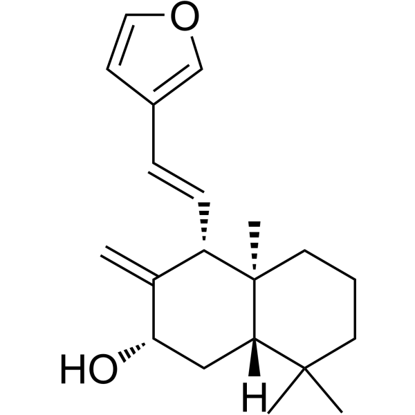
Coronarin A

Based on 1 Customer Validation
Coronarin A is an orally active natural compound that inhibits mTORC1 and S6K1 to increase IRS1 activity. Coronarin A shows anti-inflammatory activity and can also be used for type 2 diabetes mellitus research.
Para uso exclusivo en investigación. No vendemos a pacientes.
- Pureza : ≥98.0%
- No. CAS: 119188-33-9
- Fòrmula: C20H28O2
- Peso molecular:300.44
-
Almacenamiento:Powder -20°C, 3 years ; In solvent -80°C, 6 months , -20°C, 1 month

To place orders, for customer services and technical support, please contact: MedChemExpress USA
Tel: 609-228-6898 E-mail: [email protected] [email protected]


-
 Actividad biológica
-
Chemical Information
-
Solvente y solubilidad
- Protocolo
- Pureza y Documentación
- Referencias
-
 Help & FAQs




Actividad biológica
Descripciòn
IC50 & Target
[1]|
mTORC1 |
S6K1 |
Cellular Effect
|
Cell Line

|
Type | Value | Description | References |
|---|---|---|---|---|
| HUVEC | IC50 |
4.22 μg/mL
Compound: 1
|
Growth inhibitory activity against human umbilical vein endothelial cells (HUVEC) proliferation using MTT colorimetric method
Growth inhibitory activity against human umbilical vein endothelial cells (HUVEC) proliferation using MTT colorimetric method
|
[PMID: 12781184] |
| V79 | IC50 |
1.65 μg/mL
Compound: coronarin A
|
Cytotoxicity against Chinese hamster V79 cells
Cytotoxicity against Chinese hamster V79 cells
|
[PMID: 9834161] |
In Vitro
Coronarin A (3-30 μM; 4 or 12 h) stimulates glycogen synthesis through activating PI3K/Akt/GSK3β signaling and inhibits gluconeogenesis by activating ERK-dependent Wnt/β-catenin/TCF7L2 pathway in rat primary hepatocytes[1].
Coronarin A (1-30 μM; 4 h) increases tyrosine phosphorylation of IRS1 through inhibiting mTOR/S6K1 signaling[1].
MedChemExpress (MCE) has not independently confirmed the accuracy of these methods. They are for reference only. Further protocols information, click here.
-
Cell Line:Primary rat hepatocytes
-
Concentration:1, 3, 10 and 30 μM
-
Incubation Time:4 h
-
Result:Increased the Akt and GSK3β phosphorylation dose-dependently. Dose-dependently stimulated the phosphorylation of both ERK1 and ERK2. Increased the phosphorylation of β-catenin and mitogen-activated protein kinase kinase (MEK). Dose-dependently enhanced the tyrosine phosphorylation of IRS1 at Tyr1222, whereas the serine phosphorylation of IRS1 was dose-dependently inhibited. Reduced the phosphorylation of mTOR, S6K1 and S6.
-
Cell Line:Primary rat hepatocytes
-
Concentration:1, 3, 10, 30, 100 and 300 μM
-
Incubation Time:5.5 h or 12 h
-
Result:Showed no toxicity at 1-30 μM, decreased cell viability after 12 h incubation at 100 μM.
In Vivo
Coronarin A (100 mg/kg; p.o.; once daily for 22 days) inhibits the mTOR/S6K1 pathway to activate PI3K/Akt and ERK/β-catenin signaling in livers of ob/ob mice[1].
Pharmacokinetic properties of Coronarin A after single administrationa in ob/ob mice[1].
| Coronarin A | t1/2 (h) | tmax (h) | Cmax (ng/mL) | AUC0-t (ng·h/mL) | AUC0-∞ (ng·h/mL) | MRT (h) |
| i.p. | 14.8 | 1.0 | 1073 | 4571 | 11045 | 21.7 |
| p.o. | 3.01 | 1.0 | 388 | 1694 | 1856 | 4.88 |
Data are presented as the mean of three mice.
aCoronarin A was intraperitoneally or orally administered at 30 mg/kg to ob/ob mice.
MedChemExpress (MCE) has not independently confirmed the accuracy of these methods. They are for reference only.
-
Animal Model:Male ob/ob mice[1]
-
Dosage:30 mg/kg (IP) or 100 mg/kg (PO)
-
Administration:Oral or intraperitoneal administration, once daily for 22 days
-
Result:Significantly decreased the non-fasting and fasting blood glucose. Significantly reduced the serum insulin concentration at 15 min after glucose loading, reduced the average daily food intake while the body weight was unaffected. Increased hepatic glycogen content and the expression levels of gluconeogenic gene Pck1 and G6pc were significantly decreased.
-
Animal Model:Female ob/ob mice[1]
-
Dosage:30 mg/kg
-
Administration:Intraperitoneal or oral administration (Pharmacokinetic Analysis)
-
Result:Intraperitoneal injection exhibited higher plasma exposure than oral gavage at the same dose of 30 mg/kg, with Cmax value of 1073 and 388 ng/mL, respectively.


Chemical Information
-
No. CAS 119188-33-9
-
Appearance Solid
-
Peso molecular 300.44
-
Fòrmula C20H28O2
-
Color White to off-white
-
SMILES
C[C@@]1([C@H]2/C=C/C3=COC=C3)[C@@](C(C)(CCC1)C)([H])C[C@H](O)C2=C
-
Structure Classification
-
Initial Source
-
Envío
Room temperature in continental US; may vary elsewhere.
-
Almacenamiento
Powder -20°C 3 years In solvent -80°C 6 months -20°C 1 month


Solvente y solubilidad
In Vitro:
DMSO : 10 mg/mL (33.28 mM; Need ultrasonic and warming; Hygroscopic DMSO has a significant impact on the solubility of product, please use newly opened DMSO)
Please refer to the solubility information to select the appropriate solvent. Once prepared, please aliquot and store the solution to prevent product inactivation from repeated freeze-thaw cycles.
Storage method and period of stock solution: -80°C, 6 months; -20°C, 1 month. When stored at -80°C, please use it within 6 months. When stored at -20°C, please use it within 1 month.
Please refer to the solubility information to select the appropriate solvent. Once prepared, please aliquot and store the solution to prevent product inactivation from repeated freeze-thaw cycles.
Storage method and period of stock solution: -80°C, 6 months; -20°C, 1 month. When stored at -80°C, please use it within 6 months. When stored at -20°C, please use it within 1 month.
Concentration (start) × Volume (start) = Concentration (final) × Volume (final)

Protocolo
-
Cotton Pellet Granuloma
Cotton pellet granuloma is a classical in vivo chronic inflammation model used to evaluate the anti-inflammatory potential of test substances by measuring their ability to inhibit granuloma tissue formation around an implanted foreign body (cotton pellet) in rodents. The method is based on the biological response to a sterile implanted material, which induces proliferative phase inflammation characterized by fibroblast proliferation and collagen-rich granuloma formation, and the final readout reflects the extent of chronic inflammatory tissue growth surrounding the pellet. In multiple preclinical pharmacological evaluations, inhibition of cotton pellet-induced granuloma formation has been used as an indicator of anti-inflammatory activity in both synthetic and natural product screening contexts.
-
Carrageenan-Induced Paw Edema
Carrageenan-induced paw edema is an acute inflammation model in which intraplantar injection of carrageenan induces localized inflammatory swelling characterized by vascular permeability, leukocyte infiltration, and production of inflammatory mediators such as prostaglandins and cytokines, making it widely used to evaluate anti-inflammatory agents in vivo. The resulting paw volume or thickness increase is quantified over time as a direct readout of inflammatory intensity and drug efficacy, typically reflecting cyclooxygenase-mediated prostaglandin-driven edema formation and immune cell recruitment in peripheral tissue[20].
-
Research Protocol for Inflammation-related Diseases
The NLRP3 inflammasome is a cytosolic innate immune signaling platform that integrates priming signals and danger-signal activation to promote caspase-1 activation, maturation of IL-1β and IL-18, and gasdermin D-mediated pyroptotic cell death. The core experimental logic is to determine whether inflammatory disease phenotypes are driven by increased NLRP3 expression, ASC-containing inflammasome assembly, caspase-1 cleavage, GSDMD cleavage, and extracellular release of IL-1β/IL-18 rather than by nonspecific cell injury alone. The pathway is strongly linked to inflammation-related disease phenotypes because monosodium urate crystals activate NALP3/NLRP3 inflammasome signaling in gout-like crystal inflammation, cholesterol crystals activate NLRP3 inflammasomes in atherogenesis models, and DSS-induced intestinal inflammation has been reported to involve NLRP3 inflammasome activity. However, experimental colitis studies also show context-dependent protective effects of NLRP3 inflammasome co
-
Research Protocol for Metabolic Diseases
AMP-activated protein kinase, AMPK, is a conserved cellular energy sensor that responds to reduced cellular energy status and coordinates metabolism by increasing ATP-generating catabolic pathways while suppressing ATP-consuming anabolic processes. In metabolic disease research, the AMPK pathway is experimentally relevant because it regulates hepatic lipid synthesis, fatty acid oxidation, glucose production, skeletal-muscle glucose disposal, mTORC1-linked biosynthesis, autophagy, mitochondrial homeostasis, and whole-body energy balance. The central pathway logic is that energy stress, metformin, exercise-like stimulation, or direct AMPK activators increase AMPKα Thr172 phosphorylation and downstream substrate phosphorylation, including ACC and RAPTOR. Phosphorylation of ACC suppresses lipogenesis and supports fatty acid oxidation, whereas phosphorylation of RAPTOR suppresses mTORC1 signaling and links cellular energy status to growth and protein synthesis control. The pathway is linked


Pureza y Documentación
-
Ficha de datos (292 KB)
-
SDS (251 KB)
- English - EN (251 KB)
- Français - FR (251 KB)
- Deutsch - DE (251 KB)
- Norwegian - NO (251 KB)
- Español - ES (251 KB)
- Swedish - SV (251 KB)
- Italian - IT (251 KB)
- Korean - KR (251 KB)
- Portuguese - PT (251 KB)
-
Instrucciones de manejo (2659 KB)
Referencias


Complete Stock Solution Preparation Table
Please refer to the solubility information to select the appropriate solvent. Once prepared, please aliquot and store the solution to prevent product inactivation from repeated freeze-thaw cycles.
Storage method and period of stock solution: -80°C, 6 months; -20°C, 1 month. When stored at -80°C, please use it within 6 months. When stored at -20°C, please use it within 1 month.
| Optional Solvent | Concentration Solvent Mass | 1 mg | 5 mg | 10 mg | 25 mg |
|---|---|---|---|---|---|
| DMSO | 1 mM | 3.3285 mL | 16.6423 mL | 33.2845 mL | 83.2113 mL |
| 5 mM | 0.6657 mL | 3.3285 mL | 6.6569 mL | 16.6423 mL | |
| 10 mM | 0.3328 mL | 1.6642 mL | 3.3285 mL | 8.3211 mL | |
| 15 mM | 0.2219 mL | 1.1095 mL | 2.2190 mL | 5.5474 mL | |
| 20 mM | 0.1664 mL | 0.8321 mL | 1.6642 mL | 4.1606 mL | |
| 25 mM | 0.1331 mL | 0.6657 mL | 1.3314 mL | 3.3285 mL | |
| 30 mM | 0.1109 mL | 0.5547 mL | 1.1095 mL | 2.7737 mL |


Coronarin A Related Classifications
HY-N3628 Related Classifications
 Powered by Bioz