



Galiximab
Based on 1 publication(s) in Google Scholar
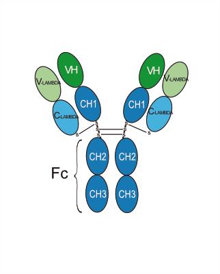
Galiximab (IDEC-114) is a primatized monoclonal IgG1 antibody targeting CD80. Galiximab variable regions are primatized, and the constant regions are humanized. Galiximab induces antibody-dependent cellular cytotoxicity by blocking CD80-CD28 binding. Galiximab has antitumor activity and can induce apoptosis in tumor cells. Galiximab can be used in the research of relapsed Hodgkin lymphoma and solid tumors.
Para uso exclusivo en investigación. No vendemos a pacientes.
- Pureza : 99.23%
- No. CAS: 357613-77-5
- Peso molecular:145.24 kDa
-
Almacenamiento:
Please store the product under the recommended conditions in the Certificate of Analysis.


To place orders, for customer services and technical support, please contact: MedChemExpress USA
Tel: 609-228-6898 E-mail: [email protected] [email protected]


-
 Actividad biológica
-
Chemical Information
- Protocolo
- Pureza y Documentación
- Referencias
-
 Help & FAQs


Publications Citing Use of MedChemExpress (MCE) Galiximab
More

Actividad biológica
Descripciòn
Isotype
Human IgG1 lambda
Recommend Isotype Controls
Species Reactivity
Human
IC50 & Target
CD28L/CD80
In Vitro
Galiximab (25-100 μg/mL; 36 h) in combination with Doxorubicin (HY-15142A) significantly enhances apoptosis in Raji cells[1].
MedChemExpress (MCE) has not independently confirmed the accuracy of these methods. They are for reference only. Further protocols information, click here.
-
Cell Line:Raji cells treated Doxorubicin (HY-15142A) (0.05-1 µg/mL)
-
Concentration:25, 50 and 100 μg/mL
-
Incubation Time:36 h
-
Result:Enhanced cell apoptosis in a dose-dependent manner.
In Vivo
MedChemExpress (MCE) has not independently confirmed the accuracy of these methods. They are for reference only.
-
Animal Model:Female CB17 SCID mice aged 6-8 weeks old treated Raji cells or SKW6.4 cells[1]
-
Dosage:Raji model: 0.1, 1, 3, 10, 30 mg/kg; once weekly; 3 times in total
SKW6.4 model: 5, 10, 30 mg/kg; once weekly; 4 times in total -
Administration:Intraperitoneal injection
-
Result:Showed dose-dependent tumor growth inhibition and survival prolongation in both models.
Ensayo clínico
| NCT Number | Sponsor | Condition | Start Date |
Phase

|
|---|---|---|---|---|
| NCT01329991 | Plexxikon| | 2011-05 | PHASE1 |
Gene ID
Accession
Conjugated
Unconjugated
Reconsititution
The product can be reconstituted/diluted with sterile PBS or saline.
Format
-
Human IgG1 lambda
Aplicación
ELISA, FACS, Functional assay
Verified Bioactivity
-
 Immobilized B7-1/CD80 Protein, Human (HEK293, HY-P70651) can bind Galiximab, The ED50 for this effect is 2.33 ng/mL.
 -
 Flow Cytometry analysis of Raji cells labelling CD28L/CD80 (red) with Galiximab (anti-CD28L/CD80) (HY-P99293). Goat Anti-Human IgG (Alexa Fluor 488) (HY-P83776) at a dilution of 1/1000 was used as the secondary antibody. Blue-Human IgG1 kappa (HY-P99001). Black-Unlabelled control, cells without incubation with primary antibody.


Chemical Information
-
No. CAS 357613-77-5
-
Appearance Liquid
-
Peso molecular 145.24 kDa
-
Color Colorless to light yellow
-
SMILES
[Galiximab]
-
Synonyms
IDEC 114; Anti-Human CD80 Recombinant Antibody
-
Envío
Shipping with dry ice.
-
Formulation
Please refer to the lot-specific COA for specific buffer information.
-
Almacenamiento
Please store the product under the recommended conditions in the Certificate of Analysis.


Publications (1)
-
Journal Impact Factor
-
Most Recent
-
Sci Rep
CD80 (B7-1) as a potential therapeutic target in Epstein-Barr virus‑associated B cell diseases. [Abstract]2026 May 26. PMID: 42191836

Protocolo
-
Apoptosis
Apoptosis, also called programmed cell death, is generally characterized by distinct morphological characteristics.
-
TUNEL staining for apoptotic DNA fragmentation
TUNEL staining detects DNA strand breaks by using terminal deoxynucleotidyl transferase to add labeled nucleotides to exposed 3′-OH DNA termini, generating either microscopic staining in fixed cells or tissue sections, or fluorescence/cytometric signal in cell suspensions. TUNEL positivity reflects DNA fragmentation but should not be interpreted alone as definitive apoptosis, because TUNEL can also label necrotic, autolytic, mechanically damaged, or DNA-repair-associated DNA breaks.
-
Cell Cytotoxicity Assay
Cytotoxicity assays are usually based on the assessment of cell membrane damage, which can also be indirectly detected by measuring cell viability. Detection methods include MTT assay, CKK-8 assay, LDH assay and ATP assay, etc.
-
Mammalian live/dead viability and cytotoxicity staining
Live/dead viability and cytotoxicity staining assays are based on the simultaneous detection of intracellular esterase activity in metabolically active (viable) cells and membrane integrity loss in non-viable cells. In commonly used dual-staining approaches, membrane-permeant fluorogenic substrates are converted by intracellular esterases into fluorescent products in live cells, while impermeant DNA-binding dyes selectively enter cells with compromised plasma membranes and label nucleic acids in dead or dying cells, enabling discrimination between viable and non-viable populations by fluorescence microscopy or flow cytometry.
-
Annexin V plus membrane-impermeant dye apoptosis staining
Annexin V-based apoptosis assays rely on the detection of phosphatidylserine (PS) externalization from the inner leaflet of the plasma membrane to the outer leaflet, an early biochemical hallmark of apoptosis. Fluorescently labeled Annexin V binds PS in a calcium-dependent manner, enabling identification of early apoptotic cells by flow cytometry or fluorescence microscopy. When combined with a membrane-impermeant DNA-binding dye (e. g. , propidium iodide), this approach allows discrimination between viable (Annexin V−/dye−), early apoptotic (Annexin V+/dye−), and late apoptotic or necrotic (Annexin V+/dye+) cell populations by assessing membrane integrity and PS exposure.
-
Apoptosis Solutions
Apoptosis is a regulated, generally non-lytic cell-death pathway that removes unwanted, damaged, infected, or abnormal cells through coordinated morphological changes, caspase activation, DNA fragmentation, and membrane remodeling. The intrinsic apoptosis pathway is controlled mainly by mitochondrial outer membrane permeabilization, BCL-2 family proteins, cytochrome c release, apoptosome formation, caspase-9 activation, and downstream executioner caspase-3/7 activation. The extrinsic apoptosis pathway is initiated by death receptors such as Fas, TNFR, and TRAIL receptors, which recruit adaptor proteins and activate caspase-8 before engaging executioner caspases or mitochondrial amplification through BID cleavage. Apoptosis is linked to many phenotypes, including cancer cell killing, tissue homeostasis, immune regulation, neurodegeneration, infection response, and treatment-induced cytotoxicity; unresolved questions include how apoptosis interacts with necroptosis, pyroptosis, ferroptos
-
Subcutaneous Cell-Line-Derived Xenograft
Subcutaneous cell-line-derived xenograft (CDX) models are established by implanting cultured human cancer cell lines into immunodeficient mice, where the injected cells form localized tumors that can be monitored in vivo as a measure of tumorigenic potential, growth kinetics, and treatment response. These models are widely used in oncology research because they allow reproducible tumor formation and enable comparative assessment of tumor growth between different cell lines or genetic manipulations in a controlled in vivo microenvironment. Subcutaneous implantation of cancer cells in immunodeficient mice is a standard approach for evaluating tumor growth behavior and therapeutic response across multiple cancer types, including prostate, esophageal, pancreatic, and colon cancer models.
-
Cell Viability Determination by MTT Colorimetric Assay
The following protocol uses the MTT colorimetric assay as a classic literature-established method for assessing cell viability/metabolic activity in cultured mammalian cells. MTT[3-(4,5-dimethylthiazol-2-yl)-2,5-diphenyltetrazolium bromide] is reduced by metabolically active cells to a colored formazan product; the amount of formazan is quantified spectrophotometrically and provides an indirect measure of metabolically active viable cells. Importantly, MTT reduction reflects cellular oxidoreductase/metabolic activity rather than an absolute direct count of living cells, so changes in cellular metabolism can alter the signal independently of cell number.


Pureza y Documentación
-
Ficha de datos (276 KB)
-
SDS (252 KB)
- English - EN (252 KB)
- Français - FR (252 KB)
- Deutsch - DE (252 KB)
- Norwegian - NO (252 KB)
- Español - ES (252 KB)
- Swedish - SV (252 KB)
- Italian - IT (252 KB)
- Korean - KR (252 KB)
- Portuguese - PT (252 KB)
-
Inhibitory Antibodies User Guide (603 KB)
Referencias
[1]. Hariharan K, et al. Galiximab (anti-CD80)-induced growth inhibition and prolongation of survival in vivo of B-NHL tumor xenografts and potentiation by the combination with fludarabine. Int J Oncol. 2013 Aug;43(2):670-6. [Content Brief]
[2]. Smith SM, et, al. The anti-CD80 primatized monoclonal antibody, galiximab, is well-tolerated but has limited activity in relapsed Hodgkin lymphoma: Cancer and Leukemia Group B 50602 (Alliance). Leuk Lymphoma. 2013 Jul;54(7):1405-10. [Content Brief]

Calculators
Concentration (start) × Volume (start) = Concentration (final) × Volume (final)


Galiximab Related Classifications
HY-P99293 Related Classifications
 Powered by Bioz