



GlcCer (d18:1/18:0)

Based on 1 Customer Validation
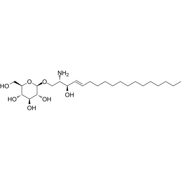
GlcCer (d18:1/18:0) (C18 Glucosyl(β) ceramide (d18:1/18:0)) is a glycosphingolipids that activates the Wnt/β-catenin pathway by targeting LRP6. GlcCer (d18:1/18:0) drives EMT, migration, invasion and GBA1-mediated liver cancer metastasis. GlcCer (d18:1/18:0) accumulates to impair lysosomal function and induce toxic α-synuclein aggregation. GlcCer (d18:1/18:0) supports growth, sporulation, germination and virulence in Penicillium digitatum. GlcCer (d18:1/18:0) is reduced in demyelinated mouse corpus callosum. GlcCer (d18:1/18:0) can be used for the research of liver cancer, synucleinopathies, fungal, Parkinson’s disease and Gaucher disease.
For research use only. We do not sell to patients.
- Purity : 99.93%
- CAS No.: 95119-86-1
- Formula: C42H81NO8
- Molecular Weight:728.09
-
Storage:Powder -20°C, 3 years ; In solvent -80°C, 6 months , -20°C, 1 month


To place orders, for customer services and technical support, please contact: MedChemExpress USA
Tel: 609-228-6898 E-mail: [email protected] [email protected]


-
 Biological Activity
-
Chemical Information
-
Solvent & Solubility
- Protocols
- Purity & Documentation
- References
-
 Help & FAQs




Biological Activity
Description
In Vitro
GlcCer (d18:1/18:0) levels are elevated in both whole cells and the plasma membrane of HepG2 cells with stable GBA1 knockdown, representing the most markedly increased GlcCer subspecies in the plasma membrane and forming an endogenous in situ interaction with the Wnt co-receptor LRP6[1].
GlcCer (d18:1/18:0) (4 days) is produced by wild-type Penicillium digitatum, is absent in PdGcs1 deletion mutants, and is reconstituted in PdGcs1 complement mutants, confirming PdGcs1 encodes the glucosylceramide synthase responsible for its biosynthesis[3].
GlcCer (d18:1/18:0) deficiency in PdGcs1-deleted Penicillium digitatum suppresses vegetative growth, sporulation, and conidial germination, and these defects are fully restored in complemented strains[3].
GlcCer (d18:1/18:0) (4 days) is produced by wild-type Penicillium digitatum PdKH8, is absent in PdGcs1 deletion mutants, and is reconstituted in PdGcs1 complement mutants[4].
GlcCer (d18:1/18:0) (5 days (radial growth and sporulation); 8, 12, 16, 24 hours (conidial germination)) is required for normal vegetative growth, sporulation, and conidial germination in Penicillium digitatum, as evidenced by impaired growth, reduced sporulation, and delayed germination in PdGcs1 deletion mutants (which cannot produce GlcCer (d18:1/18:0)) and restoration of these traits in complement mutants[4].
MedChemExpress (MCE) has not independently confirmed the accuracy of these methods. They are for reference only. Further protocols information, click here.
In Vivo
MedChemExpress (MCE) has not independently confirmed the accuracy of these methods. They are for reference only.


Chemical Information
-
CAS No. 95119-86-1
-
Appearance Solid
-
Molecular Weight 728.09
-
Formula C42H81NO8
-
Color White to off-white
-
SMILES
CCCCCCCCCCCCC/C=C/[C@@H](O)[C@@H](NC(CCCCCCCCCCCCCCCCC)=O)CO[C@@H]1O[C@@H]([C@H]([C@@H]([C@H]1O)O)O)CO
-
Synonyms
C18 Glucosyl(β) ceramide (d18:1/18:0); D-glucosyl-β-1,1' N-stearoyl-D-erythro-sphingosine
-
Shipping
Room temperature in continental US; may vary elsewhere.
-
Storage
Powder -20°C 3 years In solvent -80°C 6 months -20°C 1 month


Solvent & Solubility
In Vitro:
DMSO : 2.5 mg/mL (3.43 mM; Need ultrasonic; Hygroscopic DMSO has a significant impact on the solubility of product, please use newly opened DMSO)
Please refer to the solubility information to select the appropriate solvent. Once prepared, please aliquot and store the solution to prevent product inactivation from repeated freeze-thaw cycles.
Storage method and period of stock solution: -80°C, 6 months; -20°C, 1 month. When stored at -80°C, please use it within 6 months. When stored at -20°C, please use it within 1 month.
Please refer to the solubility information to select the appropriate solvent. Once prepared, please aliquot and store the solution to prevent product inactivation from repeated freeze-thaw cycles.
Storage method and period of stock solution: -80°C, 6 months; -20°C, 1 month. When stored at -80°C, please use it within 6 months. When stored at -20°C, please use it within 1 month.
Concentration (start) × Volume (start) = Concentration (final) × Volume (final)

Protocols
-
Cell migration
Cell migration is a method that plays an important role in wound healing, cell differentiation, embryonic development, etc.
-
Cell invasion
Cell invasion is the ability of cells to migrate from one area to another via the extracellular matrix. Cell invasion is the response of normal and cancer cells to chemical and mechanical stimuli. Before migrating to a new region, the extracellular matrix is degraded by proteases within the cell. Cell invasion often occurs during wound repair, vascularization and inflammation, abnormal tissue invasion, and tumor cell metastasis.
-
Filamentous Fungal Mold Culture and Sporulation
Filamentous fungal mold culture and sporulation assays grow hyphae under defined nutritional and environmental conditions until asexual spores, commonly conidia, are produced; the main readouts are colony growth, sporulation onset, conidial yield, conidial morphology, viability, and, when relevant, downstream infectivity or stress phenotype.
-
Liver Cancer Modeling
Liver cancer can be classified into primary liver cancer and secondary liver cancer. Secondary liver cancer is the metastatic liver cancer. Primary liver cancer includes hepatocellular carcinoma (HCC), intrahepatic cholangiocarcinoma (ICC) and fibrolamellar HCC, of which HCC is the most common form, accounting for approximately 90% of primary liver cancers[1]. HCC mouse models include chemical agent-induced models, transplanted tumor models, and genetic engineered models.
-
Lysosome and acidic-vesicle live-cell staining
Lysosome and acidic-vesicle live-cell staining detects acidic intracellular compartments by using membrane-permeant acidotropic probes that accumulate in low-pH vesicles, including lysosomes, late endosomes, autolysosomes, and acidic phagosomes. LysoTracker staining is commonly used as an intensity-based readout of acidic lysosomal compartment abundance or enlargement, while acridine orange produces green fluorescence in less concentrated compartments and red fluorescence after concentration-dependent accumulation in acidic vesicular organelles. Loss or reduction of acridine-orange red signal can be used as a readout of lysosomal membrane permeabilization or reduced acidic-vesicle integrity. This protocol is designed for live cultured cells and can be adapted for fluorescence microscopy, high-content imaging, plate-reader readout, or flow cytometry when the selected literature supports the readout. Because these dyes report acidotropic accumulation rather than lysosome identity alone,
-
Orthotopic Cell-Line Xenograft
Orthotopic cell-line xenograft models involve implantation of human cancer cell lines into the anatomically corresponding organ of immunodeficient mice to reproduce tumor growth within a native microenvironment, enabling more clinically relevant tumor behavior compared with subcutaneous models. These models are widely used because orthotopic placement better recapitulates tumor progression, including invasion and metastatic spread, which are often underrepresented in heterotopic implantation systems. Compared with conventional xenografts, orthotopic implantation is described as more technically complex but provides improved simulation of tumor-microenvironment interactions and metastatic behavior, making it particularly valuable for translational oncology research. Surgical orthotopic implantation approaches have been emphasized as enabling faithful reproduction of clinical cancer features, including metastasis and disease progression patterns that align with the tumor’s organ of origi


Purity & Documentation
-
Data Sheet (274 KB)
-
SDS (254 KB)
- English - EN (254 KB)
- Français - FR (254 KB)
- Deutsch - DE (254 KB)
- Norwegian - NO (254 KB)
- Español - ES (254 KB)
- Swedish - SV (254 KB)
- Italian - IT (254 KB)
- Korean - KR (254 KB)
- Portuguese - PT (254 KB)
-
Handling Instructions (2659 KB)
References
[1]. Qiu Z, et al. GBA1-dependent membrane glucosylceramide reprogramming promotes liver cancer metastasis via activation of the Wnt/β-catenin signalling pathway. Cell Death Dis. 2022;13(5):508. Published 2022 May 30. [Content Brief]
[2]. Lerche S, et al. CSF d18:1 sphingolipid species in Parkinson disease and dementia with Lewy bodies with and without GBA1 variants. NPJ Parkinsons Dis. 2024;10(1):198. Published 2024 Oct 24. [Content Brief]
[3]. Zhu C, et al. Glucosylceramides are required for mycelial growth and full virulence in Penicillium digitatum. Biochem Biophys Res Commun. 2014;455(3-4):165-171. [Content Brief]
[4]. Zhao Z, et al. Untargeted Metabolomic Profiling of Cuprizone-Induced Demyelination in Mouse Corpus Callosum by UPLC-Orbitrap/MS Reveals Potential Metabolic Biomarkers of CNS Demyelination Disorders. Oxid Med Cell Longev. 2021;2021:7093844. Published 2021 Sep 14. [Content Brief]


Complete Stock Solution Preparation Table
Please refer to the solubility information to select the appropriate solvent. Once prepared, please aliquot and store the solution to prevent product inactivation from repeated freeze-thaw cycles.
Storage method and period of stock solution: -80°C, 6 months; -20°C, 1 month. When stored at -80°C, please use it within 6 months. When stored at -20°C, please use it within 1 month.
| Optional Solvent | Concentration Solvent Mass | 1 mg | 5 mg | 10 mg | 25 mg |
|---|---|---|---|---|---|
| DMSO | 1 mM | 1.3735 mL | 6.8673 mL | 13.7346 mL | 34.3364 mL |


GlcCer (d18:1/18:0) Related Classifications
HY-170035 Related Classifications
 Powered by Bioz
Keywords
- GlcCer (d18:1/18:0)
- 95119-86-1
- C18 Glucosyl(β) ceramide (d18:1/18:0)
- D-glucosyl-β-1,1' N-stearoyl-D-erythro-sphingosine
- Fungal
- Wnt
- β-catenin
- epithelial-mesenchymal transition
- HepG2 liver cancer cells
- CNS demyelinating disorders
- glucocerebrosidase
- alpha-synuclein
- LRP6
- Penicillium digitatum
- liver cancer cells
- GBA1
- Wnt/β-catenin signalling pathway
- Inhibitor
- inhibitor
- inhibit