



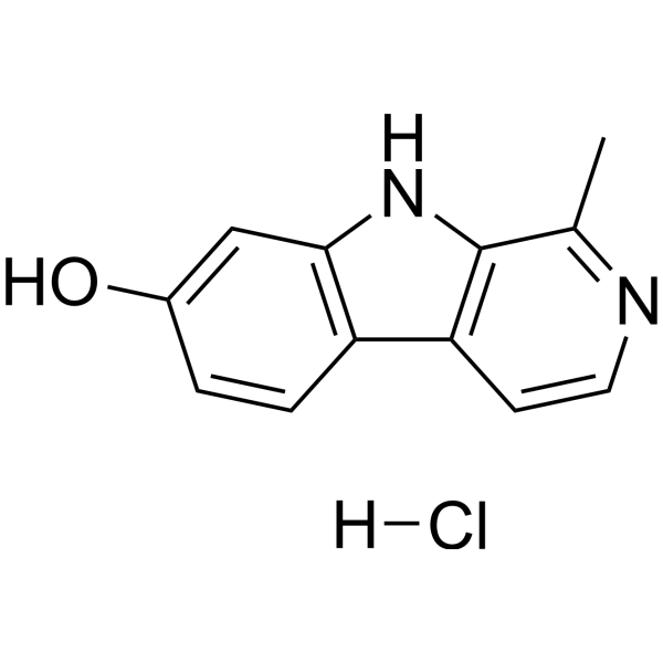
Harmol hydrochloride
Based on 1 publication(s) in Google Scholar
Harmol hydrochloride is an orally active β-carboline alkaloid. Harmol hydrochloride is a TFEB activator and monoamine oxidase inhibitor. Harmol hydrochloride can induce cell mitosis, Autophagy and Apoptosis. Harmol hydrochloride promotes the degradation of α-synuclein by regulating the autophagy-lysosomal pathway. Harmol hydrochloride has anti-tumor, anti-depressant and anti-aging activities. Harmol hydrochloride improves motor impairment in a mouse Parkinson's disease model.
Nur für Forschungszwecke. Wir verkaufen nicht an Patienten.
- CAS. Nr.: 40580-83-4
- Formel: C12H11ClN2O
- Molecular Weight:234.68
-
Speicherung:
Please store the product under the recommended conditions in the Certificate of Analysis.

To place orders, for customer services and technical support, please contact: MedChemExpress USA
Tel: 609-228-6898 E-mail: [email protected] [email protected]


-
 Biologische Aktivität
-
Chemical Information
- Protokoll
- Reinheit & Dokumentation
- Verweise
-
 Help & FAQs


Publications Citing Use of MedChemExpress (MCE) Harmol hydrochloride
More Customer Validation & Images

-
Cell Proliferation/Viability Assay

-
Cell Imaging/Staining

-
RT-PCR

-
WB

-
In Vivo Efficacy Study

Alle α-synuclein Isoform-spezifische Produkte anzeigen
More

Biologische Aktivität
Beschreibung
Cellular Effect
|
Cell Line

|
Type | Value | Description | References |
|---|---|---|---|---|
| Platelet | IC50 |
>130 μM
Compound: Harmol
|
Antiplatelet aggregation activity against collagen-induced platelet
Antiplatelet aggregation activity against collagen-induced platelet
|
[PMID: 20329729] |
| Platelet | IC50 |
130 μM
Compound: Harmol
|
Antiplatelet activity in rabbit platelets assessed as inhibition of collagen-induced platelet aggregation
Antiplatelet activity in rabbit platelets assessed as inhibition of collagen-induced platelet aggregation
|
[PMID: 21983333] |
In Vitro
Harmol (3-30 μM, 6-24 h) hydrochloride reduces α-syn levels in PC12 cells in a dose- and time-dependent manner[1].
Harmol (30 μM, 24 h) hydrochloride promotes the nuclear translocation of exogenous TFEB (transcription factor EB) in HeLa cells and enhances the nuclear translocation of endogenous TFEB in N2a cells, along with a restoration of autophagic flux and lysosomal biogenesis[1].
Harmol (0-100 μM, 24 and 48 h) hydrochloride shows time- and dose-dependent inhibition of U251MG cell proliferation and induces cell death[2].
Harmol (0-100 μM, 0-48 h) hydrochloride inhibits the expression of survivin in U251MG cells and promotes the expression of LC3-I and LC3-II proteins, suppressing the Akt/mTOR pathway and inducing cell autophagy and apoptosis[2].
Harmol (0-100 μM, 0-24 h) hydrochloride exhibits slight cytotoxicity in A549 and H226 cells, but shows strong cytotoxicity in H596, inhibiting tumor proliferation[3].
Harmol (60 μM, 3-6 h) hydrochloride enhances the activity of caspase-3, caspase-6, caspase-8, and caspase-9, increases the cleavage of RAPA (an endogenous substrate for caspase-3), inducing apoptosis in H596 cells[3].
Harmol (1.3 μg/mL, 1-3 h) hydrochloride activates autophagy in C2C12 cells[4].
Harmol (1.3 μg/mL, 45-60 min) hydrochloride activates mitochondria-specific autophagy in C2C12 cells, occurring only in inactive TMRM-negative mitochondria[4].
Harmol (1.3 μg/mL, 16 h) hydrochloride upregulates the expression of markers for mitochondrial function in C2C12 cells[4].
Harmol (1.3 μg/mL, 0-20 h) hydrochloride has a specific inhibitory effect on MAO-B[4].
Harmol hydrochloride reduces GABAergic neurotransmission but does not completely inhibit the binding of GABA to GABAARs; GABAAR activity is crucial for Harmol-mediated mitochondrial depolarization and relies on mitochondrial function[4].
MedChemExpress (MCE) has not independently confirmed the accuracy of these methods. They are for reference only.
-
Cell Line:Tet-on inducible PC12 cells[1]; Hela, N2a[1]; C2C12[4]
-
Concentration:3, 10, 30 μM; 30 μM; 1.3 μg/mL; 1.3 μg/mL
-
Incubation Time:6, 12, 24 h; 24 h; 16 h; 24 h
-
Result:Reduced the phosphorylation (p-) and total α-syn levels in PC12 cells in a dose-dependent and time-dependent manner, showing pro-degradation activity between 6 and 24 hours.
Enhanced the LC3B-Ⅱ/LC3B-Ⅰ ratio in PC12 cells, promoted TFEB expression, and increased AMPK phosphorylation at the THr172 site. It facilitated the nuclear translocation of exogenous TFEB in HeLa cells and the translocation of endogenous TFEB to the nucleus in N2a cells. This also increased the expression of the lysosomal marker LAMP1 (lysosomal-associated membrane protein 1) precursor and the mature form of CTSD (cathepsin D).
Showed in C2C12, the protein expression of the mitochondrial transcription and replication factor TFAM and components of complex III (UQCRC2) and complex V (ATP5a) was significantly elevated, along with an increase in polyamine spermidine levels, while DYRK1A phosphorylation remained unchanged.
-
Cell Line:Hela, PC12
-
Concentration:30 μM
-
Incubation Time:24 h
-
Result:Caused 60% of Flag-TFEB to undergo nuclear translocation and significantly increased the lysosomal content.
In Vivo
Harmol (100 mg/kg, administered by gavage, once daily for three weeks) hydrochloride has a mild anxiety-reducing effect in mice[4].
Harmol (100 mg/kg, orally, for three months) hydrochloride improves insulin, glucose homeostasis, and metabolic adaptations in obese mice, with no impact on kidney function[4].
Harmol (15 μg/kg, orally, from day 0 to 50) hydrochloride extends the lifespan of invertebrates[4].
Harmol (100 mg/kg, orally, for two months) hydrochloride delays frailty in aging mice[4].
MedChemExpress (MCE) has not independently confirmed the accuracy of these methods. They are for reference only.
-
Animal Model:A53T α-syn mice[1]
-
Dosage:10, 20, 40 mg/kg; twice a day; one month
-
Administration:i.g.
-
Result:Made mice climb the rod faster, spent more time on the spinning rod, travelled farther in open areas, increased their step frequency and standing posture, reduced posture width, stride, step length, swing, and showed recovery of autonomous movement behavior.
Reduced the expression of α-syn and p62 in the brain’s substantia nigra and prefrontal cortex.
Increased the phosphorylation levels of AMPK Thr172 and TFEB in the substantia nigra, while decreasing the phosphorylation level of mTOR Ser2448.Increased the expression of LC3B-II/LC3B-I, LAMP1, pro-CTSD, and mature ctsd in the substantia nigra, while reducing the expression of p62 and p-ULK1(Ser757)/ULK1.
-
Animal Model:Male mice[4]
-
Dosage:100 mg/kg, single dose; 100 mg/kg, daily, 3 weeks
-
Administration:i.g.
-
Result:Showed no change in blood glucose levels, with no change in fasting-mediated ketone body increase, and low permeability of the blood-brain barrier. High levels in the liver and plasma, but low levels in the brain. The mitochondrial autophagy marker PINK1 was elevated, while the phosphorylation levels of AMPK target ACC1 or the autophagy marker LC3-II/LC3-I were unaffected.
Showed no change in the time mice spent in the aversive area (center) versus the non-aversive area (border), with no differences in elevated plus maze tests, and significantly less time spent in the dark/light box.
Increased phosphorylation of ACC2 in the liver and BAT, and levels of the mitochondrial autophagy marker PINK1 were also significantly elevated in the liver, BAT, and soleus.
- Detection of Apoptosis
- TUNEL staining for apoptotic DNA fragmentation
- Annexin V plus membrane-impermeant dye apoptosis staining
- Human pluripotent stem cell midbrain dopaminergic neuron differentiation
- Autophagy
- Apoptosis Solutions
- Lysosome and acidic-vesicle live-cell staining
- Macroautophagy Solutions
- How to Choose the Right Model Animal


Chemical Information
-
CAS. Nr. 40580-83-4
-
Molecular Weight 234.68
-
Formel C12H11ClN2O
-
SMILES
OC1=CC2=C(C=C1)C3=C(C(C)=NC=C3)N2.[H]Cl
-
Versand
Room temperature in continental US; may vary elsewhere.
-
Speicherung
Please store the product under the recommended conditions in the Certificate of Analysis.


Publications (1)
-
Journal Impact Factor
-
Most Recent
-
Virol J
2024 May 27;21(1):118. PMID: 38802860
Harmol hydrochloride purchased from MedChemExpress. Usage Cited in: Virol J. 2024 May 27;21(1):118. [Abstract]
Vero cells in 96-well plate were cultured with Harmol (0, 6.25, 12.5, 25.0, 50.0, 100.0, 150.0, 200.0, 250.0 µM) for 48 h. Cell viability was measured using a CCK-8 assay.
Harmol hydrochloride purchased from MedChemExpress. Usage Cited in: Virol J. 2024 May 27;21(1):118. [Abstract]
Vero cells were infected with HSV-1 F (MOI = 0.5) and then they were treated with Harmol (12.5 µM) or ACV (1 µM) for 24 h. Images were obtained by inverted microscope.
Harmol hydrochloride purchased from MedChemExpress. Usage Cited in: Virol J. 2024 May 27;21(1):118. [Abstract]
The expression of gD-1 mRNA was detected by qPCR analysis treated with Harmol (0, 3.125, 6.25, 12.5 μM).
Harmol hydrochloride purchased from MedChemExpress. Usage Cited in: Virol J. 2024 May 27;21(1):118. [Abstract]
An MOI of 0.5 of HSV-1 F-infected Vero cells were treated with 0.02% DMSO, Harmol (0, 3.12, 6.25, 12.5 µM), ACV (1.0 µM) and Harmol (3.12, 6.25, 12.5 µM) + ACV (1.0 µM) for 48 h. The gD-1 protein expression was detected by Western Blot assay.
Harmol hydrochloride purchased from MedChemExpress. Usage Cited in: Virol J. 2024 May 27;21(1):118. [Abstract]
Corneal images at 3 dpi and 5 dpi were obtained following HSV-1 infection treated with Harmol (0.01 mg/kg, 5 µL/mouse).


Reinheit & Dokumentation
Verweise
[1]. Jie Xu,et al. Harmol promotes α-synuclein degradation and improves motor impairment in Parkinson's models via regulating autophagy-lysosome pathway. NPJ Parkinsons Dis. 2022 Aug 6;8(1):100. [Content Brief]
[2]. Akihisa Abe ,et al. Harmol induces autophagy and subsequent apoptosis in U251MG human glioma cells through the downregulation of surviving. Oncol Rep. 2013 Apr;29(4):1333-42. [Content Brief]
[3]. Akihisa Abe,et al. Harmol induces apoptosis by caspase-8 activation independently of Fas/Fas ligand interaction in human lung carcinoma H596 cells. Anticancer Drugs. 009 Jun;20(5):373-81. [Content Brief]
[4]. Luis Filipe Costa-Machado, et al. Peripheral modulation of antidepressant targets MAO-B and GABAAR by harmol induces mitohormesis and delays aging in preclinical models. Nat Commun. 2023 May 15;14(1):2779. [Content Brief]

Calculators
Konzentration (Stammlösung) × Volumen (Stammlösung) = Konzentration (Ziellösung) × Volumen (Ziellösung)


Harmol hydrochloride Related Classifications
HY-107811A Related Classifications
 Powered by Bioz