



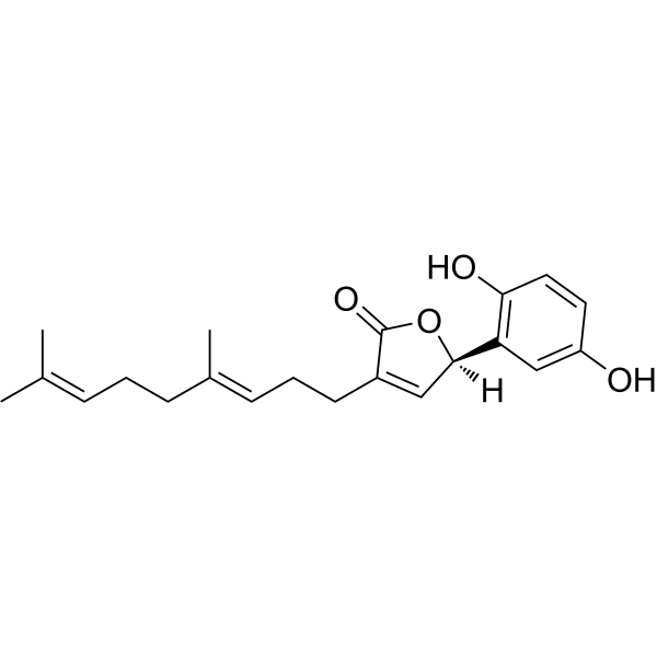
Ganomycin I
Ganomycin I is a non-competitive dual inhibitor of α-glucosidase and HMG-CoA reductase, with IC50 values of 0.3 μM and 12.3 μM, respectively. Ganomycin I inhibits HIV-1 protease, with an IC50 of 7.5 μg/mL. Ganomycin I also suppresses RANKL-induced osteoclast differentiation and bone resorption by inhibiting the activation of ERK, JNK and p38 MAPK, as well as downregulating the c-Fos/NFATc1 signaling pathway. Ganomycin I exhibits antibacterial activity against Gram-positive bacteria, and shows weak activity against Mycobacterium tuberculosis H37Ra. It can be used in research related to metabolic diseases, bone metabolism and anti-infection.
For research use only. We do not sell to patients.
- CAS No.: 1191255-15-8
- Formula: C21H26O4
- Molecular Weight:342.43
-
Storage:
Please store the product under the recommended conditions in the Certificate of Analysis.

To place orders, for customer services and technical support, please contact: MedChemExpress USA
Tel: 609-228-6898 E-mail: [email protected] [email protected]


-
 Biological Activity
-
Chemical Information
- Protocols
- Purity & Documentation
- References
-
 Help & FAQs


-
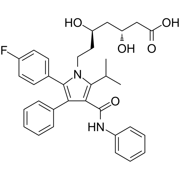
HY-B0589
AtorvastatinHMG-CoA Reductase Inhibitor -
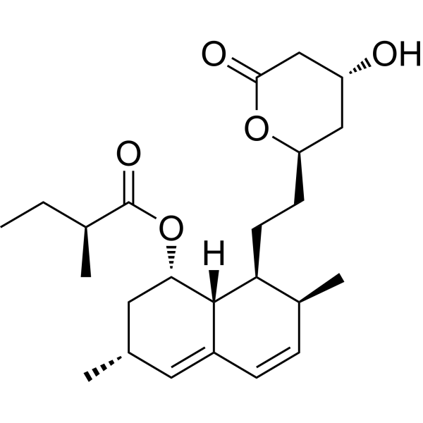
HY-N0504
LovastatinHMG-CoA Inhibitor -
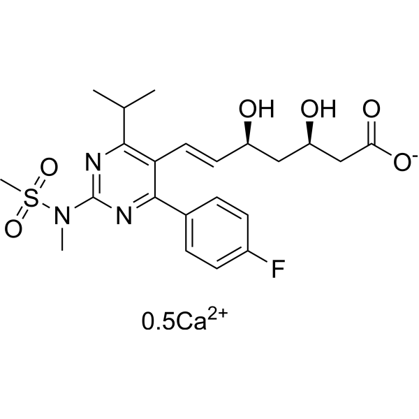
HY-17504
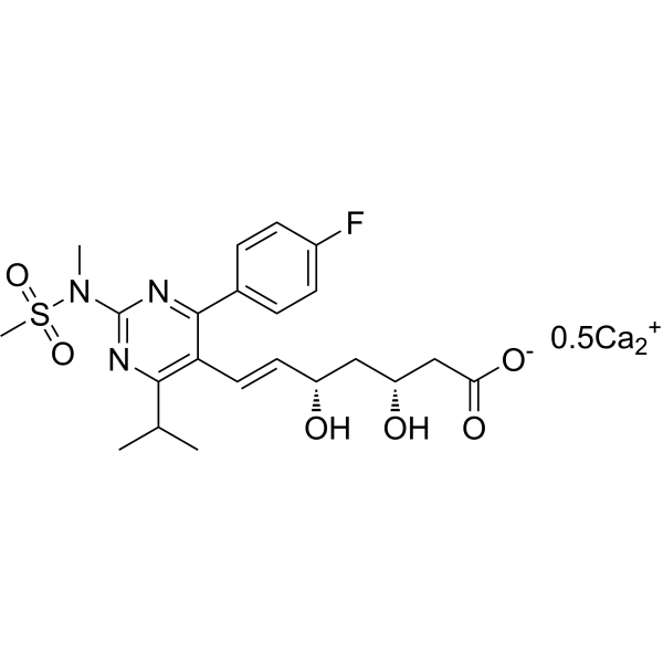
Rosuvastatin CalciumHMGCR Inhibitor -
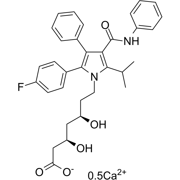
HY-17379
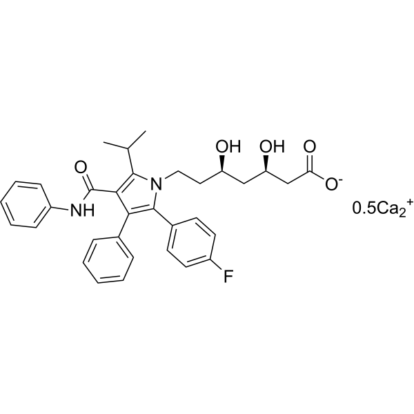
Atorvastatin hemicalcium saltHMGCR Inhibitor -
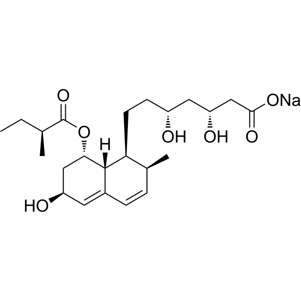
HY-B0165A
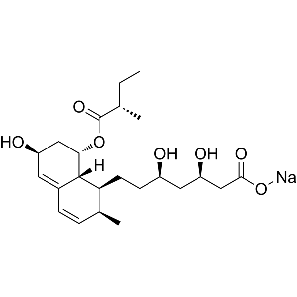
Pravastatin sodiumHMG-CoA Reductase (HMGCR) Inhibitor -
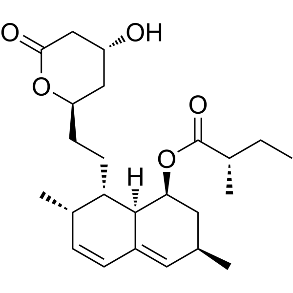
HY-N0504R
Lovastatin (Standard) -
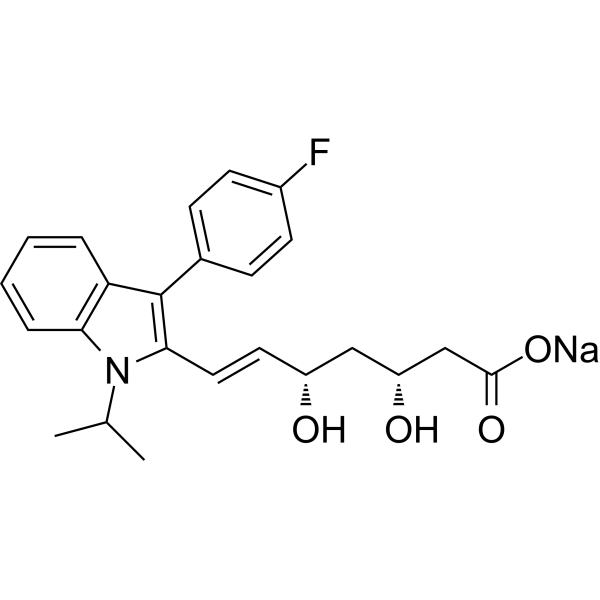
HY-14664A
Fluvastatin sodiumHMG-CoA Reductase Inhibitor -
HY-B1189
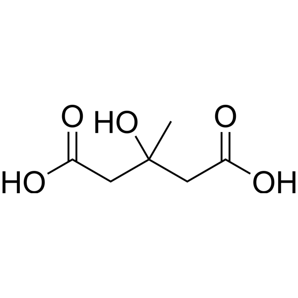
MeglutolEndogenous Metabolite -
HY-B0165AR
Pravastatin sodium (Standard) -
HY-17504R
Rosuvastatin Calcium (Standard) -
HY-13528
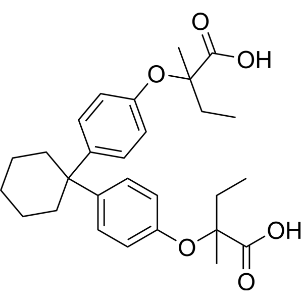
ClinofibrateHMG-CoA Reductase (HMGCR) Inhibitor -
HY-17379R
Atorvastatin hemicalcium salt (Standard)HMGCR Inhibitor


Biological Activity
Description
|
α‑glucosidase 0.38 μM (IC50, rat small intestinal mucosa) |
α‑glucosidase 0.3 μM (IC50, Baker's yeast) |
α‑glucosidase 0.4 μM (IC50, rat small intestinal mucosa) |
HMG-CoA reductase 12.3 μM (IC50) |
HIV-1 Protease 7.5 μg/mL (IC50) |
In Vitro
Ganomycin I inhibits Baker's yeast α-glucosidase and rat small intestinal mucosa α-glucosidase with IC50 values of 0.3 μM and 0.4 μM, respectively; it also inhibits rat intestinal sucrase and maltase with IC50 values of 67.6 μM and 3.6 μM, respectively[3].
Ganomycin I (3-30 μM; 6 days) inhibits RANKL-induced osteoclastogenesis in mouse bone marrow macrophages (BMMs) in a dose-dependent manner by reducing the number of mature osteoclasts and downregulating the expression of the osteoclast marker proteins c-Src and CtsK[2].
Ganomycin I (3-30 μM; 4 days) inhibits RANKL-induced osteoclast formation in mouse RAW264.7 cells in a dose-dependent manner[2].
Ganomycin I (3-30 μM; 72 h) increases the cell viability of mouse bone marrow macrophages (BMMs) after incubation at concentrations of 3, 10, and 30 μM for 72 h, indicating that its anti-osteoclastogenic activity does not originate from cytotoxicity[2].
Ganomycin I (3-30 μM; 7 days) dose-dependently inhibits RANKL-induced bone resorptive activity in mouse bone marrow macrophages (BMMs) cultured on OsteoAssay Surface plates[2].
Ganomycin I (3-30 μM; 7 days) dose-dependently inhibits RANKL-induced actin ring formation in mouse bone marrow macrophages (BMMs), where actin ring serves as a marker of mature osteoclasts[2].
Ganomycin I (3-30 μM; pretreated prior to RANKL stimulation) dose-dependently inhibits RANKL-induced activation of ERK, JNK and p38 MAPKs, as well as the downstream expression of c-Fos and NFATc1, without affecting the activation of the NF-κB pathway in mouse RAW264.7 cells[2].
Ganomycin I (3-30 μM; pretreated prior to RANKL stimulation) dose-dependently inhibits RANKL-induced nuclear translocation of NFATc1 in mouse RAW264.7 cells[2].
Ganomycin I (3-30 μM; 4 days) dose-dependently reduces the expression of RANKL-induced NFATc1 target genes DC-STAMP, OSCAR, MMP-9 and TRAP in mouse bone marrow-derived macrophages (BMMs)[2].
Ganomycin I (compound 2) shows no activity against porcine pancreatic lipase[5].
Ganomycin I (50 μg/mL) exhibits no cytotoxicity toward non-malignant Vero cells[6].
Ganomycin I exhibits cytotoxicity against E-PR293 cells, so its anti-HIV-1 protease activity cannot be determined in this cell system[4].
Ganomycin I inhibits recombinant HIV-1 protease expressed in E. coli, with an IC50 of 7.5 µg/mL, and forms hydrogen bonds with the Asp30 residue at the active site of this enzyme[7].
Ganomycin I exhibits weak activity against Mycobacterium tuberculosis H37Ra with an MIC of 50 μg/mL; its MIC values against Gram-positive bacteria Bacillus cereus, Enterococcus faecium and Staphylococcus aureus are 25, 25 and 12.5 μg/mL, respectively[6].
Ganomycin I inhibits HMG-CoA reductase with an IC50 of 12.3 μM[3].
Ganomycin I exhibits non-competitive inhibition against both α-glucosidase and HMG-CoA reductase; kinetic assays use Ganomycin I at 0.3-1 μM for α-glucosidase detection and 5-20 μM for HMG-CoA reductase detection[3].
MedChemExpress (MCE) has not independently confirmed the accuracy of these methods. They are for reference only.
-
Cell Line:mouse BMMs
-
Concentration:3, 10, 30 μM
-
Incubation Time:72 h
-
Result:Did not decrease cell viability of BMMs.
Increased cell viability at the tested concentrations.
-
Cell Line:mouse RAW264.7 cells
-
Concentration:3, 10, 30 μM
-
Incubation Time:10 min, 15 min,24 h, 48 h, 4 d
-
Result:Significantly decreased RANKL-induced phosphorylation levels of ERK, JNK, and p38 MAPKs in a concentration-dependent manner.
Attenuated RANKL-induced expression of c-Fos and NFATc1.
Did not block RANKL-induced degradation of IκBα.
-
Cell Line:mouse RAW264.7 cells
-
Concentration:30 μM
-
Incubation Time:48 h
-
Result:Significantly suppressed RANKL-induced nuclear translocation of NFATc1.
-
Cell Line:mouse BMMs
-
Concentration:3, 10, 30 μM
-
Incubation Time:4 days
-
Result:Significantly inhibited the expression of DC-STAMP, OSCAR, MMP-9, and TRAP genes in a concentration-dependent manner during RANKL-induced osteoclastogenesis.
In Vivo
Ganomycin I (1-5 mg/kg; p.o.; daily; 21-28 days) exerts potent hypoglycemic, hypolipidemic, insulin-sensitizing, and serum AST-lowering effects in KK-Ay mice; the insulin-sensitizing efficacy of the 5 mg/kg dose is superior to that of thiazolidinedione insulin sensitizers, both doses increase hepatic glycogen levels after 4 weeks of administration, and no reduction in serum ALT levels is observed[3].
No obvious abnormalities in behavior, feces and urine, convulsions, sleep or coma are observed, and no deaths are recorded within the 24 h and subsequent 14-day observation period after a single oral dose of Ganomycin I up to 3 g/kg[3].
MedChemExpress (MCE) has not independently confirmed the accuracy of these methods. They are for reference only.
-
Animal Model:C57BL/6J mice (male; 8 weeks old; high-fat diet-induced obesity)[1]
-
Dosage:3.0 mg/kg
-
Administration:p.o.; daily; 5 weeks
-
Result:Exhibited similar efficacy to compound 7d in improving hyperglycemia and hyperlipidemia in diet-induced obese mice.
-
Animal Model:8-week-old insulin-resistant male KK-Ay mice; C57BL/6J normal control[3]
-
Dosage:1, 5 mg/kg
-
Administration:p.o.; daily; 21 days
-
Result:Reduced fasting/free-feeding glucose and HbA1c.
Improved OSTT, OGTT, ITT, serum insulin and ISI.
Reduced NEFA, TG, TC and LDL-C.
Increased hepatic glycogen.
Decreased AST.
Improved hepatic steatosis and reduced WAT adipocyte size.


Chemical Information
-
CAS No. 1191255-15-8
-
Molecular Weight 342.43
-
Formula C21H26O4
-
SMILES
OC1=C(C=C(C=C1)O)[C@@]2([H])C=C(C(O2)=O)CC/C=C(C)/CC/C=C(C)/C
-
Structure Classification
-
Initial Source
Ganoderma colossum
-
Shipping
Room temperature in continental US; may vary elsewhere.
-
Storage
Please store the product under the recommended conditions in the Certificate of Analysis.


Purity & Documentation
References
[1]. Wang K, et al. Structural Modification of Natural Product Ganomycin I Leading to Discovery of a α-Glucosidase and HMG-CoA Reductase Dual Inhibitor Improving Obesity and Metabolic Dysfunction in Vivo. Journal of medicinal chemistry. 2018 Apr 26;61(8):3609-3625. [Content Brief]
[2]. Tran PT, et al. Ganomycin I from Ganoderma lucidum attenuates RANKL-mediated osteoclastogenesis by inhibiting MAPKs and NFATc1. Phytomedicine : international journal of phytotherapy and phytopharmacology. 2019 Mar 01;55:1-8. [Content Brief]

Calculators
Concentration (start) × Volume (start) = Concentration (final) × Volume (final)


Ganomycin I Related Classifications
HY-N10076 Related Classifications
 Powered by Bioz