



Isosaponarin
Based on 1 publication(s) in Google Scholar
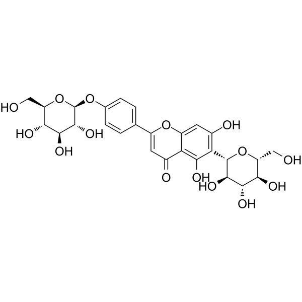
Isosaponarin is a flavone glycoside isolated from wasabi leaves. Isosaponarin is a P4HA2 enzymatic agonist. Isosaponarin increases collagen synthesis via up-regulated TGF-β type II receptor (TβR-II) and prolyl 4-hydroxylase (P4H) proteins production, promoting skin health and wound healing. Isosaponarin-rich plants exhibit strong antimicrobial, antioxidant, anti-hyaluronidase, antiplatelet, anti-atopic dermatitis, and anti-tumor effects.
For research use only. We do not sell to patients.
- Purity : 99.46%
- CAS No.: 19416-87-6
- Formula: C27H30O15
- Molecular Weight:594.52
-
Storage:
4°C, protect from light
* In solvent : -80°C, 6 months; -20°C, 1 month (protect from light)

To place orders, for customer services and technical support, please contact: MedChemExpress USA
Tel: 609-228-6898 E-mail: [email protected] [email protected]


-
 Biological Activity
-
Chemical Information
-
Solvent & Solubility
- Protocols
- Purity & Documentation
- References
-
 Help & FAQs


-
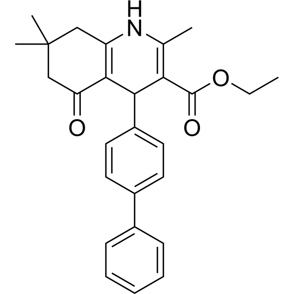
HY-12704
ITD-1TGF-β Receptor Inhibitor -
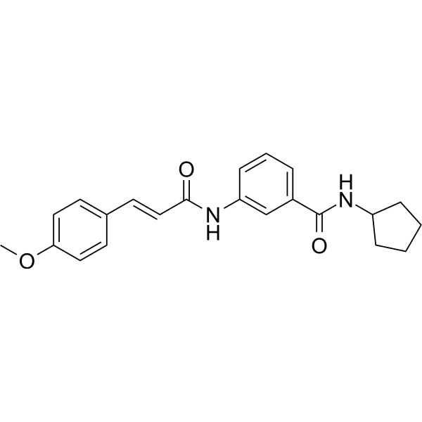
HY-151427
TGFβ1-IN-1TGFβ1 Inhibitor -
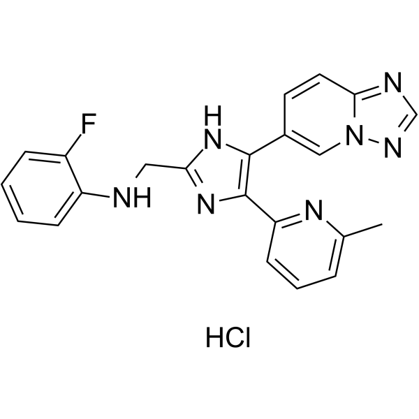
HY-19928A
Vactosertib HydrochlorideALK5 Inhibitor -
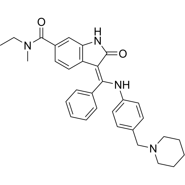
HY-13783
BIBF0775TGF-β Receptor Inhibitor -
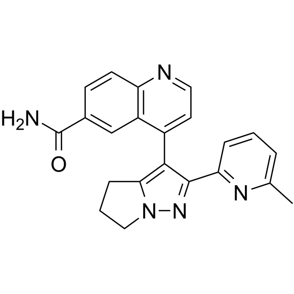
HY-13226
GalunisertibTGFβRI Inhibitor -
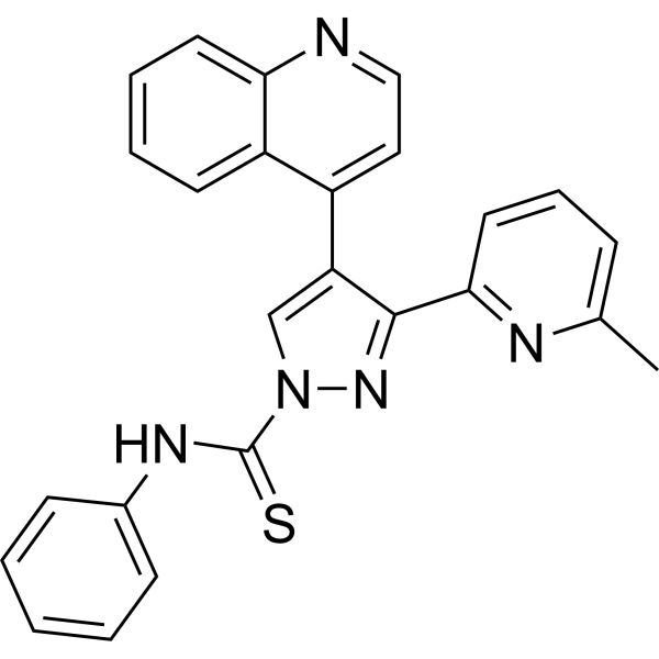
HY-10432
A 83-01ALK4/5/7 Inhibitor -
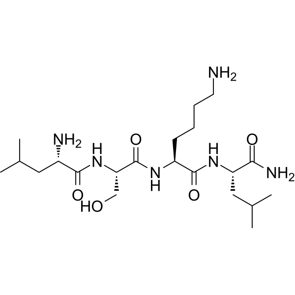
HY-P0299
LSKL, Inhibitor of Thrombospondin (TSP-1)TGF-β1 Antagonist -
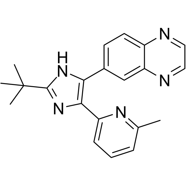
HY-12043
SB 525334ALK5 Inhibitor -
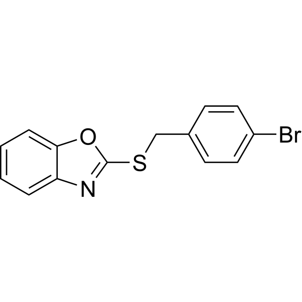
HY-124697
BMP signaling agonist sb4BMP Agonist -
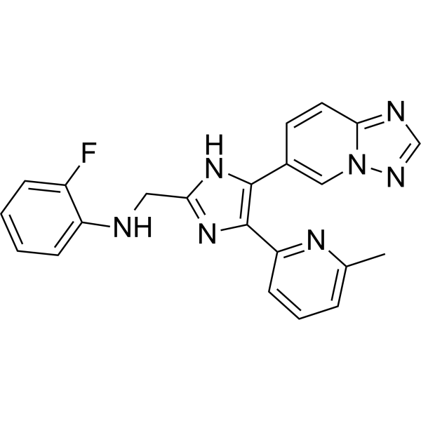
HY-19928
VactosertibALK5 Inhibitor -
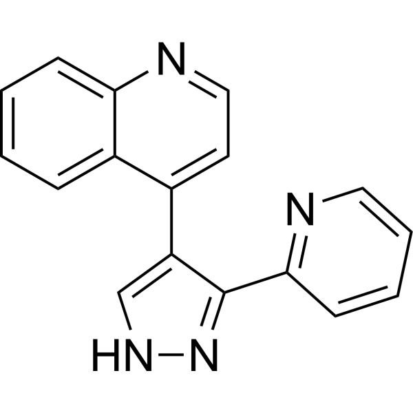
HY-13462
LY-364947TGFβR-I Inhibitor -
HY-10326
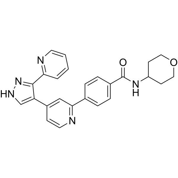
GW788388ALK5 Inhibitor
Publications Citing Use of MedChemExpress (MCE) Isosaponarin
More

Biological Activity
Description
IC50 & Target
Collagen synthesis[1]
In Vitro
Isosaponarin (0.001-0.1 μg/mL, 24 h) increases collagen synthesis in human fibroblasts, caused by up-regulated TβR-II and P4H production[1].
Isosaponarin (10-100 μM, added 10 min before 4-Aminopyridine (HY-B0604) addition) suppresses the Ca2+-dependent PKC/SNAP-25 and MARCKS pathways in synaptosomes, causing a decrease in the number of available synaptic vesicles, which inhibits vesicular glutamate release from synaptosomes[2].
MedChemExpress (MCE) has not independently confirmed the accuracy of these methods. They are for reference only. Further protocols information, click here.
-
Cell Line:Human skin fibroblast cells (neonatal foreskin)
-
Concentration:0.001, 0.01, 0.1 μg/mL
-
Incubation Time:24 h
-
Result:Significantly increased TβR-II protein level at the concentrations of 0.001-0.01 μg/mL.
P4Hα (I) production was significantly increased at the concentrations of 0.01 μg/mL.
-
Cell Line:Human skin fibroblast cells (neonatal foreskin)
-
Concentration:0.001, 0.01, 0.1 μg/mL
-
Incubation Time:24 h
-
Result:Significantly up-regulated the expression of procollagen α1 (I) mRNA at the concentration of 0.001 μg/mL.
significantly up-regulated TβR-II mRNA level at the concentration of 0.001 μg/mL.
P4Hα (I) mRNA was increased at the concentrations of 0.001-0.1 μg/mL.
In Vivo
MedChemExpress (MCE) has not independently confirmed the accuracy of these methods. They are for reference only.


Chemical Information
-
CAS No. 19416-87-6
-
Appearance Solid
-
Molecular Weight 594.52
-
Formula C27H30O15
-
Color White to yellow
-
SMILES
O=C1C=C(C2=CC=C(O[C@H]3[C@@H]([C@H]([C@@H]([C@@H](CO)O3)O)O)O)C=C2)OC4=CC(O)=C([C@H]5[C@@H]([C@H]([C@@H]([C@@H](CO)O5)O)O)O)C(O)=C14
-
Structure Classification
-
Initial Source
-
Shipping
Room temperature in continental US; may vary elsewhere.
-
Storage
4°C, protect from light
* In solvent : -80°C, 6 months; -20°C, 1 month (protect from light)


Publications (1)
-
Journal Impact Factor
-
Most Recent


Solvent & Solubility
In Vitro:
DMSO : 125 mg/mL (210.25 mM; Need ultrasonic; Hygroscopic DMSO has a significant impact on the solubility of product, please use newly opened DMSO)
Please refer to the solubility information to select the appropriate solvent. Once prepared, please aliquot and store the solution to prevent product inactivation from repeated freeze-thaw cycles.
Storage method and period of stock solution: -80°C, 6 months; -20°C, 1 month (protect from light). When stored at -80°C, please use it within 6 months. When stored at -20°C, please use it within 1 month.
Please refer to the solubility information to select the appropriate solvent. Once prepared, please aliquot and store the solution to prevent product inactivation from repeated freeze-thaw cycles.
Storage method and period of stock solution: -80°C, 6 months; -20°C, 1 month (protect from light). When stored at -80°C, please use it within 6 months. When stored at -20°C, please use it within 1 month.
Concentration (start) × Volume (start) = Concentration (final) × Volume (final)

Protocols
-
Research Protocol for Infectious Diseases
Infectious-disease experiments test how pathogens interact with host barriers, innate immune receptors, inflammatory signaling, pathogen replication, and tissue injury; pattern-recognition receptors such as TLRs, RIG-I-like receptors, NOD-like receptors, and inflammasomes detect microbial molecules and activate NF-κB, interferon, and cytokine responses. The central hypothesis is that infection severity reflects the balance between pathogen burden and host response: protective inflammation restricts pathogen growth, whereas excessive or mislocalized inflammation contributes to tissue damage and disease phenotype. Unresolved questions include which host pathways are protective versus pathogenic, why some infection models fail to translate to human disease, and which combined readouts best predict clinically relevant infection outcomes.
-
Cell migration
Cell migration is a method that plays an important role in wound healing, cell differentiation, embryonic development, etc.
-
TPA/Croton Oil Ear Edema and Dermatitis
The TPA (12-O-tetradecanoylphorbol-13-acetate) and croton oil-induced mouse ear edema model is a well-established acute cutaneous inflammation system used to evaluate topical anti-inflammatory activity by measuring edema formation, neutrophil infiltration, vascular permeability, and cytokine-mediated skin responses in vivo. The inflammatory response is triggered by topical application of phorbol esters (TPA) or croton oil constituents, leading to rapid activation of protein kinase C signaling, leukocyte recruitment, and increased vascular permeability, which can be quantified by ear thickness, weight, dye extravasation, and biochemical markers such as myeloperoxidase (MPO) activity and pro-inflammatory mediators in ear tissue homogenates. This model is widely used for screening anti-inflammatory agents, where reductions in edema and inflammatory biomarkers reflect suppression of acute dermal inflammation and immune cell infiltration. Histological evaluation typically confirms epidermal
-
Cell-Exclusion Zone Migration Assay
The Cell-Exclusion Zone (CEZ) migration assay is an in vitro 2D cell migration method in which a defined cell-free area is created using removable physical barriers such as silicone stoppers, allowing cells to be seeded around the barrier and subsequently migrate into the cleared zone after barrier removal. This approach enables quantification of collective cell migration by monitoring repopulation of the initially cell-free region over time using microscopy-based imaging. Compared with scratch-based wound healing assays, barrier-based exclusion methods are designed to avoid mechanical damage to the extracellular matrix and reduce injury-induced effects on boundary cells, thereby improving interpretability of migration behavior in vitro. The assay readout is typically the progressive reduction in the cell-free area or the number of cells invading the exclusion zone, reflecting coordinated cell motility relevant to physiological processes such as wound healing, epithelial repair, and ca


Purity & Documentation
-
Data Sheet (275 KB)
-
SDS (252 KB)
- English - EN (252 KB)
- Français - FR (252 KB)
- Deutsch - DE (252 KB)
- Norwegian - NO (252 KB)
- Español - ES (252 KB)
- Swedish - SV (252 KB)
- Italian - IT (252 KB)
- Korean - KR (252 KB)
- Portuguese - PT (252 KB)
-
Handling Instructions (2659 KB)
References
[1]. Masashi Nagai, et al. The effect of isosaponarin isolated from wasabi leaf on collagen synthesis in human fibroblasts and its underlying mechanism. J Nat Med. 2010 Jul;64(3):305-12. [Content Brief]
[2]. Lu CW, et al. The Effect of Isosaponarin Derived from Wasabi Leaves on Glutamate Release in Rat Synaptosomes and Its Underlying Mechanism. Int J Mol Sci. 2022 Aug 6;23(15):8752. [Content Brief]
[4]. Chi Z, et al. P4HA2 promotes proliferation, invasion, and metastasis through regulation of the PI3K/AKT signaling pathway in oral squamous cell carcinoma. Sci Rep. 2024 Jul 1;14(1):15023. [Content Brief]


Complete Stock Solution Preparation Table
Please refer to the solubility information to select the appropriate solvent. Once prepared, please aliquot and store the solution to prevent product inactivation from repeated freeze-thaw cycles.
Storage method and period of stock solution: -80°C, 6 months; -20°C, 1 month (protect from light). When stored at -80°C, please use it within 6 months. When stored at -20°C, please use it within 1 month.
| Optional Solvent | Concentration Solvent Mass | 1 mg | 5 mg | 10 mg | 25 mg |
|---|---|---|---|---|---|
| DMSO | 1 mM | 1.6820 mL | 8.4101 mL | 16.8203 mL | 42.0507 mL |
| 5 mM | 0.3364 mL | 1.6820 mL | 3.3641 mL | 8.4101 mL | |
| 10 mM | 0.1682 mL | 0.8410 mL | 1.6820 mL | 4.2051 mL | |
| 15 mM | 0.1121 mL | 0.5607 mL | 1.1214 mL | 2.8034 mL | |
| 20 mM | 0.0841 mL | 0.4205 mL | 0.8410 mL | 2.1025 mL | |
| 25 mM | 0.0673 mL | 0.3364 mL | 0.6728 mL | 1.6820 mL | |
| 30 mM | 0.0561 mL | 0.2803 mL | 0.5607 mL | 1.4017 mL | |
| 40 mM | 0.0421 mL | 0.2103 mL | 0.4205 mL | 1.0513 mL | |
| 50 mM | 0.0336 mL | 0.1682 mL | 0.3364 mL | 0.8410 mL | |
| 60 mM | 0.0280 mL | 0.1402 mL | 0.2803 mL | 0.7008 mL | |
| 80 mM | 0.0210 mL | 0.1051 mL | 0.2103 mL | 0.5256 mL | |
| 100 mM | 0.0168 mL | 0.0841 mL | 0.1682 mL | 0.4205 mL |


Isosaponarin Related Classifications
HY-N2589 Related Classifications
 Powered by Bioz