



Cefoselis
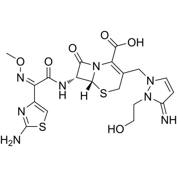
Cefoselis, the fourth gen-eration of cephalosporin, is a β-lactam antibiotic. Cefoselis exhibits good activity against a wide range of Gram-positive and Gram-negative organisms. Cefoselis penetrates the blood-brain barrier.
For research use only. We do not sell to patients.
- CAS No.: 122841-10-5
- Formula: C19H22N8O6S2
- Molecular Weight:522.56
-
Storage:
Please store the product under the recommended conditions in the Certificate of Analysis.

To place orders, for customer services and technical support, please contact: MedChemExpress USA
Tel: 609-228-6898 E-mail: [email protected] [email protected]


-
 Biological Activity
-
Chemical Information
- Protocols
- Purity & Documentation
- References
-
 Help & FAQs


All Antibiotic Isoforms
More

Biological Activity
Description
IC50 & Target
|
β-lactam |
In Vitro
Cefoselis, a new parenteral cephalosporin, was active against clinical isolates of both gram-positive and gram-negative aerobic bacteria. The activity of Cefoselis was similar to that of cefpirome and cefepime and generally superior to that of ceftazidime. Cefoselis showed potent antibacterial activity against Hemophilus influenzae and Moraxella catarrhalis.Cefoselis was highly active against MSSA and MSCNS. Cefoselis was poor in the activity against MRSA,MRCNS,PRSP and Enterococcus faecalis,and no activity for Enterococcus faecium.
MedChemExpress (MCE) has not independently confirmed the accuracy of these methods. They are for reference only.


Chemical Information
-
CAS No. 122841-10-5
-
Molecular Weight 522.56
-
Formula C19H22N8O6S2
-
SMILES
O=C(C(N12)=C(CN3N(CCO)C(C=C3)=N)CS[C@]2([H])[C@H](NC(/C(C4=CSC(N)=N4)=N\OC)=O)C1=O)O
-
Shipping
Room temperature in continental US; may vary elsewhere.
-
Storage
Please store the product under the recommended conditions in the Certificate of Analysis.


Purity & Documentation
References
[1]. K Ohtaki, et al. Cefoselis, a beta-lactam antibiotic, easily penetrates the blood-brain barrier and causes seizure independently by glutamate release. J Neural Transm (Vienna). 2004 Dec;111(12):1523-35. [Content Brief]
[3]. King, A., L. Bethune, and I. Phillips, The Comparative In Vitro Activity of FK-037 (Cefoselis), a New Broad-Spectrum Cephalosporin. Clin Microbiol Infect, 1995. 1(1): p. 13-17. [Content Brief]

Calculators
Concentration (start) × Volume (start) = Concentration (final) × Volume (final)


Cefoselis Related Classifications
HY-B0186 Related Classifications
 Powered by Bioz