



Flixebrutinib
Based on 1 publication(s) in Google Scholar
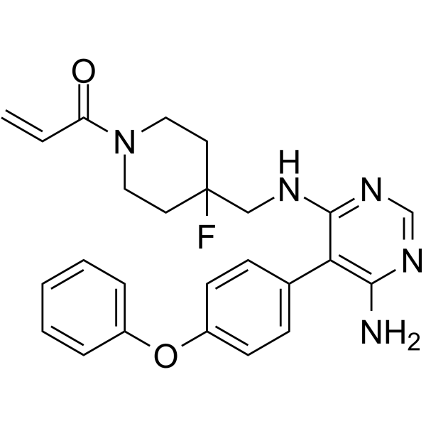
Flixebrutinib (TL-895) is a potent, orally active, ATP-competitive, and highly selective irreversible BTK inhibitor. Flixebrutinib is active against recombinant BTK (average IC50: 1.5 nM) and inhibits only three additional kinases BLK, BMX (IC50 = 1.6 nM) and TXK with IC50 within tenfold of BTK activity. Flixebrutinib inhibits BTK auto-phosphorylation at the Y223 phosphorylation site (IC50: 1-10 nM). The Flixebrutinib effectively inhibits the production of inflammatory factors such as IL-8, IL-1β, MCP-1 and TNF-α by monocytes or macrophages, and reduces the chemotactic migration of MF cells towards SDF-1. Flixebrutinib is used be for studies of chronic lymphocytic leukemia (CLL), myelofibrosis (MF), and B-cell malignancies.
For research use only. We do not sell to patients.
- Purity : 99.86%
- CAS No.: 1415823-49-2
- Formula: C25H26FN5O2
- Molecular Weight:447.50
-
Storage:Powder -20°C, 3 years , 4°C, 2 years ; In solvent -80°C, 6 months , -20°C, 1 month

To place orders, for customer services and technical support, please contact: MedChemExpress USA
Tel: 609-228-6898 E-mail: [email protected] [email protected]


-
 Biological Activity
-
Chemical Information
-
Solvent & Solubility
- Protocols
- Purity & Documentation
-
 Help & FAQs


-
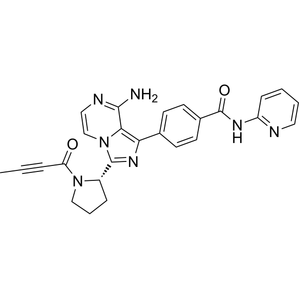
HY-17600
AcalabrutinibBTK Inhibitor -
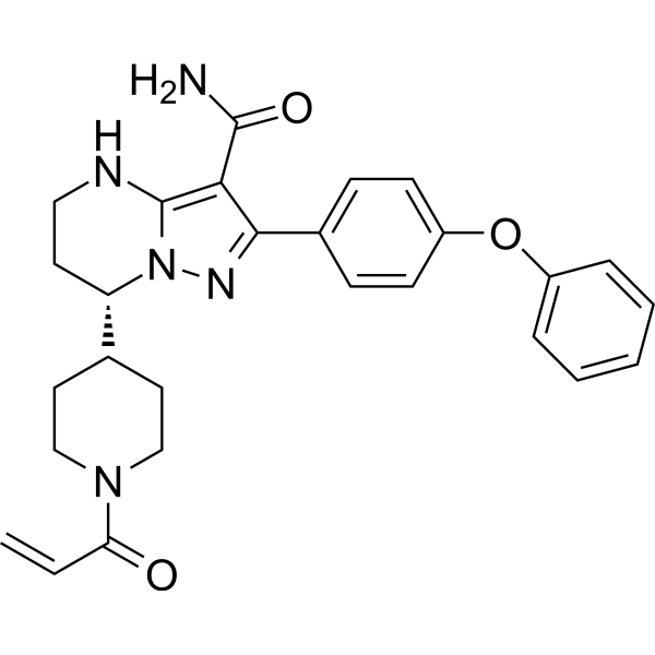
HY-101474A
ZanubrutinibBtk Inhibitor -
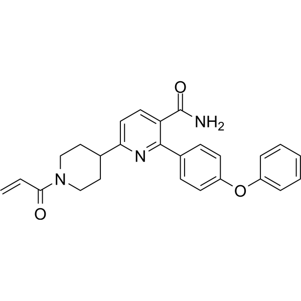
HY-129390
OrelabrutinibBTK Inhibitor -
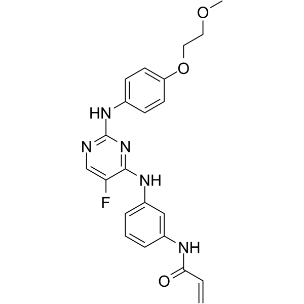
HY-18012
Spebrutinib -
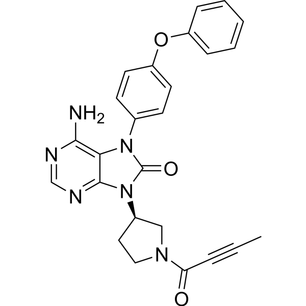
HY-15771
TirabrutinibBTK Inhibitor -
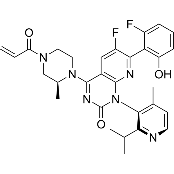
HY-114277
SotorasibKRAS G12C Inhibitor -
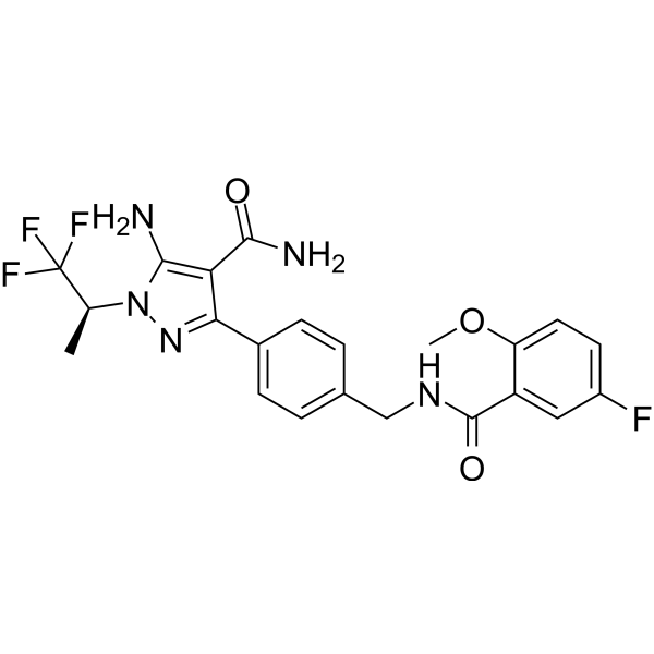
HY-131328
PirtobrutinibBTK Inhibitor -
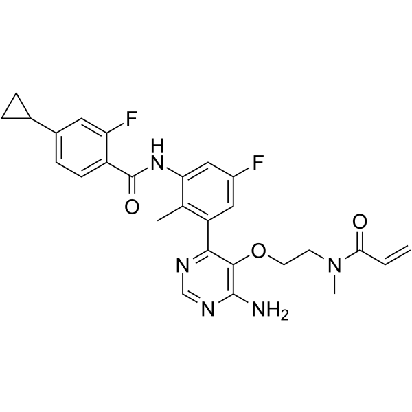
HY-128757
RemibrutinibBTK Inhibitor -
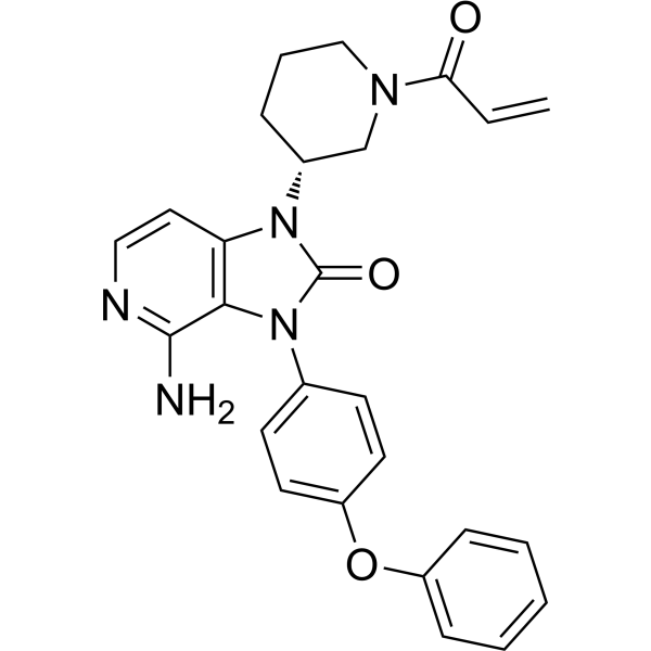
HY-109192
TolebrutinibBTK Inhibitor -
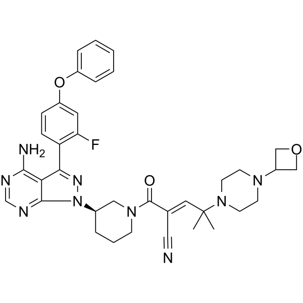
HY-112166
RilzabrutinibBTK Inhibitor -
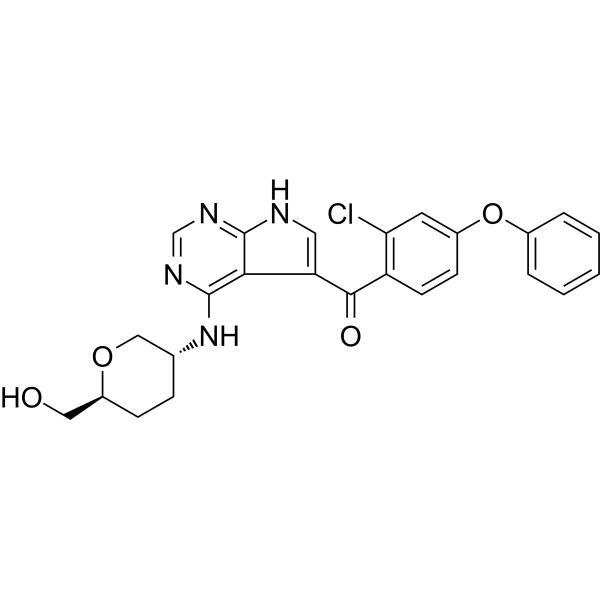
HY-112215
NemtabrutinibBTK Inhibitor -
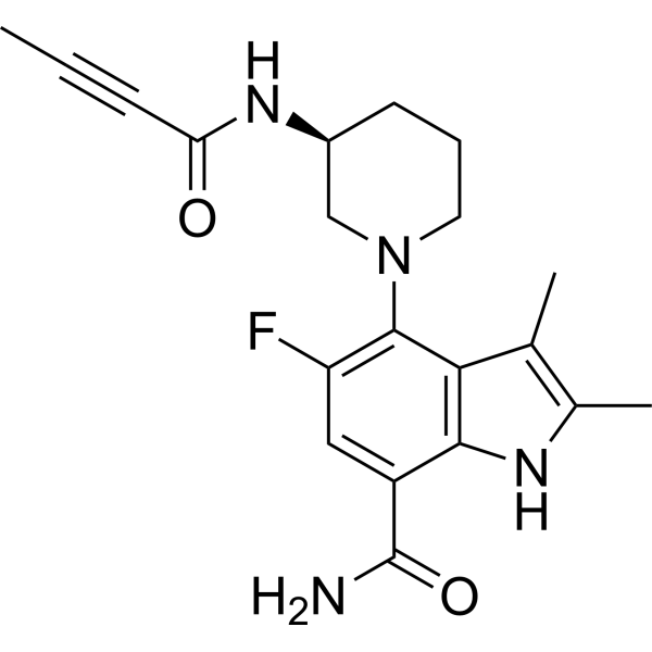
HY-112161
BranebrutinibBTK Inhibitor
Publications Citing Use of MedChemExpress (MCE) Flixebrutinib
More

Biological Activity
Description
IC50 & Target
[3]|
IL-1β |
IL-8 |
IL-6 |
In Vitro
Flixebrutinib (TL-895) in Ramos cells blocks BCR activation with an IC50 of 45.75 nM and also inhibits FcR activation in basophils with an IC50 of 1.01 μM[1].
Flixebrutinib (0-10 μM, 17-24 h) inhibits the production of TNF-α by macrophages, but does not inhibit the production of IFN in PBMCs, and blocks the activation of macrophages activated by FcR stimulation[1].
Flixebrutinib (TL-895) (0.1 nM-10 μM) effectively inhibits pBTK in B cells, Ramos cells, monocytes and Hel-92 cells[2].
Flixebrutinib reduces the chemotaxis of Hel-92 cells and inhibits their migration towards SDF-1[2].
Flixebrutinib reduces the expression of CD69 (an activation marker) on B cells in PBMCs and whole blood stimulated by B-cell receptor (BCR) (EC₅₀ values are 12 nM and 21 nM respectively)[2].
Flixebrutinib inhibits the production of IL-8, IL-1β, MCP-1, MIP-1α and IL-6 in healthy monocytes[2].
Flixebrutinib (TL-895) (0.01-50 μM, 1-4 days) effectively inhibits the proliferation of primary CLL cells, and at the effective concentration, it shows no significant toxicity to primary liver cells and HepG2 cells[3].
Flixebrutinib (0.05-3 μM) does not inhibit antibody-dependent cellular cytotoxicity (ADCC) and is suitable for use in combination with therapeutic antibodies[3].
MedChemExpress (MCE) has not independently confirmed the accuracy of these methods. They are for reference only. Further protocols information, click here.
In Vivo
Flixebrutinib (25 mg/kg feed, average 3.35 mg/kg daily, p.o., for 4 months) alleviates arthritis by inhibiting FcR signaling, and it suppresses local inflammation rather than systemic immune regulation in the Pristane (HY-N7819)-induced DBA/1 lupus model in mice[1].
Flixebrutinib (TL-895) (25 mg/kg, once daily, i.g., for 14-27 days) significantly inhibits tumor growth in the Mino model, but has limited effect in the Jeko-1, Maver-1, and Granta519 models in mice[3].
Flixebrutinib (once daily, i.g., for 13-25 days) shows significant anti-tumor activity in the Diffuse large B-cell lymphoma (DLBCL) patient-derived xenograft (PDX) models mice model[3].
MedChemExpress (MCE) has not independently confirmed the accuracy of these methods. They are for reference only.
-
Animal Model:BXSB-Yaa model established in C57BL/6mice[1]
-
Dosage:25 mg/kg feed, average 3.7 mg/kg daily
-
Administration:Oral administration (p.o.), for 11 weeks
-
Result:Significantly reduced the urine albumin/creatinine ratio (UACR). Significantly increased survival rate. Significantly reduced the scores of glomerular crescents, protein casts, interstitial inflammation and vasculitis.
Significantly reduced the levels of anti-dsDNA, anti-histone, and anti-Ro/SSA antibodies.
Reduced the expression of CD69 on B and T cells, and decreased the number of plasma cells (B220⁻).
-
Animal Model:Pristane-DBA/1 model established in C57BL/6mice[1]
-
Dosage:25 mg/kg feed, average 3.7 mg/kg daily
-
Administration:Oral administration (p.o.), for 4 months
-
Result:Significantly reduced the clinical arthritis score and the paw inflammation and injury score. Reduced some of the autoantibodies (anti-dsDNA, anti-histone, anti-Ro/SSA). Did not significantly affect the expression of IFN-regulating genes.
Partially reduced anti-dsDNA, anti-histone, and anti-Ro/SSA antibodies, but did not affect anti-Sm/RNP antibodies.
Increased the expression of CD69 on B cells, significantly reducing B220⁺ cells.
-
Animal Model:MCL xenograft model induced by Maver-1, Granta519, Mino, Jeko-1 established in 6-week-old female nude (NMRI:nu/nu) or CB17-SCID mice[3]
-
Dosage:25 mg/kg
-
Administration:Oral gavage (i.g.), once daily for 14 (Maver-1 and Granta519), 20 (Jeko-1) and 27 (Mino) days
-
Result:Showed significant anti-tumor activity in a specific MCL model.
Clinical Trial
| NCT Number | Sponsor | Condition | Start Date |
Phase

|
|---|---|---|---|---|
| NCT01329991 | Plexxikon| | 2011-05 | PHASE1 |


Chemical Information
-
CAS No. 1415823-49-2
-
Appearance Solid
-
Molecular Weight 447.50
-
Formula C25H26FN5O2
-
Color White to off-white
-
SMILES
O=C(N1CCC(CNC2=NC=NC(N)=C2C3=CC=C(C=C3)OC4=CC=CC=C4)(CC1)F)C=C
-
Synonyms
TL-895
-
Shipping
Room temperature in continental US; may vary elsewhere.
-
Storage
Powder -20°C 3 years 4°C 2 years In solvent -80°C 6 months -20°C 1 month


Publications (1)
-
Journal Impact Factor
-
Most Recent
-
Cancer Res Commun
2025 Aug 22. PMID: 40844833


Solvent & Solubility
In Vitro:
DMSO : 250 mg/mL (558.66 mM; Need ultrasonic; Hygroscopic DMSO has a significant impact on the solubility of product, please use newly opened DMSO)
Please refer to the solubility information to select the appropriate solvent. Once prepared, please aliquot and store the solution to prevent product inactivation from repeated freeze-thaw cycles.
Storage method and period of stock solution: -80°C, 6 months; -20°C, 1 month. When stored at -80°C, please use it within 6 months. When stored at -20°C, please use it within 1 month.
Please refer to the solubility information to select the appropriate solvent. Once prepared, please aliquot and store the solution to prevent product inactivation from repeated freeze-thaw cycles.
Storage method and period of stock solution: -80°C, 6 months; -20°C, 1 month. When stored at -80°C, please use it within 6 months. When stored at -20°C, please use it within 1 month.
Concentration (start) × Volume (start) = Concentration (final) × Volume (final)
In Vivo:
Select the appropriate dissolution method based on your experimental animal and administration route.
- For the following dissolution methods, please ensure to first prepare a clear stock solution using an In Vitro approach and then sequentially add co-solvents:
- To ensure reliable experimental results, the clarified stock solution can be appropriately stored based on storage conditions. As for the working solution for In Vivo experiments, it is recommended to prepare freshly and use it on the same day.
- The percentages shown for the solvents indicate their volumetric ratio in the final prepared solution. If precipitation or phase separation occurs during preparation, heat and/or sonication can be used to aid dissolution.
Add each solvent one by one: 10% DMSO 40% PEG300 5% Tween-80 45% Saline
Solubility: ≥ 2.08 mg/mL (4.65 mM); Clear solution
This protocol yields a clear solution of ≥ 2.08 mg/mL (saturation unknown).
Taking 1 mL working solution as an example, add 100 μL DMSO stock solution (20.8 mg/mL) to 400 μL PEG300, and mix evenly; then add 50 μL Tween-80 and mix evenly; then add 450 μL Saline to adjust the volume to 1 mL.
Preparation of Saline: Dissolve 0.9 g sodium chloride in ddH₂O and dilute to 100 mL to obtain a clear Saline solution.
Add each solvent one by one: 10% DMSO 90% (20% SBE-β-CD in Saline)
Solubility: ≥ 2.08 mg/mL (4.65 mM); Clear solution
This protocol yields a clear solution of ≥ 2.08 mg/mL (saturation unknown).
Taking 1 mL working solution as an example, add 100 μL DMSO stock solution (20.8 mg/mL) to 900 μL 20% SBE-β-CD in Saline, and mix evenly.
Preparation of 20% SBE-β-CD in Saline (4°C, storage for one week): 2 g SBE-β-CD powder is dissolved in 10 mL Saline, completely dissolve until clear.
In Vivo Dissolution Calculator
Please enter the basic information of animal experiments:
-
-
-
-
Recommended: Prepare an additional quantity of animals to account for potential losses during experiments.
Please enter your animal formula composition:
-
%DMSO +
Recommended: Keep the proportion of DMSO in working solution below 2% if your animal is weak.
-
%+
-
+%Tween-80 + +
-
%Saline +
The co-solvents required include: DMSO, . All of co-solvents are available by MedChemExpress (MCE). , Tween 80. All of co-solvents are available by MedChemExpress (MCE).
Working solution concentration: 0.22 mg/mL
Method for preparing stock solution: mg drug dissolved in μL DMSO. Stock solution concentration: mg/mL.
1. Take μL DMSO stock solution;
2. Add μL .
μL , mix evenly;
3. Then add μL Tween 80, mix evenly;
4. Then add μL
Please ensure that the stock solution in the first step is dissolved to a clear state, and add co-solvents in sequence. You can use ultrasonic heating (ultrasonic cleaner, recommended frequency 20-40 kHz), vortexing, etc. to assist dissolution.

Protocols
-
Kinase activity and phosphorylation assays
Kinase activity assays measure the ability of kinases to transfer phosphate groups from ATP to specific substrates, while phosphorylation assays detect the presence and levels of phosphorylated proteins. Common methods include radiolabeled ATP incorporation (e. g. ,), ADP release detection via bioluminescence (e. g. ,[3]), enzyme-linked immunosorbent assays (ELISA) for phospho-specific epitopes (e. g. ,[6]), and microtiter-based formats for high-throughput screening (e. g. ,[8]). The ADP-Glo assay quantifies kinase activity by measuring ADP produced during phosphorylation using a luciferase-based system. Radiometric assays involve autoradiography or scintillation counting after incorporation of 32P-labeled ATP into substrate proteins. ELISA-based approaches rely on phospho-specific antibodies to detect activated kinases in cell lysates or purified samples.
-
Western Blot
Western blotting (WB) is a commonly used experimental method in molecular biology, biochemistry, and immunogenetics for identifying and quantifying target proteins. It combines gel electrophoresis with immunoassay, enabling researchers to analyze protein expression, post-translational modifications, and molecular weight.
-
Cell Cytotoxicity Assay
Cytotoxicity assays are usually based on the assessment of cell membrane damage, which can also be indirectly detected by measuring cell viability. Detection methods include MTT assay, CKK-8 assay, LDH assay and ATP assay, etc.
-
Chemotaxis Gradient Chamber Assay 1
The chemotaxis gradient chamber assay is based on the principle of observing directional cell migration in response to a stable, linear or quasi-linear concentration gradient of a chemoattractant formed between two fluid reservoirs separated by a narrow observation chamber. Cells placed within the chamber respond to the gradient by polarized movement toward higher chemoattractant concentrations, allowing quantification of chemotactic behavior in real time under microscopy. The classic Zigmond chamber design enables simultaneous visualization of gradient formation and individual cell trajectories, making it suitable for studying leukocyte chemotaxis and other motile cell types in vitro.
-
Cell migration
Cell migration is a method that plays an important role in wound healing, cell differentiation, embryonic development, etc.
-
Protocol for Kinase activity and phosphorylation assays
Kinase activity assays measure transfer of phosphate from ATP to a protein or peptide substrate, generating phosphorylated substrate, ADP, or incorporated radiolabeled phosphate as the readout; phosphorylation assays measure site-specific phosphorylation in cells or tissues as a proxy for kinase-pathway activation, inhibition, or substrate regulation. Phosphorylation can be detected by phospho-specific Western blot, immunoprecipitation kinase assay, phospho-immunofluorescence, phospho-flow cytometry, luminescent ADP detection, radiolabeled ATP incorporation, or reporter-based pathway assays, and these readouts can be applied to cancer cells, primary neurons, mouse tumors, organoids, inflammatory macrophages, ferroptosis studies, and mitophagy studies when the kinase target is biologically relevant.
-
Chemotaxis Gradient Chamber Assay 2
Chemotaxis gradient chamber assays measure directional cell migration in response to a soluble chemical gradient by imaging cells as they move across a defined observation region; the readout is generated from time-lapse cell trajectories, displacement toward the gradient, forward migration index, trajectory plots, rose/polar plots, and statistical tests of non-random directionality. The Dunn chamber is a direct-viewing glass chamber in which cells migrate across a bridge between control and chemoattractant wells, allowing observation of cells in a linear concentration gradient; related direct-viewing formats include the Insall chamber, which supports defined unidirectional gradients and high numerical-aperture microscopy, and the μ-Slide Chemotaxis chamber, which supports long-term live-cell imaging and gradient characterization with fluorescent dye.
-
Pyroptosis Solutions
Pyroptosis is a lytic inflammatory cell-death pathway executed by gasdermin pores, most classically through inflammasome-mediated activation of caspase-1, cleavage of gasdermin D, membrane pore formation, LDH release, and secretion of IL-1β and IL-18. The canonical pathway is commonly modeled by priming cells with an inflammatory signal such as LPS to induce pro-IL-1β and inflammasome components, followed by an activation signal such as ATP or nigericin to activate NLRP3, ASC speck formation, caspase-1 cleavage, GSDMD cleavage, cytokine release, and pyroptotic membrane rupture. The non-canonical pathway is triggered when cytosolic LPS activates mouse caspase-11 or human caspase-4/5, leading to GSDMD cleavage and pyroptosis, and this can secondarily activate NLRP3-dependent IL-1β release. Pyroptosis is linked to inflammatory injury, infection, cancer, liver disease, ocular disease, placental inflammation, and other disease phenotypes, but unresolved questions include which gasdermin fam


Purity & Documentation
-
Data Sheet (258 KB)
-
SDS (251 KB)
- English - EN (251 KB)
- Français - FR (251 KB)
- Deutsch - DE (251 KB)
- Norwegian - NO (251 KB)
- Español - ES (251 KB)
- Swedish - SV (251 KB)
- Italian - IT (251 KB)
- Korean - KR (251 KB)
- Portuguese - PT (251 KB)
-
Handling Instructions (2659 KB)


Complete Stock Solution Preparation Table
Please refer to the solubility information to select the appropriate solvent. Once prepared, please aliquot and store the solution to prevent product inactivation from repeated freeze-thaw cycles.
Storage method and period of stock solution: -80°C, 6 months; -20°C, 1 month. When stored at -80°C, please use it within 6 months. When stored at -20°C, please use it within 1 month.
| Optional Solvent | Concentration Solvent Mass | 1 mg | 5 mg | 10 mg | 25 mg |
|---|---|---|---|---|---|
| DMSO | 1 mM | 2.2346 mL | 11.1732 mL | 22.3464 mL | 55.8659 mL |
| 5 mM | 0.4469 mL | 2.2346 mL | 4.4693 mL | 11.1732 mL | |
| 10 mM | 0.2235 mL | 1.1173 mL | 2.2346 mL | 5.5866 mL | |
| 15 mM | 0.1490 mL | 0.7449 mL | 1.4898 mL | 3.7244 mL | |
| 20 mM | 0.1117 mL | 0.5587 mL | 1.1173 mL | 2.7933 mL | |
| 25 mM | 0.0894 mL | 0.4469 mL | 0.8939 mL | 2.2346 mL | |
| 30 mM | 0.0745 mL | 0.3724 mL | 0.7449 mL | 1.8622 mL | |
| 40 mM | 0.0559 mL | 0.2793 mL | 0.5587 mL | 1.3966 mL | |
| 50 mM | 0.0447 mL | 0.2235 mL | 0.4469 mL | 1.1173 mL | |
| 60 mM | 0.0372 mL | 0.1862 mL | 0.3724 mL | 0.9311 mL | |
| 80 mM | 0.0279 mL | 0.1397 mL | 0.2793 mL | 0.6983 mL | |
| 100 mM | 0.0223 mL | 0.1117 mL | 0.2235 mL | 0.5587 mL |


Flixebrutinib Related Classifications
HY-139481 Related Classifications
 Powered by Bioz