



W6134

Based on 1 Customer Validation
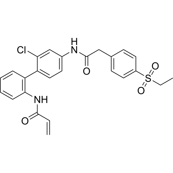
W6134 is highly potent and selective RORγ covalent inhibitor with an IC50 of 0.21 μM. W6134 exhibits excellent selectivity for RORγ over RORα, RXRγ, and ERRγ. W6134 significantly inhibits RORγ transcriptional activity W6134 inhibits the proliferation and colony formation and induces apoptosis in castration-resistant prostate cancer cells (CRPC). W6134 can be used for the research of castration-resistant prostate cancers (CRPC).
For research use only. We do not sell to patients.
- Purity : 99.90%
- CAS No.: 2983540-55-0
- Formula: C25H23ClN2O4S
- Molecular Weight:482.98
-
Storage:Powder -20°C, 3 years ; In solvent -80°C, 6 months , -20°C, 1 month

To place orders, for customer services and technical support, please contact: MedChemExpress USA
Tel: 609-228-6898 E-mail: [email protected] [email protected]


-
 Biological Activity
-
Chemical Information
-
Solvent & Solubility
- Protocols
- Purity & Documentation
- References
-
 Help & FAQs


-
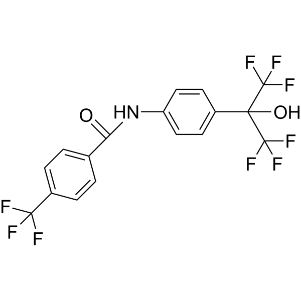
HY-14422
SR1078RORα/RORγ Agonist -
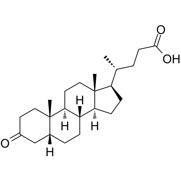
HY-125801
3-Oxo-5β-cholanoic acidTH17 Cell Differentiation Inhibitor -
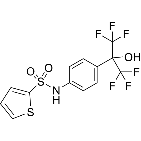
HY-14413
SR3335RORα Inverse Agonist -
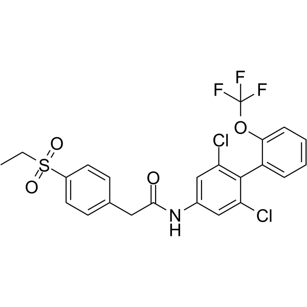
HY-12776
GSK805RORγt Inhibitor -
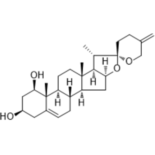
HY-N2253
NeoruscogeninRORα Agoinst -
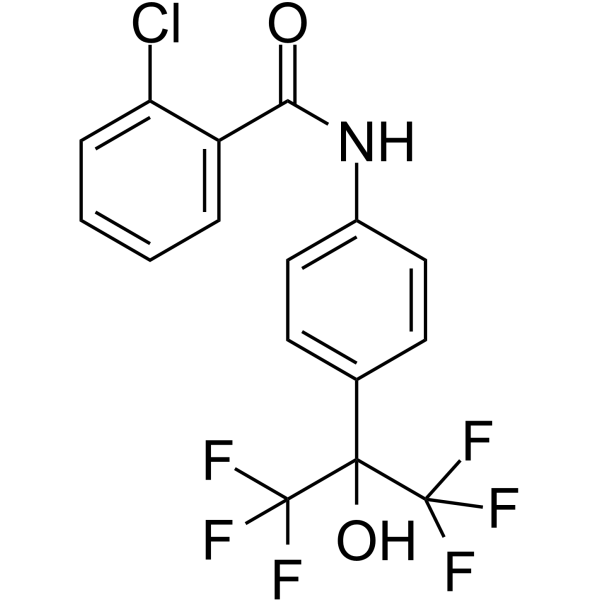
HY-101454
SR0987RORγt Agonist -
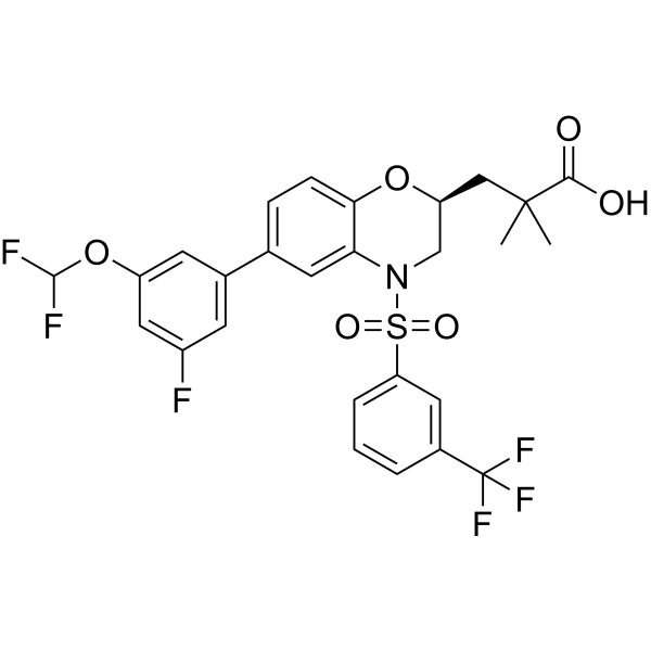
HY-104037
CintirorgonRORγ Agonist -
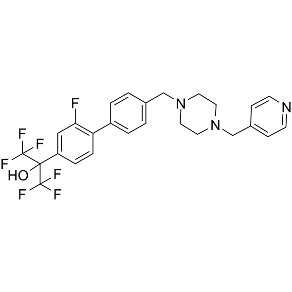
HY-16998
SR2211RORγ Inverse Agonist -
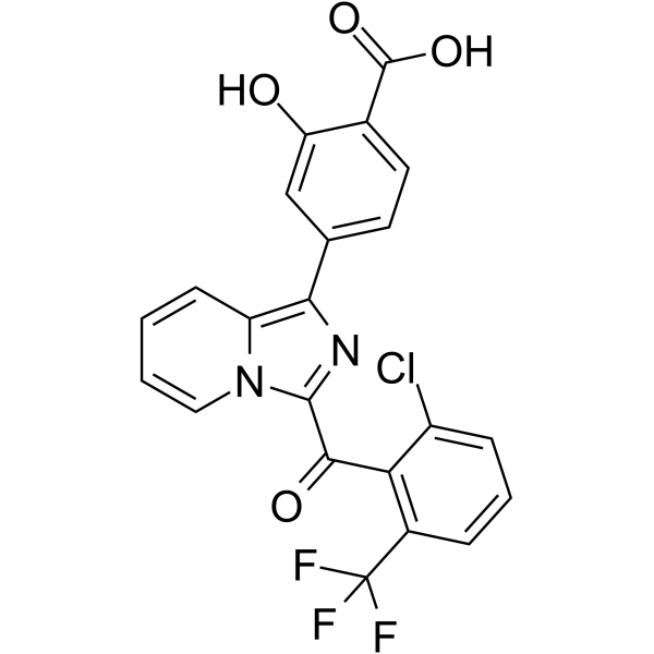
HY-19774
GNE-0946RORγ/RORc Agonist -
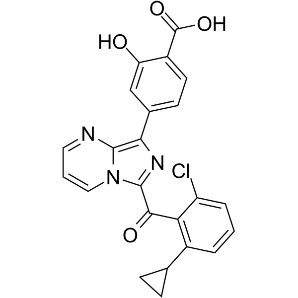
HY-19775
GNE-6468RORγ (RORc) Inverse Agonist -
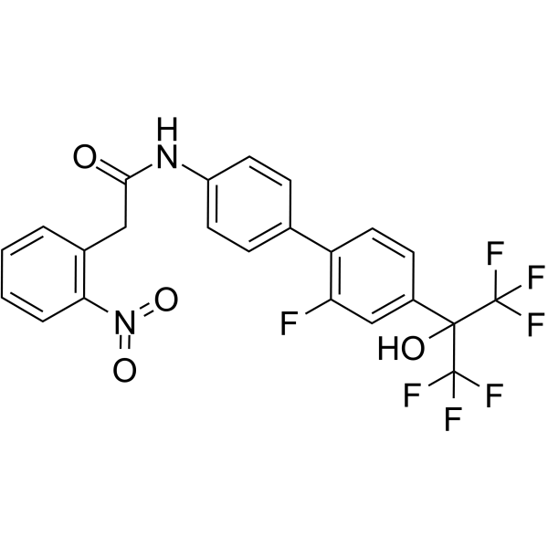
HY-120210
XY018ROR-γ Antagonist -
HY-120384
AZD-0284RORγ Inverse Agonist


Biological Activity
Description
IC50 & Target
|
RORγ 0.21 μM (IC50) |
Cellular Effect
|
Cell Line

|
Type | Value | Description | References |
|---|---|---|---|---|
| CWR22R | IC50 |
0.77 μM
Compound: 29
|
Antiproliferative activity against human 22Rv1 cells incubated for 4 days by CCK8 assay
Antiproliferative activity against human 22Rv1 cells incubated for 4 days by CCK8 assay
|
[PMID: 38227771] |
| LNCaP | IC50 |
0.63 μM
Compound: 29
|
Antiproliferative activity against human LNCaP cells incubated for 4 days by CCK8 assay
Antiproliferative activity against human LNCaP cells incubated for 4 days by CCK8 assay
|
[PMID: 38227771] |
| LNCaP C4-2B | IC50 |
0.47 μM
Compound: 29
|
Antiproliferative activity against human LNCaP C4-2B cells incubated for 4 days by CCK8 assay
Antiproliferative activity against human LNCaP C4-2B cells incubated for 4 days by CCK8 assay
|
[PMID: 38227771] |
In Vitro
W6134 (compound 29) has antiproliferative potencies on androgen receptor (AR)-positive human CRPC cell lines, including C4-2B, 22Rv1, and LNCaP, with IC50 values of 0.47 μM, 0.77 μM, and 0.63 μM, respectively[1].
W6134 (1.25-5 μM; 14 days) significantly suppresses the colony formation in C4-2B and 22Rv1 cell lines. W6134 dramatically induces apoptosis of C4-2B and 22Rv1 cells[1].
W6134 (1.25-5 μM; 48 h) inhibits the mRNA levels of AR-regulated genes, such as KLK2, KLK3, and NKX3-1[1].
W6134 (1.25-5 μM; 24-48 h) effectively suppresses the levels of AR or AR-V7 in a dose- and time-dependent manner[1].
MedChemExpress (MCE) has not independently confirmed the accuracy of these methods. They are for reference only.
-
Cell Line:C4-2B and 22Rv1 cell lines
-
Concentration:1.25 μM, 2.5 μM, 5 μM
-
Incubation Time:14 days
-
Result:Significantly suppressed the colony formation in C4-2B and 22Rv1 cell lines in a dose-dependent manner.
-
Cell Line:C4-2B and 22Rv1 cell lines
-
Concentration:1.25 μM, 2.5 μM, 5 μM
-
Incubation Time:48 h
-
Result:Significantly decreased the expression of AR and its variant AR-V7 in a dose-dependent manner.
The mRNA levels of AR-regulated genes, such as KLK2, KLK3, and NKX3-1, were also dramatically suppressed.
-
Cell Line:C4-2B and 22Rv1 cell lines
-
Concentration:1.25 μM, 2.5 μM, 5 μM
-
Incubation Time:24 h, 48 h, 72 h
-
Result:Effectively suppressed the levels of AR or AR-V7 in a dose- and time-dependent manner.
In Vivo
MedChemExpress (MCE) has not independently confirmed the accuracy of these methods. They are for reference only.
-
Animal Model:Male NOD/SCID mice (~18 g; 4-week-old) were subcutaneously 22Rv1 cells[1]
-
Dosage:5 mg/kg or 10 mg/kg
-
Administration:i.p; five times per week; for 2 weeks
-
Result:Significantly inhibited the tumor growth in a dose-dependent manner with tumor growth inhibition (TGI) values of 41.90 and 62.58% at doses of 5 and 10 mg/kg.
- Dual Luciferin reporter gene assay
- Detection of Apoptosis
- TUNEL staining for apoptotic DNA fragmentation
- Annexin V plus membrane-impermeant dye apoptosis staining
- Apoptosis Solutions
- Soft Agar Colony Formation Assay
- Colony Formation (Clonogenic) Assay
- Cell Viability Determination by MTT Colorimetric Assay


Chemical Information
-
CAS No. 2983540-55-0
-
Appearance Solid
-
Molecular Weight 482.98
-
Formula C25H23ClN2O4S
-
Color White to off-white
-
SMILES
O=C(NC1=CC=C(C2=C(C=CC=C2)NC(C=C)=O)C(Cl)=C1)CC3=CC=C(C=C3)S(=O)(CC)=O
-
Shipping
Room temperature in continental US; may vary elsewhere.
-
Storage
Powder -20°C 3 years In solvent -80°C 6 months -20°C 1 month


Solvent & Solubility
In Vitro:
DMSO : 83.33 mg/mL (172.53 mM; Need ultrasonic; Hygroscopic DMSO has a significant impact on the solubility of product, please use newly opened DMSO)
Please refer to the solubility information to select the appropriate solvent. Once prepared, please aliquot and store the solution to prevent product inactivation from repeated freeze-thaw cycles.
Storage method and period of stock solution: -80°C, 6 months; -20°C, 1 month. When stored at -80°C, please use it within 6 months. When stored at -20°C, please use it within 1 month.
Please refer to the solubility information to select the appropriate solvent. Once prepared, please aliquot and store the solution to prevent product inactivation from repeated freeze-thaw cycles.
Storage method and period of stock solution: -80°C, 6 months; -20°C, 1 month. When stored at -80°C, please use it within 6 months. When stored at -20°C, please use it within 1 month.
Concentration (start) × Volume (start) = Concentration (final) × Volume (final)


Purity & Documentation
-
Data Sheet (275 KB)
-
SDS (251 KB)
- English - EN (251 KB)
- Français - FR (251 KB)
- Deutsch - DE (251 KB)
- Norwegian - NO (251 KB)
- Español - ES (251 KB)
- Swedish - SV (251 KB)
- Italian - IT (251 KB)
- Korean - KR (251 KB)
- Portuguese - PT (251 KB)
-
Handling Instructions (2659 KB)
References


Complete Stock Solution Preparation Table
Please refer to the solubility information to select the appropriate solvent. Once prepared, please aliquot and store the solution to prevent product inactivation from repeated freeze-thaw cycles.
Storage method and period of stock solution: -80°C, 6 months; -20°C, 1 month. When stored at -80°C, please use it within 6 months. When stored at -20°C, please use it within 1 month.
| Optional Solvent | Concentration Solvent Mass | 1 mg | 5 mg | 10 mg | 25 mg |
|---|---|---|---|---|---|
| DMSO | 1 mM | 2.0705 mL | 10.3524 mL | 20.7048 mL | 51.7620 mL |
| 5 mM | 0.4141 mL | 2.0705 mL | 4.1410 mL | 10.3524 mL | |
| 10 mM | 0.2070 mL | 1.0352 mL | 2.0705 mL | 5.1762 mL | |
| 15 mM | 0.1380 mL | 0.6902 mL | 1.3803 mL | 3.4508 mL | |
| 20 mM | 0.1035 mL | 0.5176 mL | 1.0352 mL | 2.5881 mL | |
| 25 mM | 0.0828 mL | 0.4141 mL | 0.8282 mL | 2.0705 mL | |
| 30 mM | 0.0690 mL | 0.3451 mL | 0.6902 mL | 1.7254 mL | |
| 40 mM | 0.0518 mL | 0.2588 mL | 0.5176 mL | 1.2940 mL | |
| 50 mM | 0.0414 mL | 0.2070 mL | 0.4141 mL | 1.0352 mL | |
| 60 mM | 0.0345 mL | 0.1725 mL | 0.3451 mL | 0.8627 mL | |
| 80 mM | 0.0259 mL | 0.1294 mL | 0.2588 mL | 0.6470 mL | |
| 100 mM | 0.0207 mL | 0.1035 mL | 0.2070 mL | 0.5176 mL |


W6134 Related Classifications
HY-163192 Related Classifications
 Powered by Bioz