
Lenaldekar
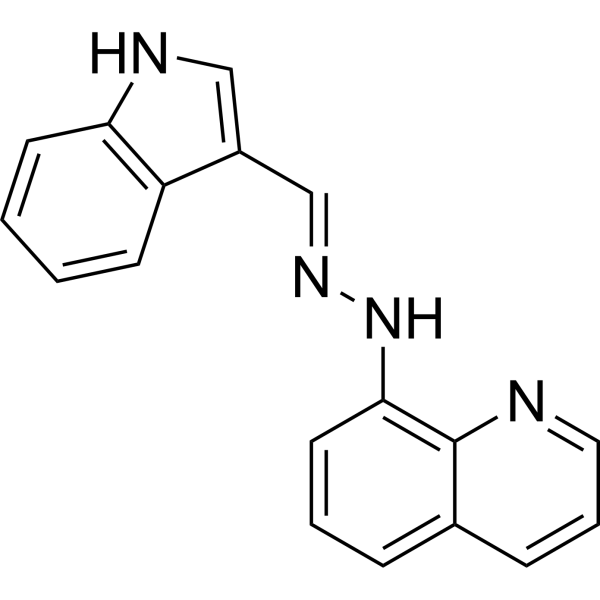
Lenaldekar (LDK) inhibits human and murine T-cell expansiomn. Lenaldekar inhibits autoimmune T cell response. Lenaldekar also induces cancer cell apoptosis. Lenaldekar can be used for T-cell mediated autoimmune diseases research.
商品は「研究用試薬」です。人や動物の医療用・臨床診断用・食品用の製品ではありません。
研究用途以外に使用した場合、当社は一切の責任を負いかねます。
- CAS 番号: 418800-15-4
- 分子式: C18H14N4
- 分子量:286.33
-
保管条件:
Please store the product under the recommended conditions in the Certificate of Analysis.
生物活性
製品説明
体外実験
Lenaldekar (4 days) inhibits CD3+ T cell proliferation in a dose-dependent manner with an IC50 of 3 μM[1].
Lenaldekar (3 μM, 4 days) reduces a memory T cell response to influenza antigen H3[1].
Lenaldekar (48 h) shows cytotoxicity in Jurkat T-ALL cell, with IC50s of 0.8 μM, and induces apoptosis (1 μM)[2].
MedChemExpress (MCE) has not independently confirmed the accuracy of these methods. They are for reference only. Further protocols information, click here.
体内実験
Lenaldekar (16 mg/kg, i.p., twice daily) inhibits tumor progession in a mouse xenograft model of T-cell acute lymphoblastic leukemia (T-ALL)[2].
MedChemExpress (MCE) has not independently confirmed the accuracy of these methods. They are for reference only.
化学情報
-
CAS 番号 418800-15-4
-
分子量 286.33
-
分子式 C18H14N4
-
SMILES
C1(/C=N/NC2=C3N=CC=CC3=CC=C2)=CNC4=C1C=CC=C4
-
別名
LDK
-
輸送条件
Room temperature in continental US; may vary elsewhere.
-
保管条件
Please store the product under the recommended conditions in the Certificate of Analysis.
プロトコル
-
Apoptosis
Apoptosis, also called programmed cell death, is generally characterized by distinct morphological characteristics.
-
TUNEL staining for apoptotic DNA fragmentation
TUNEL staining detects DNA strand breaks by using terminal deoxynucleotidyl transferase to add labeled nucleotides to exposed 3′-OH DNA termini, generating either microscopic staining in fixed cells or tissue sections, or fluorescence/cytometric signal in cell suspensions. TUNEL positivity reflects DNA fragmentation but should not be interpreted alone as definitive apoptosis, because TUNEL can also label necrotic, autolytic, mechanically damaged, or DNA-repair-associated DNA breaks.
-
Annexin V plus membrane-impermeant dye apoptosis staining
Annexin V-based apoptosis assays rely on the detection of phosphatidylserine (PS) externalization from the inner leaflet of the plasma membrane to the outer leaflet, an early biochemical hallmark of apoptosis. Fluorescently labeled Annexin V binds PS in a calcium-dependent manner, enabling identification of early apoptotic cells by flow cytometry or fluorescence microscopy. When combined with a membrane-impermeant DNA-binding dye (e. g. , propidium iodide), this approach allows discrimination between viable (Annexin V−/dye−), early apoptotic (Annexin V+/dye−), and late apoptotic or necrotic (Annexin V+/dye+) cell populations by assessing membrane integrity and PS exposure.
-
Apoptosis Solutions
Apoptosis is a regulated, generally non-lytic cell-death pathway that removes unwanted, damaged, infected, or abnormal cells through coordinated morphological changes, caspase activation, DNA fragmentation, and membrane remodeling. The intrinsic apoptosis pathway is controlled mainly by mitochondrial outer membrane permeabilization, BCL-2 family proteins, cytochrome c release, apoptosome formation, caspase-9 activation, and downstream executioner caspase-3/7 activation. The extrinsic apoptosis pathway is initiated by death receptors such as Fas, TNFR, and TRAIL receptors, which recruit adaptor proteins and activate caspase-8 before engaging executioner caspases or mitochondrial amplification through BID cleavage. Apoptosis is linked to many phenotypes, including cancer cell killing, tissue homeostasis, immune regulation, neurodegeneration, infection response, and treatment-induced cytotoxicity; unresolved questions include how apoptosis interacts with necroptosis, pyroptosis, ferroptos
-
Research Protocol for Cancer Immunology
Cancer immunology studies how the immune system recognizes, suppresses, edits, or fails to eliminate malignant cells through tumor antigen release, antigen presentation, T-cell priming, immune trafficking, tumor-cell killing, and feedback inhibition in the tumor microenvironment. The cancer-immunity cycle links tumor antigenicity, dendritic-cell priming, CD8+ T-cell infiltration, cytotoxic function, and immune-checkpoint regulation to tumor rejection or immune escape. Immune-checkpoint pathways such as PD-1/PD-L1 and CTLA-4 suppress antitumor T-cell activity and can be therapeutically blocked, but many tumors remain resistant because of poor antigen presentation, weak T-cell infiltration, suppressive myeloid cells, regulatory T cells, and tumor-intrinsic immune-exclusion programs. Unresolved questions include which immune-cell states predict response, how tumor-intrinsic pathways exclude immune cells, how myeloid suppression limits checkpoint blockade, and which combination strategies
純度とドキュメンテーション
参考文献
[1]. Cusick MF, et al. Human T cell expansion and experimental autoimmune encephalomyelitis inhibited by Lenaldekar, a small molecule discovered in a zebrafish screen. J Neuroimmunol. 2012 Mar;244(1-2):35-44. [Content Brief]
[2]. Ridges S, et al, et al. Zebrafish screen identifies novel compound with selective toxicity against leukemia. Blood. 2012 Jun 14;119(24):5621-31. [Content Brief]
Calculators
濃度 (開始) × 体積 (開始) = 濃度 (終了) × 体積 (終了)

