



Inulin
Based on 5 publication(s) in Google Scholar
Inulin is an orally active prebiotic targeting the intestinal microbiota, selectively promoting the proliferation and activity of beneficial bacteria such as bifidobacteria and lactic acid bacteria, and playing a role in regulating the intestinal microecology. The functions of Inulin include: Fermentation by probiotics in the colon to produce short-chain fatty acids (such as butyrate and propionate), lowering the intestinal pH and inhibiting the overgrowth of harmful bacteria; Enhancing the intestinal barrier function and reducing endotoxin translocation; Directly scavenging free radicals (such as superoxide free radicals, hydroxyl free radicals) and activating antioxidant enzymes (SOD, CAT) to reduce oxidative stress. Inulin can also be used in the study of intestinal diseases (constipation, IBD), metabolic syndrome (diabetes, obesity) and liver damage by regulating glucose and lipid metabolism (such as reducing triglycerides, improving insulin sensitivity) and immune response (enhancing NK cell activity, inhibiting inflammatory factors)
연구목적의 판매만을 진행합니다. 환자를 대상으로 한 판매는 하지 않습니다.
- CAS No.: 9005-80-5
-
보관:Powder -20°C, 3 years , 4°C, 2 years ; In solvent -80°C, 6 months , -20°C, 1 month


To place orders, for customer services and technical support, please contact: MedChemExpress USA
Tel: 609-228-6898 E-mail: [email protected] [email protected]


-
 Biological Activity
-
Chemical Information
-
용액&용해도
- Protocol
- 순도&문서
- References
-
 Help & FAQs


-
HY-W145518
PectinAntibacterial Agent -

HY-107846
XylanActive Compound -
HY-13753
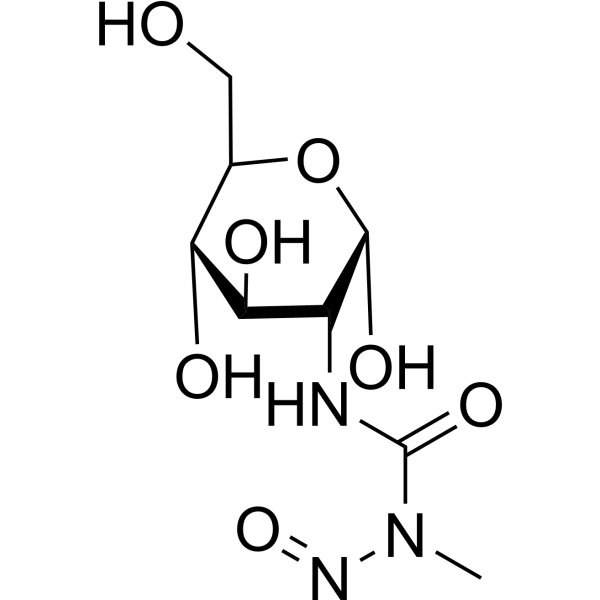
StreptozotocinDiabetes Modeling Agent -
HY-N0055
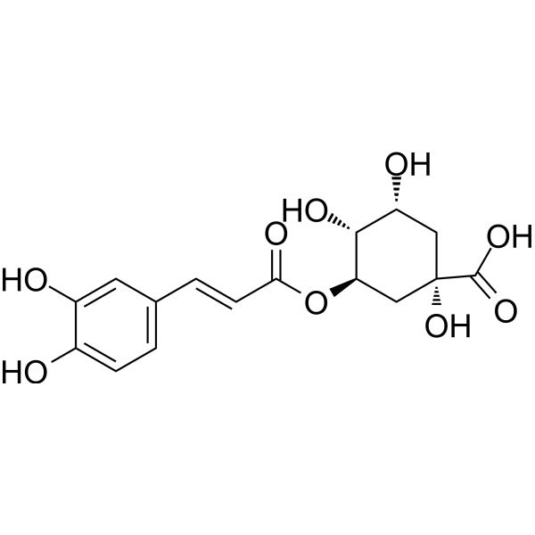
Chlorogenic acidAntioxidant -
HY-W145521
β-1,3-GlucanPolysaccharide -
HY-107863
FructooligosaccharidesPrebiotics -

HY-W350004
(+)-Arabinogalactan,from larch wood, 80%Sugar -

HY-N6664
Gum arabicComplex Branched Polysaccharide -
HY-139413
β-D-GlucanNon-digestible Polysaccharide -

HY-W145516
Guar gumNatural Polymer -
HY-W127758
Alginic acidHistamine Release Inhibitor -

HY-W145665
AmyloseBiochemical Assay Reagent
Publications Citing Use of MedChemExpress (MCE) Inulin
More Customer Validation & Images

-
Histological Imaging/Staining

-
RT-PCR

-
WB

All Endogenous Metabolite Isoforms
More

Biological Activity
제품 설명
In Vitro
Antioxidant activity assay: Inulin (0.025-1 mg/mL; 5 min) exhibited moderate scavenging ability in vitro, promoted the scavenging of superoxide radicals, hydroxyl radicals and H2O2, and inhibited lipid peroxidation[2].
Cytotoxicity assay: Inulin (2.0%; 24 h) significantly increased the survival rate of HT-29 cells and inhibited the cytotoxicity induced by Deoxycholic acid (HY-N0593) (100% protection), Lithocholic acid (HY-B0172) (30% protection) and fecal water (40% protection)[3].
Genotoxicity assay: Inulin (2%; 90 min) significantly reduced the genotoxicity of 4-NQO and fecal water by SOS chromotest and Ames test, with SOS induction potency (SOSIP) reduced by 29-74% and 57-70%, respectively[3].
MedChemExpress (MCE) has not independently confirmed the accuracy of these methods. They are for reference only.
In Vivo
Inulin (100-400 mg/kg; gavage; once a day; 3 weeks) significantly reduces 0.3% CCl4 (olive oil)-induced acute liver injury in male ICR mice, reduced serum ALT, AST, ALP levels, and increased liver tissue antioxidant enzyme activities (SOD, CAT, GSH-Px)[2].
MedChemExpress (MCE) has not independently confirmed the accuracy of these methods. They are for reference only.
Clinical Trial
| NCT Number | Sponsor | Condition | Start Date |
Phase

|
|---|---|---|---|---|
| NCT01329991 | Plexxikon| | 2011-05 | PHASE1 |


Chemical Information
-
CAS No. 9005-80-5
-
Appearance Solid
-
Color White to off-white
-
SMILES
[Inulin]
-
Structure Classification
-
선적
Room temperature in continental US; may vary elsewhere.
-
보관
Powder -20°C 3 years 4°C 2 years In solvent -80°C 6 months -20°C 1 month


Publications (5)
-
Journal Impact Factor
-
Most Recent
-
-
Int J Mol Sci
2026 Jan 27;27(3):1262. PMID: 41683691 -
Front Pharmacol
Inulin ameliorates metabolic syndrome in high-fat diet-fed mice by regulating gut microbiota and bile acid excretion. [Abstract]2023 Jul 24:14:1226448. PMID: 37554983 -
FASEB J
Improvement in glucose metabolism in adult male offspring of maternal mice fed diets supplemented with inulin via regulation of the hepatic long noncoding RNA profile. [Abstract]2021 Nov;35(11):e22003. PMID: 34706105 -
Am J Physiol Regul Integr Comp Physiol
Inulin Alleviates Intestinal Barrier Dysfunction Induced by Chronic Intermittent Hypoxia by modulating intestinal microbiota in Mice. [Abstract]2025 Oct 15. PMID: 41092384
Inulin purchased from MedChemExpress. Usage Cited in: Am J Physiol Regul Integr Comp Physiol. 2025 Oct 15. [Abstract]
Hematoxylin and eosin (H&E) were employed to stain paraffin-embedded sections of mouse colon tissue and analyzed via histology score treated with Inulin.
Inulin purchased from MedChemExpress. Usage Cited in: Am J Physiol Regul Integr Comp Physiol. 2025 Oct 15. [Abstract]
Effects of Inulin supplementation on expression of cytokines at mRNA levels. Expression of inflammatory cytokines (TGF-β, TNF-α, IL-23, -10, -6 and -1β) at mRNA levels in intestinal tissues were measured using real-time quantitative polymerase chain reaction.
Inulin purchased from MedChemExpress. Usage Cited in: Am J Physiol Regul Integr Comp Physiol. 2025 Oct 15. [Abstract]
Effects on levels of Occludin, Claudin-1, -2, -4, -5 and ZO-1 expressions treated with Inulin.


용액&용해도
In Vitro:
DMSO : 100 mg/mL (Need ultrasonic; Hygroscopic DMSO has a significant impact on the solubility of product, please use newly opened DMSO)
H2O : ≥ 50 mg/mL
* "≥" means soluble, but saturation unknown.
In Vivo:
Select the appropriate dissolution method based on your experimental animal and administration route.
- For the following dissolution methods, please ensure to first prepare a clear stock solution using an In Vitro approach and then sequentially add co-solvents:
- To ensure reliable experimental results, the clarified stock solution can be appropriately stored based on storage conditions. As for the working solution for In Vivo experiments, it is recommended to prepare freshly and use it on the same day.
- The percentages shown for the solvents indicate their volumetric ratio in the final prepared solution. If precipitation or phase separation occurs during preparation, heat and/or sonication can be used to aid dissolution.
Add each solvent one by one: 10% DMSO 40% PEG300 5% Tween-80 45% Saline
Solubility: ≥ 2.5 mg/mL; Clear solution
This protocol yields a clear solution of ≥ 2.5 mg/mL (saturation unknown).
Taking 1 mL working solution as an example, add 100 μL DMSO stock solution (25.0 mg/mL) to 400 μL PEG300, and mix evenly; then add 50 μL Tween-80 and mix evenly; then add 450 μL Saline to adjust the volume to 1 mL.
Preparation of Saline: Dissolve 0.9 g sodium chloride in ddH₂O and dilute to 100 mL to obtain a clear Saline solution.
Add each solvent one by one: 10% DMSO 90% (20% SBE-β-CD in Saline)
Solubility: ≥ 2.5 mg/mL; Clear solution
This protocol yields a clear solution of ≥ 2.5 mg/mL (saturation unknown).
Taking 1 mL working solution as an example, add 100 μL DMSO stock solution (25.0 mg/mL) to 900 μL 20% SBE-β-CD in Saline, and mix evenly.
Preparation of 20% SBE-β-CD in Saline (4°C, storage for one week): 2 g SBE-β-CD powder is dissolved in 10 mL Saline, completely dissolve until clear.
For the following dissolution methods, please prepare the working solution directly:
It is recommended to prepare fresh solutions and use them promptly within a short period of time.
The percentages shown for the solvents indicate their volumetric ratio in the final prepared solution. If precipitation or phase separation occurs during preparation, heat and/or sonication can be used to aid dissolution.
Add each solvent one by one: PBS
Solubility: 27.5 mg/mL; Clear solution; Need ultrasonic
In Vivo Dissolution Calculator
Please enter the basic information of animal experiments:
-
-
-
-
Recommended: Prepare an additional quantity of animals to account for potential losses during experiments.
Working solution concentration: 0.22 mg/mL
This product has good water solubility, please refer to the measured solubility data in water/PBS/Saline for details.


순도&문서
-
Data Sheet (270 KB)
-
SDS (251 KB)
- English - EN (251 KB)
- Français - FR (251 KB)
- Deutsch - DE (251 KB)
- Norwegian - NO (251 KB)
- Español - ES (251 KB)
- Swedish - SV (251 KB)
- Italian - IT (251 KB)
- Korean - KR (251 KB)
- Portuguese - PT (251 KB)
-
Handling Instructions (2659 KB)
References
[1]. Shoaib M, et al. Inulin: Properties, health benefits and food applications. Carbohydr Polym. 2016 Aug 20;147:444-454. [Content Brief]
[2]. Liu J, et al. Antioxidant and protective effect of inulin and catechin grafted inulin against CCl4-induced liver injury. Int J Biol Macromol. 2015 Jan;72:1479-84. [Content Brief]

Calculators
Concentration (start) × Volume (start) = Concentration (final) × Volume (final)


Inulin Related Classifications
HY-N7075 Related Classifications
 Powered by Bioz