



JGB1741
Based on 1 publication(s) in Google Scholar
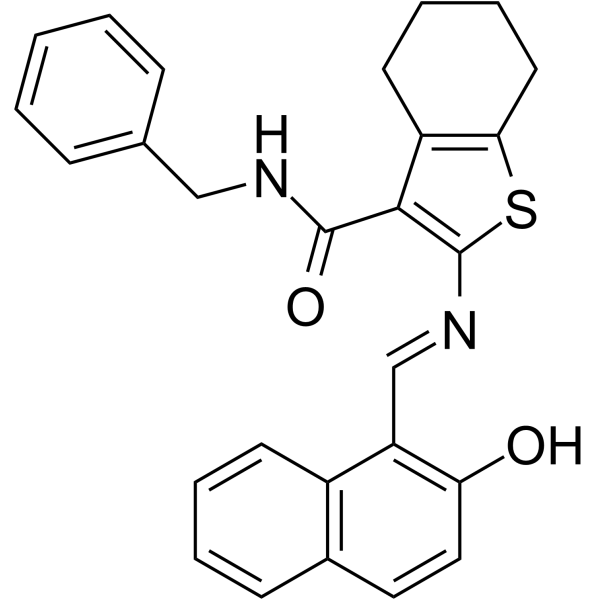
JGB1741 (ILS-JGB-1741) is a potent and specific SIRT1 activity inhibitor with an IC50 of ∼15 μM. JGB1741 is a weak SIRT2 and SIRT3 inhibitor with an all IC50>100 μM. JGB1741 increases the acetylated p53 levels leading to p53-mediated apoptosis with modulation of Bax/Bcl2 ratio, cytochrome c release and PARP cleavage. JGB1741 has the potential for breast cancer research.
연구목적의 판매만을 진행합니다. 환자를 대상으로 한 판매는 하지 않습니다.
- Purity : 99.37%
- CAS No.: 1256375-38-8
- 화학식: C27H24N2O2S
- 분자량:440.56
-
보관:
4°C, protect from light
* In solvent : -80°C, 6 months; -20°C, 1 month (protect from light)

To place orders, for customer services and technical support, please contact: MedChemExpress USA
Tel: 609-228-6898 E-mail: [email protected] [email protected]


-
 Biological Activity
-
Chemical Information
-
용액&용해도
- Protocol
- 순도&문서
- References
-
 Help & FAQs


-
HY-16691
AK-7Sirtuin Inhibitor -
HY-15452
SelisistatSirT1/Sir2 Inhibitor -
HY-108331
3-TYPSIRT3 Inhibitor -
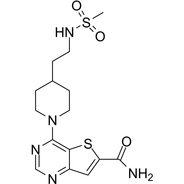
HY-16616
SIRT-IN-2SIRT1/2/3 Inhibitor -
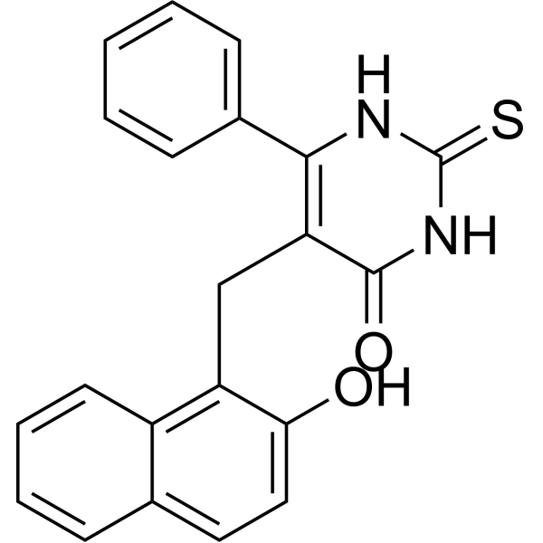
HY-100732
CambinolSIRT1/2 Inhibitor -
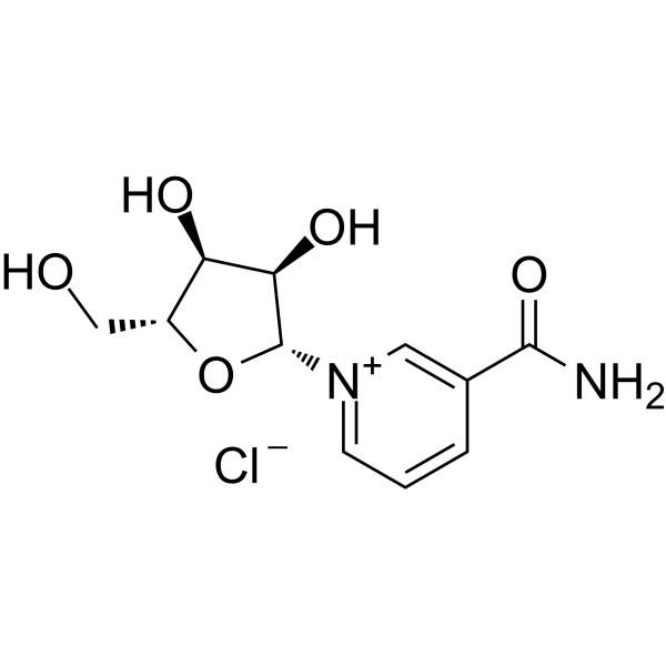
HY-123033A
Nicotinamide riboside chlorideEndogenous Metabolite -
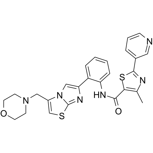
HY-15262
SRT 2104SIRT1 Activator -
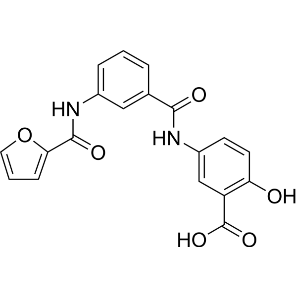
HY-107454
OSS_128167SIRT6 Inhibitor -
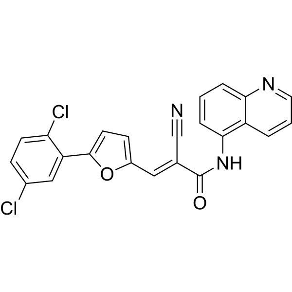
HY-100578
AGK2SIRT2 Inhibitor -
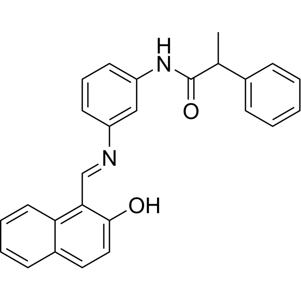
HY-101073
SalermideSirt1/Sirt2 Inhibitor -

HY-123033AR
Nicotinamide riboside chloride (Standard)Endogenous Metabolite -
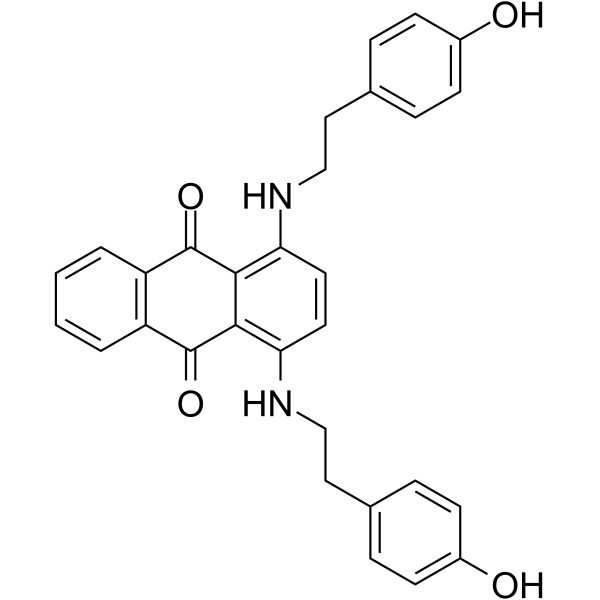
HY-108986
JFD00244SIRT2 Inhibitor
Publications Citing Use of MedChemExpress (MCE) JGB1741
More

Biological Activity
제품 설명
IC50 & Target
[1]|
SIRT1 ∼15 μM (IC50) |
SIRT2 >100 μM (IC50) |
SIRT3 >100 μM (IC50) |
In Vitro
JGB1741 (ILS-JGB-1741; 1-10000 nM; 24 h) inhibits MDA-MB 231 cell proliferation[1].
JGB1741 (0.01-1 μM; 24 h) induces apoptosis of MDA-MB 231 cells[1].
JGB1741 (0.01-1 μM; 24 h) shows a cell cycle arrest at G1 phase with more and more cells entering into sub G0/G1 phase[1].
JGB1741 (0.01-1 μM; 24 h) shows an increase in the global acetylation of H3K9, p53 expression and acetylated p53K382 levels[1].
MedChemExpress (MCE) has not independently confirmed the accuracy of these methods. They are for reference only.
-
Cell Line:K562, HepG2 and MDA-MB 231 cell lines
-
Concentration:1, 10, 50, 100, 500, 1000, 10000 nM
-
Incubation Time:24 hours
-
Result:Inhibited MDA-MB 231 cell proliferation more potently with an IC50 of 0.5 μM than K562 and HepG2 cell proliferation (IC50>1 μM).
-
Cell Line:MDA-MB 231 cells
-
Concentration:0.01, 0.1, 0.5, 1 μM
-
Incubation Time:
-
Result:Showed an increase in the percent apoptotic cells in a dose-dependent fashion with ∼70% apoptosis at 1 μM concentration.
-
Cell Line:MDA-MB 231 cells
-
Concentration:0.01, 0.1, 0.5, 1 μM
-
Incubation Time:
-
Result:Showed a cell cycle arrest at G1 phase with more and more cells entering into sub G0/G1 phase, the apoptotic phase, in a dose-dependent fashion.
-
Cell Line:MDA-MB 231 cells
-
Concentration:0.01, 0.1, 0.5, 1 μM
-
Incubation Time:
-
Result:Caused a dose-dependent increase in the global acetylation of H3K9.
Showed an increase in both p53 expression and acetylated p53K382 levels.


Chemical Information
-
CAS No. 1256375-38-8
-
Appearance Solid
-
분자량 440.56
-
화학식 C27H24N2O2S
-
Color Light yellow to yellow
-
SMILES
OC1=C(C2=C(C=C1)C=CC=C2)/C=N/C3=C(C4=C(S3)CCCC4)C(NCC5=CC=CC=C5)=O
-
Synonyms
ILS-JGB-1741
-
선적
Room temperature in continental US; may vary elsewhere.
-
보관
4°C, protect from light
* In solvent : -80°C, 6 months; -20°C, 1 month (protect from light)


Publications (1)
-
Journal Impact Factor
-
Most Recent
-
Lipids Health Dis
Dehydrodiisoeugenol alleviates palmitate-induced mitochondrial dysfunction in human vascular smooth muscle cells through the activation of SIRT1-mediated Drp1 deacetylation. [Abstract]2025 May 24;24(1):187. PMID: 40413480


용액&용해도
In Vitro:
DMSO : 5 mg/mL (11.35 mM; Need ultrasonic; Hygroscopic DMSO has a significant impact on the solubility of product, please use newly opened DMSO)
Please refer to the solubility information to select the appropriate solvent. Once prepared, please aliquot and store the solution to prevent product inactivation from repeated freeze-thaw cycles.
Storage method and period of stock solution: -80°C, 6 months; -20°C, 1 month (protect from light). When stored at -80°C, please use it within 6 months. When stored at -20°C, please use it within 1 month.
Please refer to the solubility information to select the appropriate solvent. Once prepared, please aliquot and store the solution to prevent product inactivation from repeated freeze-thaw cycles.
Storage method and period of stock solution: -80°C, 6 months; -20°C, 1 month (protect from light). When stored at -80°C, please use it within 6 months. When stored at -20°C, please use it within 1 month.
Concentration (start) × Volume (start) = Concentration (final) × Volume (final)


순도&문서
-
Data Sheet (285 KB)
-
SDS (252 KB)
- English - EN (252 KB)
- Français - FR (252 KB)
- Deutsch - DE (252 KB)
- Norwegian - NO (252 KB)
- Español - ES (252 KB)
- Swedish - SV (252 KB)
- Italian - IT (252 KB)
- Korean - KR (252 KB)
- Portuguese - PT (252 KB)
-
Handling Instructions (2659 KB)
References


Complete Stock Solution Preparation Table
Please refer to the solubility information to select the appropriate solvent. Once prepared, please aliquot and store the solution to prevent product inactivation from repeated freeze-thaw cycles.
Storage method and period of stock solution: -80°C, 6 months; -20°C, 1 month (protect from light). When stored at -80°C, please use it within 6 months. When stored at -20°C, please use it within 1 month.
| Optional Solvent | Concentration Solvent Mass | 1 mg | 5 mg | 10 mg | 25 mg |
|---|---|---|---|---|---|
| DMSO | 1 mM | 2.2698 mL | 11.3492 mL | 22.6984 mL | 56.7460 mL |
| 5 mM | 0.4540 mL | 2.2698 mL | 4.5397 mL | 11.3492 mL | |
| 10 mM | 0.2270 mL | 1.1349 mL | 2.2698 mL | 5.6746 mL |


JGB1741 Related Classifications
HY-111329 Related Classifications
 Powered by Bioz