



ROCK-IN-4
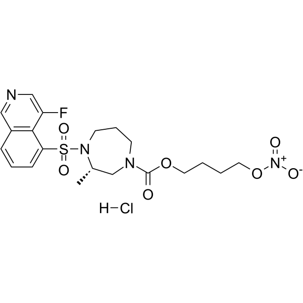
ROCK-IN-4 is a potent ROCK inhibitor maintaining NO releasing ability. ROCK-IN-4 reversibly depolymerizes F-actin, and suppresses mitochondrial respiration in human trabecular meshwork (HTM) cells. ROCK-IN-4 can be used for glaucoma or ocular hypertension research.
연구목적의 판매만을 진행합니다. 환자를 대상으로 한 판매는 하지 않습니다.
- CAS No.: 2488395-07-7
- 화학식: C20H26ClFN4O7S
- 분자량:520.96
-
보관:
Please store the product under the recommended conditions in the Certificate of Analysis.

To place orders, for customer services and technical support, please contact: MedChemExpress USA
Tel: 609-228-6898 E-mail: [email protected] [email protected]


-
 Biological Activity
-
Chemical Information
- Protocol
- 순도&문서
- References
-
 Help & FAQs




Biological Activity
제품 설명
In Vitro
ROCK-IN-4 (RNO-6) (10 μM; 1 h) decreases p-MLC level and induces reversible F-actin depolymerization[1].
ROCK-IN-4 (10 μM; 24 h) involves in cGMP activation, increases cGMP concentration in human trabecular meshwork (HTM) cells[1].
ROCK-IN-4 (10 μM; 1 h) suppresses mitochondrial respiration by releasing NO and remarkably decreases the basal respiration, maximal respiration, and ATP production[1].
MedChemExpress (MCE) has not independently confirmed the accuracy of these methods. They are for reference only. Further protocols information, click here.
-
Cell Line:Human trabecular meshwork (HTM) cells
-
Concentration:10 μM
-
Incubation Time:1 hour
-
Result:Inhibited the phosphorylation of MLC, reduced the level of p-MLC.
-
Cell Line:Human trabecular meshwork (HTM) cells
-
Concentration:10 μM
-
Incubation Time:1 hour
-
Result:Exhibited conspicuous F-actin depolymerization, and after recovery for 4 h, recovered F-actin to the polymerization morphology, indicating a reversible depolymerization effect without permanent damage to cells.
In Vivo
ROCK-IN-4 (0.579% w/v for 25 μL; o.p. in left eye; single dose) shows low hyperemic effect and eye irritation in New Zealand White rabbits[1].
MedChemExpress (MCE) has not independently confirmed the accuracy of these methods. They are for reference only.
-
Animal Model:Chronic Ocular Hypertension Mouse Model induced by Microbead[1]
-
Dosage:0.26% (w/v), 10 μL
-
Administration:Ophthalmic drop; singel dose; administration in right eye 7 days after modeling, and measured IOP prior to and at 1, 2, 3, 4, and 6 h after instillation
-
Result:Reduced IOP (mmHg) to 3.68 ± 0.5 mmHg (19.9%) and 1.36 ± 0.6 mmHg (7.4%) at 1 and 4 h after instillation, respectively.
-
Animal Model:New Zealand White rabbits[1]
-
Dosage:0.579% (w/v); 25 μL
-
Administration:Ophthalmic drop; singel dose; administration in left eye before and at 1, 2, and 4 h after the first instillation
-
Result:Showed low side effects in conjunctival hyperemia.


Chemical Information
-
CAS No. 2488395-07-7
-
분자량 520.96
-
화학식 C20H26ClFN4O7S
-
SMILES
C[C@H]1CN(C(OCCCCO[N+]([O-])=O)=O)CCCN1S(=O)(C2=CC=CC3=C2C(F)=CN=C3)=O.[H]Cl
-
선적
Room temperature in continental US; may vary elsewhere.
-
보관
Please store the product under the recommended conditions in the Certificate of Analysis.

Protocol
-
Research Protocol for Cardiovascular Diseases
Cardiovascular disease can be modeled as maladaptive cardiac remodeling, where ischemic injury or pressure overload activates inflammatory signaling, fibroblast activation, extracellular-matrix deposition, cardiomyocyte hypertrophy, vascular remodeling, and progressive ventricular dysfunction. The TGF-β/SMAD axis is a central profibrotic pathway after myocardial injury and pressure overload, while innate immune and cytokine pathways regulate leukocyte recruitment, scar formation, and adverse remodeling. Key unresolved questions include which inflammatory signals are reparative versus harmful, when fibrosis is protective versus maladaptive, and whether pathway inhibition improves function without weakening necessary infarct healing or compensatory remodeling.
-
Phalloidin F-actin cytoskeleton staining
Phalloidin F-actin staining detects polymerized filamentous actin in fixed and permeabilized specimens by using fluorescent phalloidin or phalloidin-derived phallotoxins that bind actin filaments and generate a fluorescence microscopy readout corresponding to F-actin organization, including stress fibers, cortical actin, filament bundles, and tissue-specific actin networks. Phalloidin stabilizes F-actin by reducing actin subunit dissociation from filament ends, and fluorescent phallotoxins were established as tools for visualizing actin-containing structures in eukaryotic cells.


순도&문서
References

Calculators
Concentration (start) × Volume (start) = Concentration (final) × Volume (final)


ROCK-IN-4 Related Classifications
HY-151189 Related Classifications
 Powered by Bioz