



KX2-361

Based on 1 Customer Validation
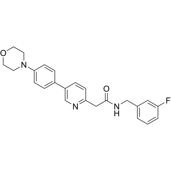
KX2-361 (KX-02) is a Src-kinase and tubulin polymerization inhibitor. KX2-361 shows good oral bioavailability and readily crosses the BBB in mice. KX2-361 shows anti-tumor activity and induces apoptosis of Glioblastoma (GBM) cell.
For research use only. We do not sell to patients.
- Purity : 99.09%
- CAS No.: 897016-26-1
- Formula: C24H24FN3O2
- Molecular Weight:405.46
-
Storage:
4°C, protect from light
* In solvent : -80°C, 6 months; -20°C, 1 month (protect from light)

To place orders, for customer services and technical support, please contact: MedChemExpress USA
Tel: 609-228-6898 E-mail: [email protected] [email protected]


-
 Biological Activity
-
Chemical Information
-
Solvent & Solubility
- Protocols
- Purity & Documentation
- References
-
 Help & FAQs


-
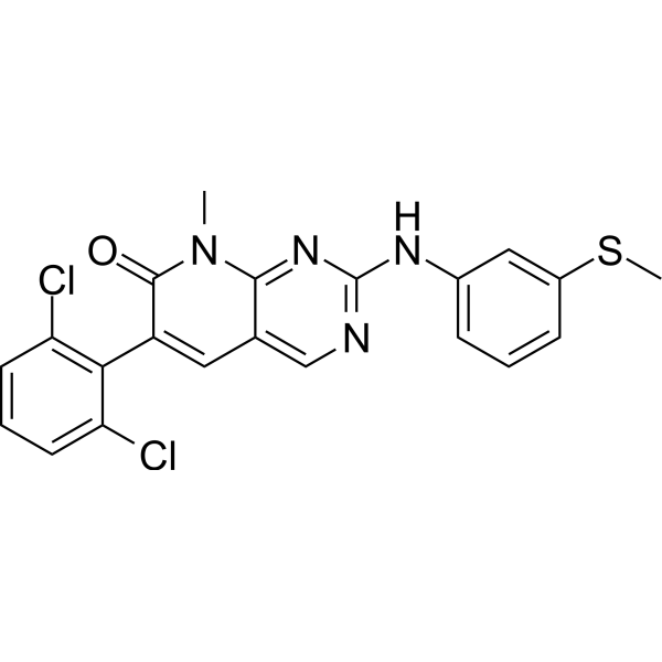
HY-10395
PD173955Src Inhibitor -

HY-10395R
PD173955 (Standard)Reference Standard -
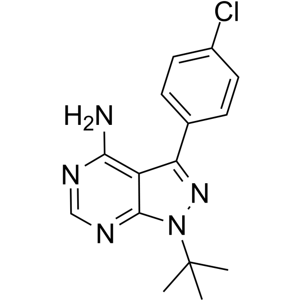
HY-13805
PP2Src Inhibitor -
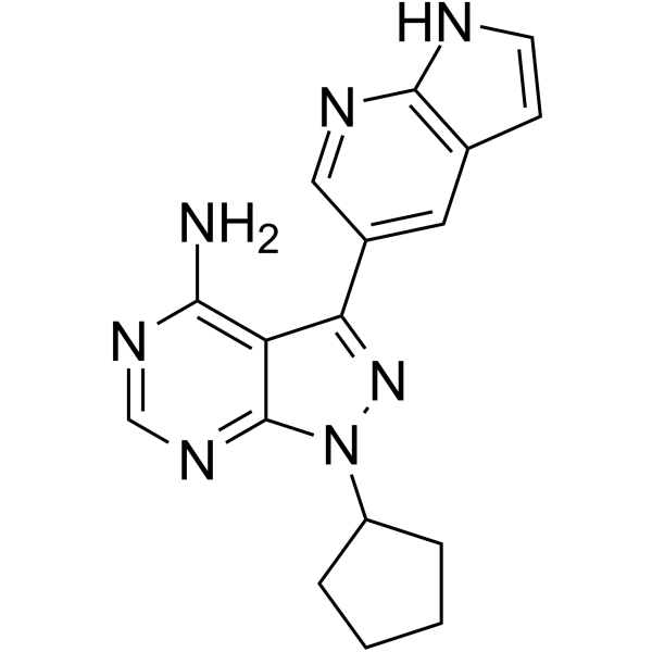
HY-10372
PP121Multi-targeted Kinase Inhibitor -
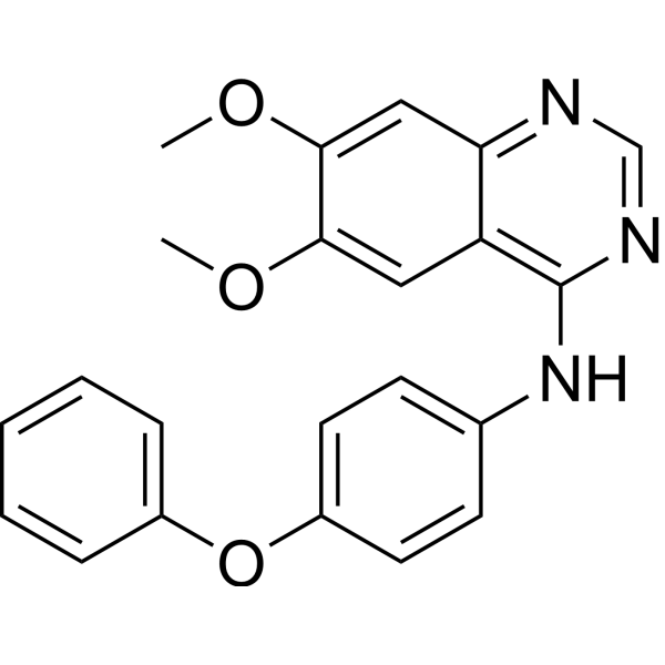
HY-101053
Src Inhibitor 1Src Inhibitor -
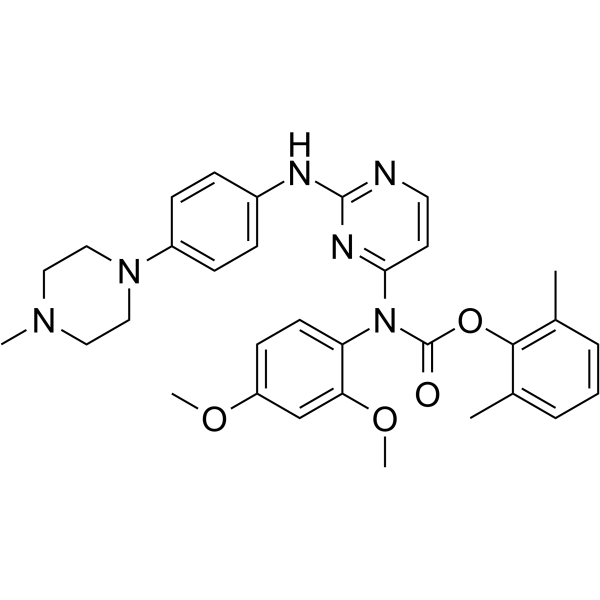
HY-12299
WH-4-023Src Inhibitor -
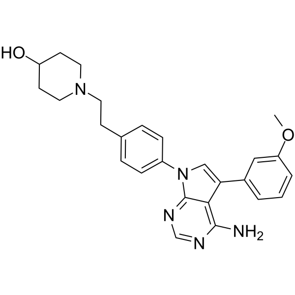
HY-W062835
CGP77675Src inhibitor -
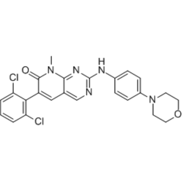
HY-122113
PD173952Tyrosine Kinases Inhibitor -
HY-10186
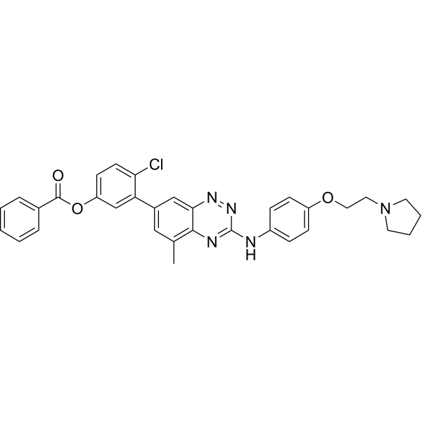
TG 100801Prodrug of TG 100572 -
HY-13942
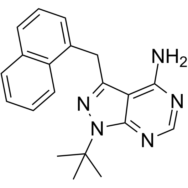
1-NM-PP1Src Inhibitor -
HY-13561
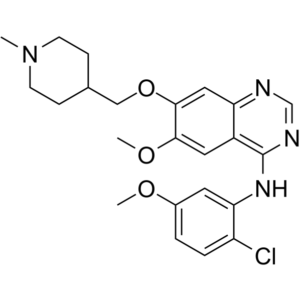
AZM475271Src Inhibitor -

HY-10372R
PP121 (Standard)Reference Standard


Biological Activity
Description
Cellular Effect
|
Cell Line

|
Type | Value | Description | References |
|---|---|---|---|---|
| Daoy | GI50 |
16 nM
Compound: 2; KX2-361; KX-02
|
Antiproliferative activity against human DaOY cells
Antiproliferative activity against human DaOY cells
|
[PMID: 29617135] |
| GL261 | IC50 |
60 nM
Compound: 2; KX2-361; KX-02
|
Inhibition of Src autophosphorylation in mouse GL261 cells
Inhibition of Src autophosphorylation in mouse GL261 cells
|
[PMID: 29617135] |
| LN-18 | GI50 |
2.9 nM
Compound: 2; KX2-361; KX-02
|
Antiproliferative activity against human LN18 cells
Antiproliferative activity against human LN18 cells
|
[PMID: 29617135] |
| SK-N-FI | GI50 |
11 nM
Compound: 2; KX2-361; KX-02
|
Antiproliferative activity against human SK-N-FI cells
Antiproliferative activity against human SK-N-FI cells
|
[PMID: 29617135] |
| SK-N-MC | GI50 |
8 nM
Compound: 2; KX2-361; KX-02
|
Antiproliferative activity against human SK-N-MC cells
Antiproliferative activity against human SK-N-MC cells
|
[PMID: 29617135] |
| SW1088 | GI50 |
26 nM
Compound: 2; KX2-361; KX-02
|
Antiproliferative activity against human SW1088 cells
Antiproliferative activity against human SW1088 cells
|
[PMID: 29617135] |
| T98G | GI50 |
14 nM
Compound: 2; KX2-361; KX-02
|
Antiproliferative activity against human T98G cells
Antiproliferative activity against human T98G cells
|
[PMID: 29617135] |
| U-118-MG | GI50 |
29 nM
Compound: 2; KX2-361; KX-02
|
Antiproliferative activity against human U118MG cells
Antiproliferative activity against human U118MG cells
|
[PMID: 29617135] |
| U138-MG | GI50 |
51 nM
Compound: 2; KX2-361; KX-02
|
Antiproliferative activity against human U138MG cells
Antiproliferative activity against human U138MG cells
|
[PMID: 29617135] |
| U-373MG ATCC | GI50 |
54 nM
Compound: 2; KX2-361; KX-02
|
Antiproliferative activity against human U373 cells
Antiproliferative activity against human U373 cells
|
[PMID: 29617135] |
| U-87MG ATCC | GI50 |
76 nM
Compound: 2; KX2-361; KX-02
|
Antiproliferative activity against human U87 cells
Antiproliferative activity against human U87 cells
|
[PMID: 29617135] |
In Vitro
KX2-361 (0-200 nM, 24-72 h) reduces autophosphorylation of Src in GL261 cells[1].
KX2-361 (0-270 nM) promotes cell cycle arrest at the G2/M phase in U87 cells. Note the dose dependent effect and virtual complete arrest at 270 nM[1].
KX2-361 (0-800 nM) induces apoptosis of U87, GL261 and T98G cell lines[1].
KX2-361 (5 µM) inhibits the in vitro assembly of tubulin polymers[1].
MedChemExpress (MCE) has not independently confirmed the accuracy of these methods. They are for reference only.
In Vivo
KX2-361 demonstrates appreciable brain penetration when dosed orally to mice (20 mg/kg), with a brain Cmax of 4025±319 ng/g observed 15 min post-dosing and an overall exposure (AUClast) of 5044±355 h ng/g[1].
MedChemExpress (MCE) has not independently confirmed the accuracy of these methods. They are for reference only.
Clinical Trial
| NCT Number | Sponsor | Condition | Start Date |
Phase
|
|---|---|---|---|---|
| NCT01329991 | 2011-05 | PHASE1 |


Chemical Information
-
CAS No. 897016-26-1
-
Appearance Solid
-
Molecular Weight 405.46
-
Formula C24H24FN3O2
-
Color White to off-white
-
SMILES
O=C(NCC1=CC=CC(F)=C1)CC2=NC=C(C3=CC=C(N4CCOCC4)C=C3)C=C2
-
Synonyms
KX-02
-
Shipping
Room temperature in continental US; may vary elsewhere.
-
Storage
4°C, protect from light
* In solvent : -80°C, 6 months; -20°C, 1 month (protect from light)


Solvent & Solubility
In Vitro:
DMSO : 10.71 mg/mL (26.41 mM; ultrasonic and warming and heat to 60°C; Hygroscopic DMSO has a significant impact on the solubility of product, please use newly opened DMSO)
Please refer to the solubility information to select the appropriate solvent. Once prepared, please aliquot and store the solution to prevent product inactivation from repeated freeze-thaw cycles.
Storage method and period of stock solution: -80°C, 6 months; -20°C, 1 month (protect from light). When stored at -80°C, please use it within 6 months. When stored at -20°C, please use it within 1 month.
Please refer to the solubility information to select the appropriate solvent. Once prepared, please aliquot and store the solution to prevent product inactivation from repeated freeze-thaw cycles.
Storage method and period of stock solution: -80°C, 6 months; -20°C, 1 month (protect from light). When stored at -80°C, please use it within 6 months. When stored at -20°C, please use it within 1 month.
Concentration (start) × Volume (start) = Concentration (final) × Volume (final)


Purity & Documentation
-
Data Sheet (270 KB)
-
SDS (252 KB)
- English - EN (252 KB)
- Français - FR (252 KB)
- Deutsch - DE (252 KB)
- Norwegian - NO (252 KB)
- Español - ES (252 KB)
- Swedish - SV (252 KB)
- Italian - IT (252 KB)
- Korean - KR (252 KB)
- Portuguese - PT (252 KB)
-
Handling Instructions (2659 KB)
References


Complete Stock Solution Preparation Table
Please refer to the solubility information to select the appropriate solvent. Once prepared, please aliquot and store the solution to prevent product inactivation from repeated freeze-thaw cycles.
Storage method and period of stock solution: -80°C, 6 months; -20°C, 1 month (protect from light). When stored at -80°C, please use it within 6 months. When stored at -20°C, please use it within 1 month.
| Optional Solvent | Concentration Solvent Mass | 1 mg | 5 mg | 10 mg | 25 mg |
|---|---|---|---|---|---|
| DMSO | 1 mM | 2.4663 mL | 12.3317 mL | 24.6633 mL | 61.6584 mL |
| 5 mM | 0.4933 mL | 2.4663 mL | 4.9327 mL | 12.3317 mL | |
| 10 mM | 0.2466 mL | 1.2332 mL | 2.4663 mL | 6.1658 mL | |
| 15 mM | 0.1644 mL | 0.8221 mL | 1.6442 mL | 4.1106 mL | |
| 20 mM | 0.1233 mL | 0.6166 mL | 1.2332 mL | 3.0829 mL | |
| 25 mM | 0.0987 mL | 0.4933 mL | 0.9865 mL | 2.4663 mL |


KX2-361 Related Classifications
HY-111187 Related Classifications
 Powered by Bioz