



Based on 1 Customer Validation
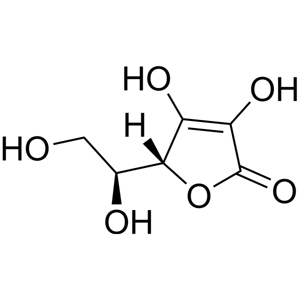
L-Ascorbic acid (GMP Like) is the GMP Like class L-Ascorbic acid (HY-B0166). L-Ascorbic acid (L-Ascorbate, Vitamin C), an electron donor, is an endogenous antioxidant agent. L-Ascorbic acid inhibits selectively Cav3.2 channels with an IC50 of 6.5 μM. L-Ascorbic acid is also a collagen deposition enhancer and an elastogenesis inhibitor. L-Ascorbic acid exhibits anti-cancer effects through the generation of reactive oxygen species (ROS) and selective damage to cancer cells.
Para uso exclusivo en investigación. No vendemos a pacientes.
- Pureza : 99.88%
- No. CAS: 50-81-7
- Fòrmula: C6H8O6
- Peso molecular:176.12
-
Almacenamiento:
Please store the product under the recommended conditions in the Certificate of Analysis.

To place orders, for customer services and technical support, please contact: MedChemExpress USA
Tel: 609-228-6898 E-mail: [email protected] [email protected]


-
 Actividad biológica
-
Chemical Information
-
Solvente y solubilidad
- Protocolo
- Pureza y Documentación
- Referencias
-
 Help & FAQs


-
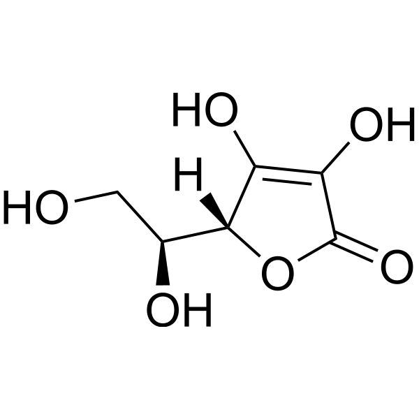
HY-B0166
L-Ascorbic acidAntioxidant -
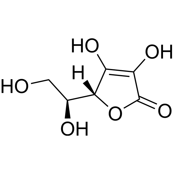
HY-B0166G
L-Ascorbic acid (GMP)Antioxidant -

HY-B0166R
L-Ascorbic acid (Standard)Antioxidant -
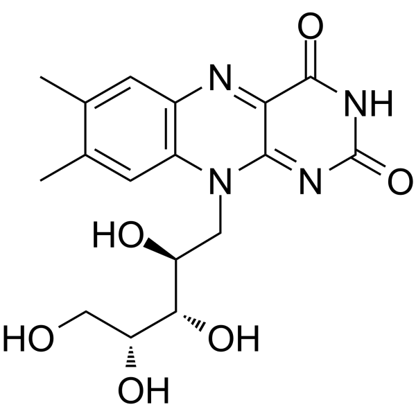
HY-B0456
RiboflavinVitamin B2 -
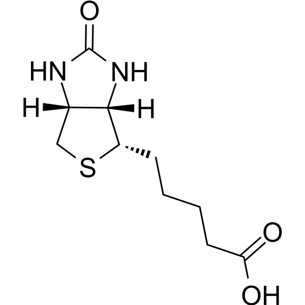
HY-B0511
BiotinVitamin B7 -
HY-B0143
NiacinB3 vitamin -
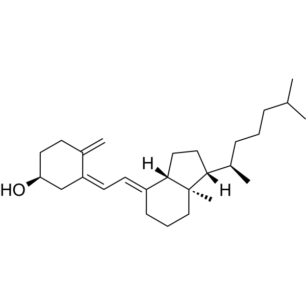
HY-15398
Vitamin D3Vitamin -
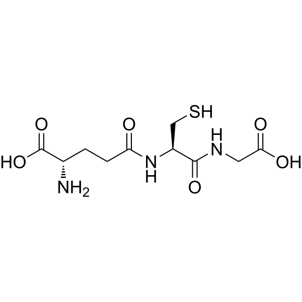
HY-D0187
L-Glutathione reducedAntioxidant -
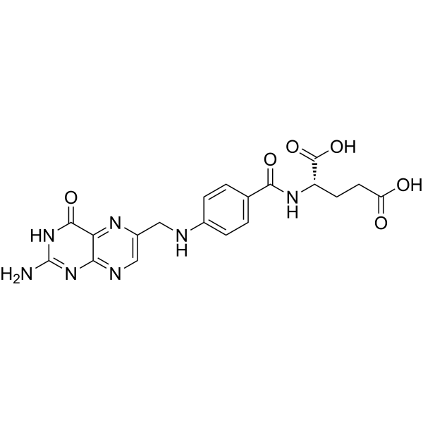
HY-16637
Folic acidB complex -
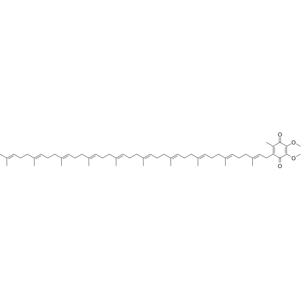
HY-N0111
Coenzyme Q10Antioxidant -
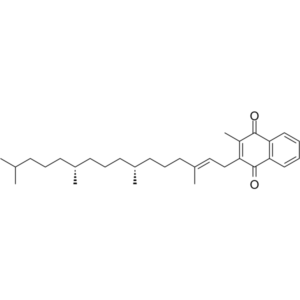
HY-N0684
Vitamin K1Endogenous Metabolite -
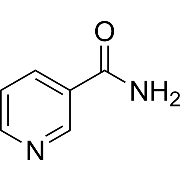
HY-B0150
NicotinamideSIRT Inhibitor
Ver todos los productos específicos de isoformas Endogenous Metabolite
MoreVer todos los productos específicos de isoformas Calcium Channel
More

Actividad biológica
Descripciòn
Cellular Effect
|
Cell Line

|
Type | Value | Description | References |
|---|---|---|---|---|
| A549 | IC50 |
>100 μM
Compound: L-ASA
|
Antiproliferative activity against human A549 cells assessed as reduction in cell viability incubated for 72 hrs by MTT assay
Antiproliferative activity against human A549 cells assessed as reduction in cell viability incubated for 72 hrs by MTT assay
|
[PMID: 31586832] |
| CFPAC-1 | IC50 |
>100 μM
Compound: L-ASA
|
Antiproliferative activity against human CFPAC-1 cells assessed as reduction in cell viability incubated for 72 hrs by MTT assay
Antiproliferative activity against human CFPAC-1 cells assessed as reduction in cell viability incubated for 72 hrs by MTT assay
|
[PMID: 31586832] |
| Erythrocyte | EC50 |
119.87 μg/mL
Compound: Ascorbic acid
|
Antioxidant activity in erythrocytes assessed as inhibition of hemolysis
Antioxidant activity in erythrocytes assessed as inhibition of hemolysis
|
[PMID: 22677320] |
| Erythrocyte | EC50 |
96.625 μg/mL
Compound: Ascorbic acid
|
Hemolysis of human erythrocytes after 30 mins by spectrophotometry
Hemolysis of human erythrocytes after 30 mins by spectrophotometry
|
[PMID: 23811092] |
| HCT-116 | IC50 |
>100 μM
Compound: L-ASA
|
Antiproliferative activity against human HCT116cells assessed as reduction in cell viability incubated for 72 hrs by MTT assay
Antiproliferative activity against human HCT116cells assessed as reduction in cell viability incubated for 72 hrs by MTT assay
|
[PMID: 31586832] |
| HEK293 | EC50 |
0.5 nM
Compound: Ascorbic acid
|
Agonist activity at human adrenergic alpha2B receptor expressed in HEK293 cells coexpressing Gqi5 protein by FLIPR assay
Agonist activity at human adrenergic alpha2B receptor expressed in HEK293 cells coexpressing Gqi5 protein by FLIPR assay
|
[PMID: 19243956] |
| HEK293 | EC50 |
156 μM
Compound: 54670067
|
Substrate uptake and inhibition of the Na(+)/L-Ascorbic Acid Transporter 1 (SVCT1, SLC23A1) as assessed by the fluorescent FLIPR membrane potential dye in HEK-293 JumpIN-SLC23A1 cells (PubChem AID: 1794810)
Substrate uptake and inhibition of the Na(+)/L-Ascorbic Acid Transporter 1 (SVCT1, SLC23A1) as assessed by the fluorescent FLIPR membrane potential dye in HEK-293 JumpIN-SLC23A1 cells (PubChem AID: 1794810)
|
10.5281/zenodo.7360349 |
| HeLa | IC50 |
>100 μM
Compound: L-ASA
|
Antiproliferative activity against human HeLa cells assessed as reduction in cell viability incubated for 72 hrs by MTT assay
Antiproliferative activity against human HeLa cells assessed as reduction in cell viability incubated for 72 hrs by MTT assay
|
[PMID: 31586832] |
| HepG2 | IC50 |
>100 μM
Compound: L-ASA
|
Antiproliferative activity against human HepG2 cells assessed as reduction in cell viability incubated for 72 hrs by MTT assay
Antiproliferative activity against human HepG2 cells assessed as reduction in cell viability incubated for 72 hrs by MTT assay
|
[PMID: 31586832] |
| HFF | IC50 |
>100 μM
Compound: L-ASA
|
Cytotoxicity against human HFF cells assessed as reduction in cell viability incubated for 72 hrs by MTT assay
Cytotoxicity against human HFF cells assessed as reduction in cell viability incubated for 72 hrs by MTT assay
|
[PMID: 31586832] |
| HL-60 | IC50 |
0.01 μM
Compound: Vitamin C
|
Antioxidant activity in human HL60 cells assessed as inhibition of relative oxygen species generation by DCFH-DA method
Antioxidant activity in human HL60 cells assessed as inhibition of relative oxygen species generation by DCFH-DA method
|
[PMID: 19555121] |
| HL-60 | IC50 |
10.2 μM
Compound: Vitamin C
|
Antioxidant activity in human HL60 cells assessed as inhibition of TPA-stimulated hydrogen peroxide induced DCFH-DA oxidation by fluorometric microplate assay
Antioxidant activity in human HL60 cells assessed as inhibition of TPA-stimulated hydrogen peroxide induced DCFH-DA oxidation by fluorometric microplate assay
|
[PMID: 16562834] |
| HL-60 | IC50 |
9.7 μM
Compound: Vitamin C
|
Antioxidant activity against TPA-induced ROS production in human HL60 cells by 2',7'-dichlorodihydrofluorescein diacetate cellular-based assay
Antioxidant activity against TPA-induced ROS production in human HL60 cells by 2',7'-dichlorodihydrofluorescein diacetate cellular-based assay
|
[PMID: 12762791] |
| J774.A1 | IC50 |
>100 μM
Compound: AA
|
Antiinflammatory activity in mouse J774.A1 cells assessed as inhibition of LPS-induced NO production incubated for 24 hrs by Griess reagent based analysis
Antiinflammatory activity in mouse J774.A1 cells assessed as inhibition of LPS-induced NO production incubated for 24 hrs by Griess reagent based analysis
|
[PMID: 33422907] |
| MCF7 | IC50 |
>100 μM
Compound: L-ASA
|
Antiproliferative activity against human MCF7 cells assessed as reduction in cell viability incubated for 72 hrs by MTT assay
Antiproliferative activity against human MCF7 cells assessed as reduction in cell viability incubated for 72 hrs by MTT assay
|
[PMID: 31586832] |
| RD | EC50 |
>2270 μM
Compound: AA
|
Antiviral activity against Herpes simplex virus type 1 infected in human RD cells assessed as inhibition of virus-induced cytopathic effect
Antiviral activity against Herpes simplex virus type 1 infected in human RD cells assessed as inhibition of virus-induced cytopathic effect
|
[PMID: 22390834] |
| SW-620 | IC50 |
>100 μM
Compound: L-ASA
|
Antiproliferative activity against human SW620 cells assessed as reduction in cell viability incubated for 72 hrs by MTT assay
Antiproliferative activity against human SW620 cells assessed as reduction in cell viability incubated for 72 hrs by MTT assay
|
[PMID: 31586832] |
In Vitro
The anti-cancer effects of L-Ascorbic acid are determined by sodium-dependent vitamin C transporter 2 (SVCT-2), a transporter of L-ascorbic acid. L-Ascorbic acid (0.1 μM-2 mM) exhibits anti-cancer effects according to SVCT-2 expression and L-ascorbic acid uptake. Human colorectal cancer cell lines displays differential responses to L-ascorbic acid, primarily depending on the expression level of SVCT-2[4].
L-Ascorbic acid (10 μg/ml, 5 days) enhances the reprogramming of mouse fibroblasts into IPSCs[5].
L-Ascorbic acid (50 μg/ml, 9 days) promotes fibroblasts conversion into cardiomyocytes[6].
L-Ascorbic acid (50 ng/ml, 4-6 days) facilitates generation of all-iPS cell mice from terminally differentiated B cells[7].
MedChemExpress (MCE) has not independently confirmed the accuracy of these methods. They are for reference only. Further protocols information, click here.
In Vivo
MedChemExpress (MCE) has not independently confirmed the accuracy of these methods. They are for reference only.


Chemical Information
-
No. CAS 50-81-7
-
Appearance Solid
-
Peso molecular 176.12
-
Fòrmula C6H8O6
-
Color White to off-white
-
SMILES
OC([C@@]1([H])[C@H](CO)O)=C(O)C(O1)=O
-
Synonyms
L-Ascorbate (GMP Like); Vitamin C (GMP Like)
-
Envío
Room temperature in continental US; may vary elsewhere.
-
Almacenamiento
Please store the product under the recommended conditions in the Certificate of Analysis.


Solvente y solubilidad
In Vitro:
DMSO : 100 mg/mL (567.79 mM; Need ultrasonic; Hygroscopic DMSO has a significant impact on the solubility of product, please use newly opened DMSO)
H2O : 100 mg/mL (567.79 mM; Need ultrasonic)
* Note: If you choose water as the stock solution, please dilute it to the working solution, then filter and sterilize it with a 0.22 μm filter before use.
* Note: If you choose water as the stock solution, please dilute it to the working solution, then filter and sterilize it with a 0.22 μm filter before use.
Concentration (start) × Volume (start) = Concentration (final) × Volume (final)

Protocolo
-
Fibrosis/Collagen Morphometry
Fibrosis and collagen morphometry is based on the quantitative visualization of fibrillar collagen deposition in tissue sections using histochemical stains such as Sirius Red (Picrosirius Red) or Masson's trichrome, followed by image-based or polarization-enhanced analysis to estimate collagen proportional area as a surrogate of extracellular matrix accumulation during fibrotic remodeling. Sirius Red combined with polarized light microscopy enhances detection of collagen fibers due to birefringence properties, enabling more specific visualization of collagen type I and III fibrils compared to conventional bright-field histology, while whole-section or region-restricted digital morphometry reduces field-selection bias in fibrosis assessment. Alternative quantitative approaches include second harmonic generation (SHG) and two-photon excited fluorescence microscopy, which enable label-free detection of fibrillar collagen and have been validated against histological staining and biochemica
-
Collagen: Sirius Red Staining
Sirius Red or picrosirius red staining is a histochemical method for visualizing collagen-rich extracellular matrix in tissue sections, and collagen fibers are detected as red-stained structures under bright-field microscopy with enhanced birefringence under polarized light. Picrosirius red is useful for assessing total collagen organization, distribution, and fibrosis burden, but polarized color should not be interpreted as a definitive collagen type I versus type III readout because color is affected by fiber orientation, thickness, and packing.
-
Cell Cytotoxicity Assay
Cytotoxicity assays are usually based on the assessment of cell membrane damage, which can also be indirectly detected by measuring cell viability. Detection methods include MTT assay, CKK-8 assay, LDH assay and ATP assay, etc.
-
ROS/oxidative-stress fluorescent staining
ROS/oxidative-stress fluorescent staining uses cell-permeant fluorogenic probes that become fluorescent after oxidation inside cells or tissues; commonly used examples include DCFH-DA/DCFDA for broad cellular oxidant detection, DHE for superoxide-related signal detection, MitoSOX for mitochondrial superoxide-related signal detection, and CellROX probes for oxidative-stress-associated fluorescence readouts. The assay detects probe oxidation rather than a single ROS species unless the probe and analysis method have been chemically validated for that species. DCFH-DA enters cells, is deacetylated by intracellular esterases to DCFH, and produces fluorescent DCF after oxidation, so the readout is used as an operational measure of total cellular oxidative stress rather than a species-specific ROS measurement. DHE and MitoSOX can report superoxide-related oxidation, but red fluorescence alone can include non-specific ethidium-like oxidation products; HPLC or optimized spectral approaches are
-
Connective Tissue: Masson's Trichrome/Collagen Trichrome Staining
Masson’s Trichrome (collagen/trichrome staining) is a histological technique that differentially stains tissue compartments using sequential acidic dyes to distinguish collagen from muscle and cytoplasmic components based on dye affinity and tissue permeability differences, enabling visualization of fibrosis and connective tissue architecture in histological sections. The classical formulation typically uses Weigert's iron hematoxylin for nuclear staining, Biebrich scarlet-acid fuchsin for cytoplasm and muscle, and aniline blue (or light green variants) for collagen, producing a characteristic blue/green collagen signal contrasted against red cytoplasm and dark nuclei. The staining principle relies on selective displacement of smaller dye molecules by larger anionic dyes in collagen-rich regions under controlled acidified conditions, which enhances collagen-specific dye retention. This property makes the method widely used for fibrosis assessment in organs such as heart, liver, lung, a


Pureza y Documentación
-
Ficha de datos (277 KB)
-
SDS (252 KB)
- English - EN (252 KB)
- Français - FR (252 KB)
- Deutsch - DE (252 KB)
- Norwegian - NO (252 KB)
- Español - ES (252 KB)
- Swedish - SV (252 KB)
- Italian - IT (252 KB)
- Korean - KR (252 KB)
- Portuguese - PT (252 KB)
-
Instrucciones de manejo (2659 KB)
Referencias
[1]. Michael T Nelson, et al. Molecular mechanisms of subtype-specific inhibition of neuronal T-type calcium channels by ascorbate. J Neurosci. 2007 Nov 14;27(46):12577-83. [Content Brief]
[2]. Aleksander Hinek, et al. Sodium L-ascorbate enhances elastic fibers deposition by fibroblasts from normal and pathologic human skin. J Dermatol Sci. 2014 Sep;75(3):173-82. [Content Brief]
[3]. Sungrae Cho, et al. Hormetic dose response to L-ascorbic acid as an anti-cancer drug in colorectal cancer cell lines according to SVCT-2 expression. Sci Rep. 2018 Jul 27;8(1):11372. [Content Brief]
[4]. Satyanarayana Sreemantula, et al. Influence of antioxidant (L- ascorbic acid) on tolbutamide induced hypoglycaemia/antihyperglycaemia in normal and diabetic rats. BMC Endocr Disord. 2005 Mar 3;5(1):2. [Content Brief]
[5]. Sebastian J Padayatty, et al. Vitamin C as an antioxidant: evaluation of its role in disease prevention. J Am Coll Nutr. 2003 Feb;22(1):18-35. [Content Brief]
[6]. Esteban MA, Wang T, Qin B, et al. Vitamin C enhances the generation of mouse and human induced pluripotent stem cells. Cell Stem Cell. 2010;6(1):71-79. doi:10.1016/j.stem.2009.12.001 [Content Brief]
[7]. Talkhabi M, Pahlavan S, Aghdami N, Baharvand H. Ascorbic acid promotes the direct conversion of mouse fibroblasts into beating cardiomyocytes. Biochem Biophys Res Commun. 2015;463(4):699-705. [Content Brief]
[8]. Stadtfeld M, Apostolou E, Ferrari F, et al. Ascorbic acid prevents loss of Dlk1-Dio3 imprinting and facilitates generation of all-iPS cell mice from terminally differentiated B cells. Nat Genet. 2012;44(4):398-S2. [Content Brief]


Complete Stock Solution Preparation Table
| Optional Solvent | Concentration Solvent Mass | 1 mg | 5 mg | 10 mg | 25 mg |
|---|---|---|---|---|---|
| DMSO / H2O | 1 mM | 5.6779 mL | 28.3897 mL | 56.7795 mL | 141.9487 mL |
| 5 mM | 1.1356 mL | 5.6779 mL | 11.3559 mL | 28.3897 mL | |
| 10 mM | 0.5678 mL | 2.8390 mL | 5.6779 mL | 14.1949 mL | |
| 15 mM | 0.3785 mL | 1.8926 mL | 3.7853 mL | 9.4632 mL | |
| 20 mM | 0.2839 mL | 1.4195 mL | 2.8390 mL | 7.0974 mL | |
| 25 mM | 0.2271 mL | 1.1356 mL | 2.2712 mL | 5.6779 mL | |
| 30 mM | 0.1893 mL | 0.9463 mL | 1.8926 mL | 4.7316 mL | |
| 40 mM | 0.1419 mL | 0.7097 mL | 1.4195 mL | 3.5487 mL | |
| 50 mM | 0.1136 mL | 0.5678 mL | 1.1356 mL | 2.8390 mL | |
| 60 mM | 0.0946 mL | 0.4732 mL | 0.9463 mL | 2.3658 mL | |
| 80 mM | 0.0710 mL | 0.3549 mL | 0.7097 mL | 1.7744 mL | |
| 100 mM | 0.0568 mL | 0.2839 mL | 0.5678 mL | 1.4195 mL |
* Note: If you choose water as the stock solution, please dilute it to the working solution, then filter and sterilize it with a 0.22 μm filter before use.


L-Ascorbic acid (GMP Like) Related Classifications
HY-B0166GL Related Classifications
 Powered by Bioz