



Liensinine Diperchlorate
Based on 22 publication(s) in Google Scholar
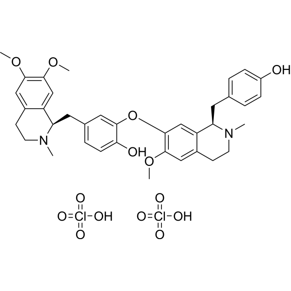
Liensinine Diperchlorate is a major isoquinoline alkaloid, extracted from the seed embryo of Nelumbo nucifera Gaertn. Liensinine Diperchlorate inhibits late-stage autophagy/mitophagy through blocking autophagosome-lysosome fusion. Liensinine Diperchlorate has a wide range of biological activities, including anti-arrhythmias, anti-hypertension, anti-pulmonary fibrosis, relaxation on vascular smooth muscle, etc.
For research use only. We do not sell to patients.
- Purity : 99.96%
- CAS No.: 5088-90-4
- Formula: C37H44Cl2N2O14
- Molecular Weight:811.66
-
Storage:
4°C, sealed storage, away from moisture
* In solvent : -80°C, 6 months; -20°C, 1 month (sealed storage, away from moisture)

To place orders, for customer services and technical support, please contact: MedChemExpress USA
Tel: 609-228-6898 E-mail: [email protected] [email protected]


-
 Biological Activity
-
Chemical Information
-
Solvent & Solubility
- Protocols
- Purity & Documentation
- References
-
 Help & FAQs


-
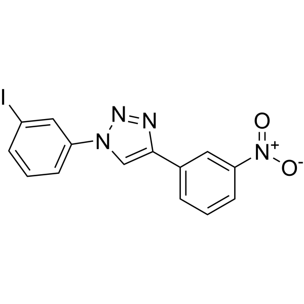
HY-115576
P62-mediated mitophagy inducerMitochondrial Autophagy Regulator -
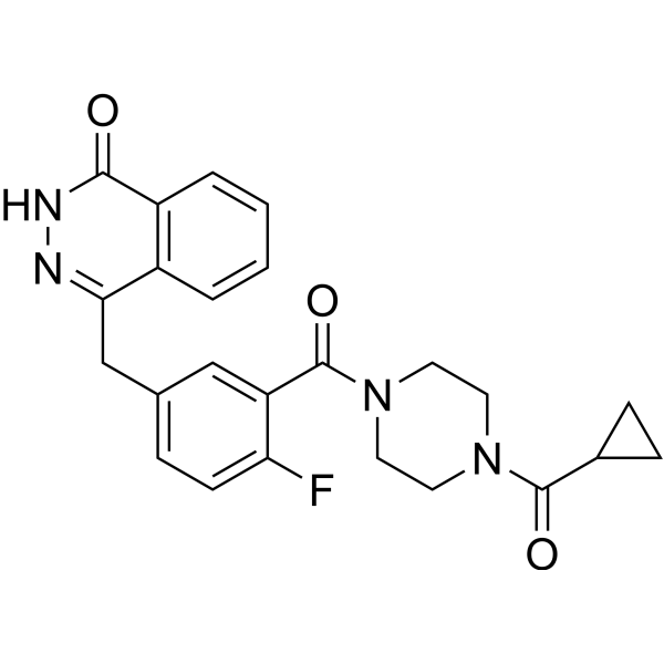
HY-10162
OlaparibPARP Inhibitor -
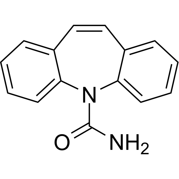
HY-B0246
CarbamazepineAnticonvulsant -
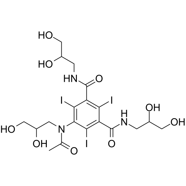
HY-B0594
IohexolContrast Agent -

HY-10162R
Olaparib (Standard)PARP Inhibitor -
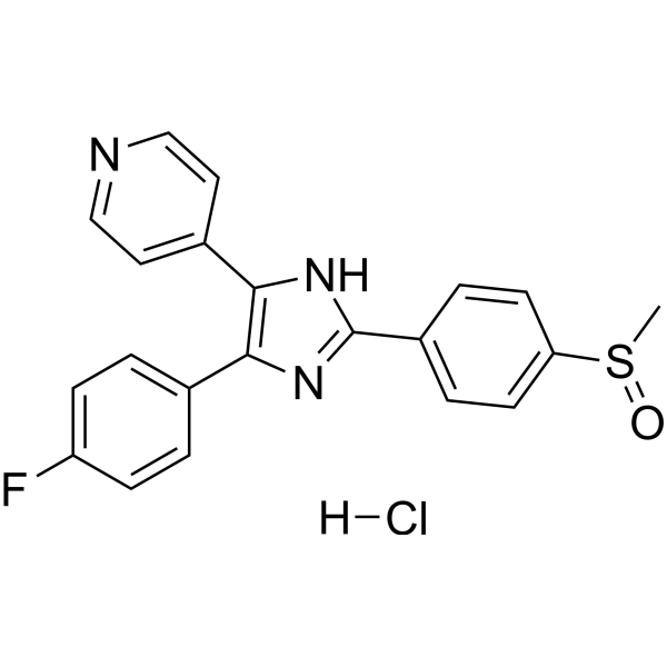
HY-10256A
Adezmapimod hydrochloridep38 MAPK Inhibitor -

HY-B0246R
Carbamazepine (Standard)Anticonvulsant -
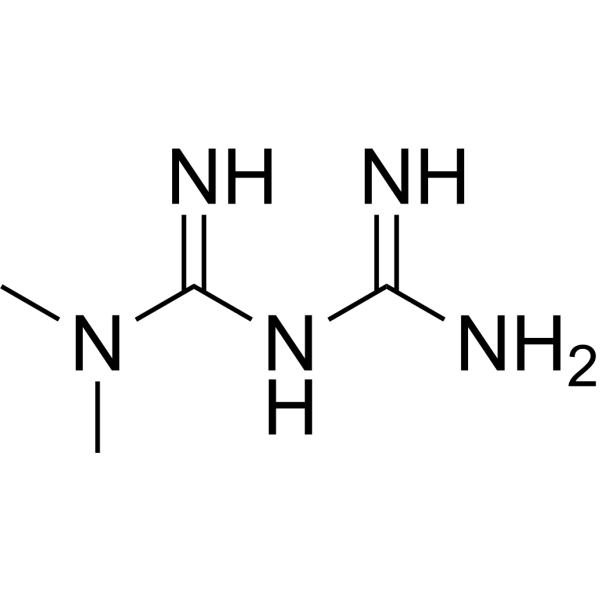
HY-B0627
MetforminAntihyperglycemic Agent -
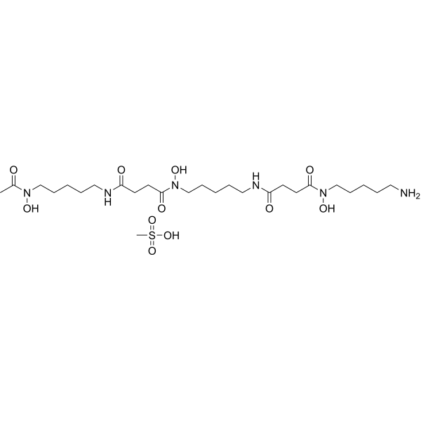
HY-B0988
Deferoxamine mesylateIron Chelator -

HY-B0594R
Iohexol (Standard)Contrast Agent -
HY-160019
MTX115325USP30 Inhibitor
Publications Citing Use of MedChemExpress (MCE) Liensinine Diperchlorate
More- Signal Transduct Target Ther. 2025 Dec 17;10(1):413. [Abstract]
- Redox Biol. 2025 Oct:86:103793. [Abstract]
- Cell Death Dis. 2023 Sep 30;14(9):645. [Abstract]
- Int J Biol Sci. 2022 Aug 8;18(13):5168-5184. [Abstract]
- Anim Nutr. 2026 Feb 6:25:160-174. [Abstract]
- Chin Med. 2026 Jan 24;21(1):51. [Abstract]
- J Ethnopharmacol. 2024 Nov 15:334:118576. [Abstract]
- Life Sci. 2023 Jun 1:322:121653. [Abstract]
- Int J Mol Sci. 2025 May 10;26(10):4566. [Abstract]
- Molecules. 2026 Mar 12;31(6):947. [Abstract]
- J Mol Med (Berl). 2025 Dec 26;104(1):13. [Abstract]
- J Biol Chem. 2024 Aug;300(8):107542. [Abstract]
- Adipocyte. 2022 Dec;11(1):202-212. [Abstract]
- Tissue Cell. 2026 Jun 12:103:103696. [Abstract]
- Genes Nutr. 2025 Dec 24;20(1):29. [Abstract]
- J Int Med Res. 2026 Feb;54(2):3000605261419604. [Abstract]
- Lett Drug Des Discov. 2025 Dec 2.
- Int J Pharmacol. 2026 Jun 28;22(3).
- SSRN. 2023 Apr 18.
- Research Square Print. 2023 Mar 24.
- J Oncol. 2022 Jul 1:2022:1533779. [Abstract]
- Oxid Med Cell Longev. 2022 Jan 25:2022:8245614. [Abstract]
 Customer Validation & Images

-
Cell Proliferation/Viability Assay
-
WB
-
Flow Cytometry
-
Cell Imaging/Staining
-
RT-PCR


Biological Activity
Description
Clinical Trial
| NCT Number | Sponsor | Condition | Start Date |
Phase

|
|---|---|---|---|---|
| NCT01329991 | Plexxikon| | 2011-05 | PHASE1 |


Chemical Information
-
CAS No. 5088-90-4
-
Appearance Solid
-
Molecular Weight 811.66
-
Formula C37H44Cl2N2O14
-
Color White to off-white
-
SMILES
O=Cl(=O)(O)=O.CN1[C@@H](C2=CC(OC3=CC(C[C@@H]4C5=CC(OC)=C(OC)C=C5CCN4C)=CC=C3O)=C(OC)C=C2CC1)CC6=CC=C(O)C=C6.O=Cl(=O)(O)=O
-
Structure Classification
-
Initial Source
-
Shipping
Room temperature in continental US; may vary elsewhere.
-
Storage
4°C, sealed storage, away from moisture
* In solvent : -80°C, 6 months; -20°C, 1 month (sealed storage, away from moisture)


Publications (22)
-
Journal Impact Factor
-
Most Recent
-
Signal Transduct Target Ther
Disruption of heme homeostasis by nuclear receptor Nur77 induces pyroptosis through granzyme B-dependent GSDMC cleavage. [Abstract]2025 Dec 17;10(1):413. PMID: 41407678 -
Redox Biol
Arylsulfatase K attenuates airway epithelial cell senescence in COPD by regulating parkin-mediated mitophagy. [Abstract]2025 Oct:86:103793. PMID: 40763656
Liensinine Diperchlorate purchased from MedChemExpress. Usage Cited in: Redox Biol. 2025 Oct:86:103793. [Abstract]
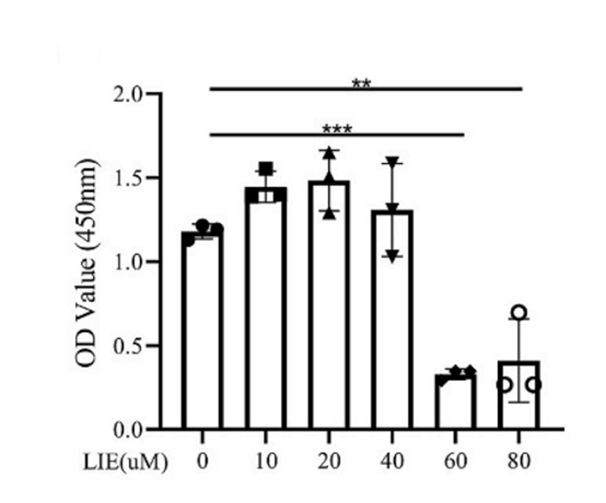
The viability of HBE cells treated with different concentration of Liensinine (LIE) (10, 20, 40, 60, 80 μM) measured by CCK8 assay.
Liensinine Diperchlorate purchased from MedChemExpress. Usage Cited in: Redox Biol. 2025 Oct:86:103793. [Abstract]
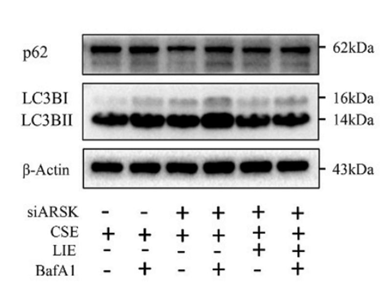
Liensinine (LIE) (40 μM) treatment effectively suppressed autophagy by Western blot.
Liensinine Diperchlorate purchased from MedChemExpress. Usage Cited in: Redox Biol. 2025 Oct:86:103793. [Abstract]
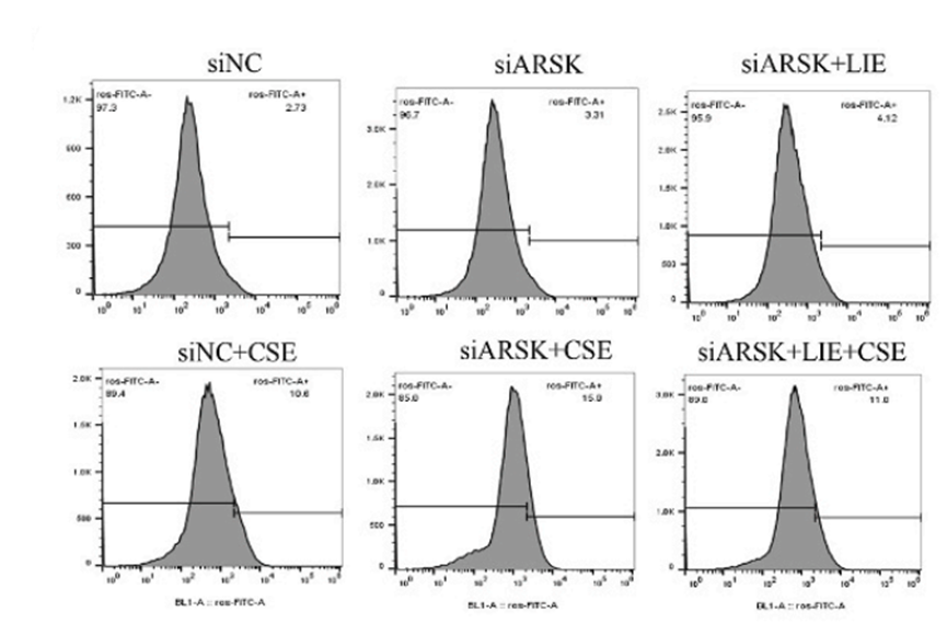
Liensinine (LIE) (40 μM) improved mitochondrial damage induced by ARSK knockdown, as evidenced by the reductions in intracellular and mitochondrial ROS levels.
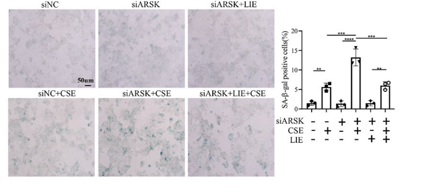
Liensinine Diperchlorate purchased from MedChemExpress. Usage Cited in: Redox Biol. 2025 Oct:86:103793. [Abstract]
SA-β-gal staining results demonstrated that Liensinine (LIE) (40 μM) treatment obviously decreased the percentage of positive cells.
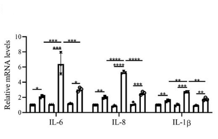
Liensinine Diperchlorate purchased from MedChemExpress. Usage Cited in: Redox Biol. 2025 Oct:86:103793. [Abstract]
The expressions of IL-6, IL-8 and IL-1β were both relived in HBE cells by Liensinine (LIE) (40 μM).
-
Cell Death Dis
Crotonylated BEX2 interacts with NDP52 and enhances mitophagy to modulate chemotherapeutic agent-induced apoptosis in non-small-cell lung cancer cells. [Abstract]2023 Sep 30;14(9):645. PMID: 37777549 -
Int J Biol Sci
Tubule-mitophagic secretion of SerpinG1 reprograms macrophages to instruct anti-septic acute kidney injury efficacy of high-dose ascorbate mediated by NRF2 transactivation. [Abstract]2022 Aug 8;18(13):5168-5184. PMID: 35982894 -
Anim Nutr
Caffeic acid phenethyl ester ameliorates high-fat diet-induced muscle textural deterioration in grass carp (Ctenopharyngodon idellus) by modulating adipose-muscle crosstalk via myostatin-taz signaling. [Abstract]2026 Feb 6:25:160-174. PMID: 41938616 -
Chin Med
Targeting G-protein-coupled receptors and gut microbiota: Ge-Lian Qi-Shen decoction elevates GLP-1 to combat non-alcoholic fatty liver disease. [Abstract]2026 Jan 24;21(1):51. PMID: 41580820 -
J Ethnopharmacol
Integrating UHPLC-Q-TOF-MS/MS, network pharmacology, bioinformatics and experimental validation to uncover the anti-cancer mechanisms of TiaoPi AnChang decoction in colorectal cancer. [Abstract]2024 Nov 15:334:118576. PMID: 39002822 -
Life Sci
Auto- and paracrine rewiring of NIX-mediated mitophagy by insulin-like growth factor-binding protein 7 in septic AKI escalates inflammation-coupling tubular damage. [Abstract]2023 Jun 1:322:121653. PMID: 37011875 -
Int J Mol Sci
Liensinine Prevents Acute Myocardial Ischemic Injury via Inhibiting the Inflammation Response Mediated by the Wnt/β-Catenin Signaling Pathway. [Abstract]2025 May 10;26(10):4566. PMID: 40429711 -
Molecules
Molecular Mechanisms Underlying the Anti-Tumor Activity of Lotus-Derived Alkaloids in Breast Cancer. [Abstract]2026 Mar 12;31(6):947. PMID: 41900047 -
J Mol Med (Berl)
Liensinine can improve vascular remodeling in hypertension through the ferroptosis-related TLR4 inflammatory pathway. [Abstract]2025 Dec 26;104(1):13. PMID: 41452356 -
J Biol Chem
Activation of Nemo-like Kinase in Diamond Blackfan Anemia suppresses early erythropoiesis by preventing mitochondrial biogenesis. [Abstract]2024 Aug;300(8):107542. PMID: 38992436 -
Adipocyte
The combination of nuclear receptor NR1D1 and ULK1 promotes mitophagy in adipocytes to ameliorate obesity. [Abstract]2022 Dec;11(1):202-212. PMID: 35410572 -
Tissue Cell
Liensinine induces autophagy and apoptosis in hepatocellular carcinoma via reactive oxygen species-mediated inhibition of the PI3K/AKT/mTOR pathway. [Abstract]2026 Jun 12:103:103696. PMID: 42314534 -
Genes Nutr
Liensinine alleviates type 2 diabetes mellitus through modulating the pancreatic β cell function and gut microbiota. [Abstract]2025 Dec 24;20(1):29. PMID: 41444518 -
J Int Med Res
RNA sequencing-based evaluation of the mechanisms underlying the liensinine-mediated inhibition of hypopharyngeal cancer proliferation. [Abstract]2026 Feb;54(2):3000605261419604. PMID: 41698653 -
-
-
-
-
J Oncol
2022 Jul 1:2022:1533779. PMID: 35813859 -
Oxid Med Cell Longev
Liensinine Inhibits Osteosarcoma Growth by ROS-Mediated Suppression of the JAK2/STAT3 Signaling Pathway. [Abstract]2022 Jan 25:2022:8245614. PMID: 35116094


Solvent & Solubility
In Vitro:
DMSO : 62.5 mg/mL (77.00 mM; Need ultrasonic; Hygroscopic DMSO has a significant impact on the solubility of product, please use newly opened DMSO)
Please refer to the solubility information to select the appropriate solvent. Once prepared, please aliquot and store the solution to prevent product inactivation from repeated freeze-thaw cycles.
Storage method and period of stock solution: -80°C, 6 months; -20°C, 1 month (sealed storage, away from moisture). When stored at -80°C, please use it within 6 months. When stored at -20°C, please use it within 1 month.
Please refer to the solubility information to select the appropriate solvent. Once prepared, please aliquot and store the solution to prevent product inactivation from repeated freeze-thaw cycles.
Storage method and period of stock solution: -80°C, 6 months; -20°C, 1 month (sealed storage, away from moisture). When stored at -80°C, please use it within 6 months. When stored at -20°C, please use it within 1 month.
Concentration (start) × Volume (start) = Concentration (final) × Volume (final)
In Vivo:
Select the appropriate dissolution method based on your experimental animal and administration route.
- For the following dissolution methods, please ensure to first prepare a clear stock solution using an In Vitro approach and then sequentially add co-solvents:
- To ensure reliable experimental results, the clarified stock solution can be appropriately stored based on storage conditions. As for the working solution for In Vivo experiments, it is recommended to prepare freshly and use it on the same day.
- The percentages shown for the solvents indicate their volumetric ratio in the final prepared solution. If precipitation or phase separation occurs during preparation, heat and/or sonication can be used to aid dissolution.
Add each solvent one by one: 10% DMSO 40% PEG300 5% Tween-80 45% Saline
Solubility: ≥ 2.08 mg/mL (2.56 mM); Clear solution
This protocol yields a clear solution of ≥ 2.08 mg/mL (saturation unknown).
Taking 1 mL working solution as an example, add 100 μL DMSO stock solution (20.8 mg/mL) to 400 μL PEG300, and mix evenly; then add 50 μL Tween-80 and mix evenly; then add 450 μL Saline to adjust the volume to 1 mL.
Preparation of Saline: Dissolve 0.9 g sodium chloride in ddH₂O and dilute to 100 mL to obtain a clear Saline solution.
Add each solvent one by one: 10% DMSO 90% (20% SBE-β-CD in Saline)
Solubility: ≥ 2.08 mg/mL (2.56 mM); Clear solution
This protocol yields a clear solution of ≥ 2.08 mg/mL (saturation unknown).
Taking 1 mL working solution as an example, add 100 μL DMSO stock solution (20.8 mg/mL) to 900 μL 20% SBE-β-CD in Saline, and mix evenly.
Preparation of 20% SBE-β-CD in Saline (4°C, storage for one week): 2 g SBE-β-CD powder is dissolved in 10 mL Saline, completely dissolve until clear.
In Vivo Dissolution Calculator
Please enter the basic information of animal experiments:
-
-
-
-
Recommended: Prepare an additional quantity of animals to account for potential losses during experiments.
Please enter your animal formula composition:
-
%DMSO +
Recommended: Keep the proportion of DMSO in working solution below 2% if your animal is weak.
-
%+
-
+%Tween-80 + +
-
%Saline +
The co-solvents required include: DMSO, . All of co-solvents are available by MedChemExpress (MCE). , Tween 80. All of co-solvents are available by MedChemExpress (MCE).
Working solution concentration: 0.22 mg/mL
Method for preparing stock solution: mg drug dissolved in μL DMSO. Stock solution concentration: mg/mL. * In solvent : -80°C, 6 months; -20°C, 1 month (sealed storage, away from moisture)
1. Take μL DMSO stock solution;
2. Add μL .
μL , mix evenly;
3. Then add μL Tween 80, mix evenly;
4. Then add μL
Please ensure that the stock solution in the first step is dissolved to a clear state, and add co-solvents in sequence. You can use ultrasonic heating (ultrasonic cleaner, recommended frequency 20-40 kHz), vortexing, etc. to assist dissolution.

Protocols
-
Fibrosis/Collagen Morphometry
Fibrosis and collagen morphometry is based on the quantitative visualization of fibrillar collagen deposition in tissue sections using histochemical stains such as Sirius Red (Picrosirius Red) or Masson's trichrome, followed by image-based or polarization-enhanced analysis to estimate collagen proportional area as a surrogate of extracellular matrix accumulation during fibrotic remodeling. Sirius Red combined with polarized light microscopy enhances detection of collagen fibers due to birefringence properties, enabling more specific visualization of collagen type I and III fibrils compared to conventional bright-field histology, while whole-section or region-restricted digital morphometry reduces field-selection bias in fibrosis assessment. Alternative quantitative approaches include second harmonic generation (SHG) and two-photon excited fluorescence microscopy, which enable label-free detection of fibrillar collagen and have been validated against histological staining and biochemica
-
Collagen: Sirius Red Staining
Sirius Red or picrosirius red staining is a histochemical method for visualizing collagen-rich extracellular matrix in tissue sections, and collagen fibers are detected as red-stained structures under bright-field microscopy with enhanced birefringence under polarized light. Picrosirius red is useful for assessing total collagen organization, distribution, and fibrosis burden, but polarized color should not be interpreted as a definitive collagen type I versus type III readout because color is affected by fiber orientation, thickness, and packing.
-
Connective Tissue: Masson's Trichrome/Collagen Trichrome Staining
Masson’s Trichrome (collagen/trichrome staining) is a histological technique that differentially stains tissue compartments using sequential acidic dyes to distinguish collagen from muscle and cytoplasmic components based on dye affinity and tissue permeability differences, enabling visualization of fibrosis and connective tissue architecture in histological sections. The classical formulation typically uses Weigert's iron hematoxylin for nuclear staining, Biebrich scarlet-acid fuchsin for cytoplasm and muscle, and aniline blue (or light green variants) for collagen, producing a characteristic blue/green collagen signal contrasted against red cytoplasm and dark nuclei. The staining principle relies on selective displacement of smaller dye molecules by larger anionic dyes in collagen-rich regions under controlled acidified conditions, which enhances collagen-specific dye retention. This property makes the method widely used for fibrosis assessment in organs such as heart, liver, lung, a
-
Autophagy
Autophagy is a process in which eukaryotic cells use lysosomes to degrade their own cytoplasmic proteins and damaged organelles under the regulation of autophagy related gene (Atg). Microtubule-associated proteins light chain 3 (LC3) is recognized as autophagy marker, which transfers from cytoplasmic LC3 (LC3-I) to membrane type (LC3-II). LC3-II/I ratio could be detected by Western Blot and fluorescence microscopy.
-
Research Protocol for Cardiovascular Diseases
Cardiovascular disease can be modeled as maladaptive cardiac remodeling, where ischemic injury or pressure overload activates inflammatory signaling, fibroblast activation, extracellular-matrix deposition, cardiomyocyte hypertrophy, vascular remodeling, and progressive ventricular dysfunction. The TGF-β/SMAD axis is a central profibrotic pathway after myocardial injury and pressure overload, while innate immune and cytokine pathways regulate leukocyte recruitment, scar formation, and adverse remodeling. Key unresolved questions include which inflammatory signals are reparative versus harmful, when fibrosis is protective versus maladaptive, and whether pathway inhibition improves function without weakening necessary infarct healing or compensatory remodeling.
-
Lysosome and acidic-vesicle live-cell staining
Lysosome and acidic-vesicle live-cell staining detects acidic intracellular compartments by using membrane-permeant acidotropic probes that accumulate in low-pH vesicles, including lysosomes, late endosomes, autolysosomes, and acidic phagosomes. LysoTracker staining is commonly used as an intensity-based readout of acidic lysosomal compartment abundance or enlargement, while acridine orange produces green fluorescence in less concentrated compartments and red fluorescence after concentration-dependent accumulation in acidic vesicular organelles. Loss or reduction of acridine-orange red signal can be used as a readout of lysosomal membrane permeabilization or reduced acidic-vesicle integrity. This protocol is designed for live cultured cells and can be adapted for fluorescence microscopy, high-content imaging, plate-reader readout, or flow cytometry when the selected literature supports the readout. Because these dyes report acidotropic accumulation rather than lysosome identity alone,
-
Mitophagy Solutions
Mitophagy is the selective autophagic degradation of mitochondria and functions as a mitochondrial quality-control pathway that removes damaged, depolarized, excess, or developmentally programmed mitochondria. The pathway links mitochondrial damage recognition, autophagosome recruitment, lysosomal delivery, and mitochondrial turnover to phenotypes such as mitochondrial homeostasis, oxidative-stress control, metabolic remodeling, differentiation, and neurodegeneration-related mitochondrial fidelity. The best-characterized damage-induced pathway is the PINK1-Parkin axis. Parkin is recruited selectively to impaired mitochondria and promotes their autophagic elimination, while mitochondrial depolarization stabilizes PINK1 on damaged mitochondria, recruits Parkin, and activates Parkin-dependent mitophagy. PINK1 also phosphorylates ubiquitin to activate Parkin E3 ubiquitin ligase activity, and PINK1-driven ubiquitin phosphorylation creates a feed-forward signal for recruiting autophagy machi
-
Macroautophagy Solutions
Macroautophagy is a conserved lysosome-dependent degradation pathway in which cytoplasmic material is sequestered into double-membrane autophagosomes and delivered to lysosomes for degradation and recycling. The pathway supports cellular homeostasis during nutrient limitation, organelle stress, protein-aggregate accumulation, infection, differentiation, and tissue remodeling by coupling cargo sequestration, autophagosome maturation, lysosomal fusion, and degradation of cargo-derived macromolecules. The core molecular sequence includes initiation by nutrient- and stress-regulated autophagy machinery, autophagosome nucleation, LC3/ATG8-family conjugation to autophagosomal membranes, cargo selection through receptors such as SQSTM1/p62, autophagosome-lysosome fusion, and lysosomal degradation. LC3 was identified as a mammalian homolog of yeast Atg8 that localizes to autophagosomal membranes after processing, and p62/SQSTM1 was shown to connect ubiquitinated cargo with autophagic degradati


Purity & Documentation
-
Data Sheet (275 KB)
-
SDS (252 KB)
- English - EN (252 KB)
- Français - FR (252 KB)
- Deutsch - DE (252 KB)
- Norwegian - NO (252 KB)
- Español - ES (252 KB)
- Swedish - SV (252 KB)
- Italian - IT (252 KB)
- Korean - KR (252 KB)
- Portuguese - PT (252 KB)
-
Handling Instructions (2659 KB)
References


Complete Stock Solution Preparation Table
Please refer to the solubility information to select the appropriate solvent. Once prepared, please aliquot and store the solution to prevent product inactivation from repeated freeze-thaw cycles.
Storage method and period of stock solution: -80°C, 6 months; -20°C, 1 month (sealed storage, away from moisture). When stored at -80°C, please use it within 6 months. When stored at -20°C, please use it within 1 month.
| Optional Solvent | Concentration Solvent Mass | 1 mg | 5 mg | 10 mg | 25 mg |
|---|---|---|---|---|---|
| DMSO | 1 mM | 1.2320 mL | 6.1602 mL | 12.3204 mL | 30.8011 mL |
| 5 mM | 0.2464 mL | 1.2320 mL | 2.4641 mL | 6.1602 mL | |
| 10 mM | 0.1232 mL | 0.6160 mL | 1.2320 mL | 3.0801 mL | |
| 15 mM | 0.0821 mL | 0.4107 mL | 0.8214 mL | 2.0534 mL | |
| 20 mM | 0.0616 mL | 0.3080 mL | 0.6160 mL | 1.5401 mL | |
| 25 mM | 0.0493 mL | 0.2464 mL | 0.4928 mL | 1.2320 mL | |
| 30 mM | 0.0411 mL | 0.2053 mL | 0.4107 mL | 1.0267 mL | |
| 40 mM | 0.0308 mL | 0.1540 mL | 0.3080 mL | 0.7700 mL | |
| 50 mM | 0.0246 mL | 0.1232 mL | 0.2464 mL | 0.6160 mL | |
| 60 mM | 0.0205 mL | 0.1027 mL | 0.2053 mL | 0.5134 mL |


Liensinine Diperchlorate Related Classifications
HY-N0485 Related Classifications
 Powered by Bioz