



Liensinine perchlorate
Based on 22 publication(s) in Google Scholar
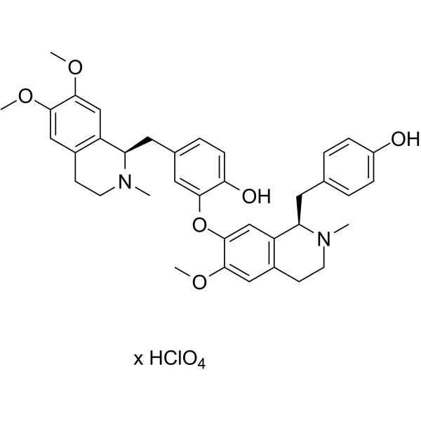
Liensinine perchlorate is a bisbenzylisoquinoline alkaloid. By inhibiting the PI3K/AKT and JNK/p38-MAPK signaling pathways, Liensinine perchlorate suppresses autophagy and apoptosis, clears Aβ, and exerts anti-inflammatory, antioxidant and neuroprotective effects. Liensinine perchlorate activates AMPK and inhibits the expression of HIF-1α and VEGF, thereby suppressing angiogenesis. Liensinine perchlorate exerts anti-tumor effects through ROS-mediated inhibition of the JAK2/STAT3 signaling pathway. Liensinine perchlorate can be used for the research of diseases such as Alzheimer's disease, hepatocellular carcinoma, osteosarcoma, sepsis-induced organ injury and stroke.
For research use only. We do not sell to patients.
- Purity : 99.67%
- CAS No.: 2385-63-9
- Formula: C37H42N2O6.xHClO4
-
Storage:
4°C, sealed storage, away from moisture and light
* In solvent : -80°C, 6 months; -20°C, 1 month (sealed storage, away from moisture and light)

To place orders, for customer services and technical support, please contact: MedChemExpress USA
Tel: 609-228-6898 E-mail: [email protected] [email protected]


-
 Biological Activity
-
Chemical Information
-
Solvent & Solubility
- Protocols
- Purity & Documentation
- References
-
 Help & FAQs


Publications Citing Use of MedChemExpress (MCE) Liensinine perchlorate
More- Signal Transduct Target Ther. 2025 Dec 17;10(1):413. [Abstract]
- Redox Biol. 2025 Oct:86:103793. [Abstract]
- Cell Death Dis. 2023 Sep 30;14(9):645. [Abstract]
- Int J Biol Sci. 2022 Aug 8;18(13):5168-5184. [Abstract]
- Anim Nutr. 2026 Feb 6:25:160-174. [Abstract]
- Chin Med. 2026 Jan 24;21(1):51. [Abstract]
- J Ethnopharmacol. 2024 Nov 15:334:118576. [Abstract]
- Life Sci. 2023 Jun 1:322:121653. [Abstract]
- Int J Mol Sci. 2025 May 10;26(10):4566. [Abstract]
- Molecules. 2026 Mar 12;31(6):947. [Abstract]
- J Mol Med (Berl). 2025 Dec 26;104(1):13. [Abstract]
- J Biol Chem. 2024 Aug;300(8):107542. [Abstract]
- Adipocyte. 2022 Dec;11(1):202-212. [Abstract]
- Tissue Cell. 2026 Jun 12:103:103696. [Abstract]
- Genes Nutr. 2025 Dec 24;20(1):29. [Abstract]
- J Int Med Res. 2026 Feb;54(2):3000605261419604. [Abstract]
- Lett Drug Des Discov. 2025 Dec 2.
- Int J Pharmacol. 2026 Jun 28;22(3).
- SSRN. 2023 Apr 18.
- Research Square Print. 2023 Mar 24.
- J Oncol. 2022 Jul 1:2022:1533779. [Abstract]
- Oxid Med Cell Longev. 2022 Jan 25:2022:8245614. [Abstract]
 Customer Validation & Images

-
Cell Proliferation/Viability Assay
-
WB
-
Flow Cytometry
-
Cell Imaging/Staining
-
RT-PCR
All VEGFR Isoforms
More

Biological Activity
Description
IC50 & Target
IC50: apoptosis[1]
In Vitro
Liensinine (5-80 μM; 24 h) perchlorate reduces the viability of SaOS-2, MG-63, 143B and U-2OS osteosarcoma cells in a dose-dependent manner, but has no effect on the viability of hFOB 1.19 normal osteoblasts[1].
Liensinine (40-80 μM; 24 h) perchlorate inhibits the proliferation and colony-forming ability of SaOS-2 and 143B osteosarcoma cells, induces apoptosis, and causes G0/G1 cell cycle arrest in a dose-dependent manner[1].
Liensinine (40-80 μM; 24 h) perchlorate increases intracellular ROS production in SaOS-2 and 143B osteosarcoma cells in a dose-dependent manner, but does not affect ROS levels in hFOB 1.19 normal osteoblasts[1].
Liensinine (40-80 μM; 24 h) perchlorate disrupts the GSH/GSSG redox balance in SaOS-2 and 143B osteosarcoma cells in a dose-dependent manner by decreasing GSH levels, increasing GSSG levels, and elevating the GSSG/GSH ratio[1].
Liensinine (40-80 μM; 24 h) perchlorate induces the loss of mitochondrial membrane potential in SaOS-2 and 143B osteosarcoma cells in a dose-dependent manner[1].
Liensinine (40-80 μM; 24 h) perchlorate dose-dependently regulates the expression of apoptosis- and cell cycle-related proteins in SaOS-2 and 143B osteosarcoma cells in vitro, and inhibits the activation of the JAK2/STAT3 pathway, but exerts no effect on the aforementioned proteins in hFOB 1.19 normal osteoblasts[1].
Liensinine (20 μM; 24 h) perchlorate enhances cell viability, reduces apoptosis and cytotoxicity, and inhibits autophagy in an in vitro oxygen-glucose deprivation/reoxygenation ischemia-reperfusion model[3].
MedChemExpress (MCE) has not independently confirmed the accuracy of these methods. They are for reference only. Further protocols information, click here.
In Vivo
Liensinine (20 mg/kg; i.p.; once daily for 16 consecutive days) perchlorate significantly inhibits orthotopic HCC tumor progression in male C57BL/6 mice, induces tumor metabolic reprogramming, reduces tumor vascular density, regulates macrophage polarization, and downregulates PD-L1 expression[5].
MedChemExpress (MCE) has not independently confirmed the accuracy of these methods. They are for reference only.


Chemical Information
-
CAS No. 2385-63-9
-
Appearance Solid
-
Formula C37H42N2O6.xHClO4
-
Color White to off-white
-
SMILES
OC1=CC=C(C[C@H]2N(C)CCC3=C2C=C(OC)C(OC)=C3)C=C1OC4=CC5=C(C=C4OC)CCN(C)[C@@H]5CC6=CC=C(O)C=C6.[x HClO4]
-
Structure Classification
-
Initial Source
-
Shipping
Room temperature in continental US; may vary elsewhere.
-
Storage
4°C, sealed storage, away from moisture and light
* In solvent : -80°C, 6 months; -20°C, 1 month (sealed storage, away from moisture and light)


Publications (22)
-
Journal Impact Factor
-
Most Recent
-
Signal Transduct Target Ther
Disruption of heme homeostasis by nuclear receptor Nur77 induces pyroptosis through granzyme B-dependent GSDMC cleavage. [Abstract]2025 Dec 17;10(1):413. PMID: 41407678 -
Redox Biol
Arylsulfatase K attenuates airway epithelial cell senescence in COPD by regulating parkin-mediated mitophagy. [Abstract]2025 Oct:86:103793. PMID: 40763656
Liensinine perchlorate purchased from MedChemExpress. Usage Cited in: Redox Biol. 2025 Oct:86:103793. [Abstract]
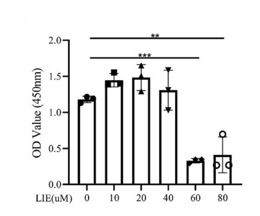
The viability of HBE cells treated with different concentration of Liensinine (LIE) (10, 20, 40, 60, 80 μM) measured by CCK8 assay.
Liensinine perchlorate purchased from MedChemExpress. Usage Cited in: Redox Biol. 2025 Oct:86:103793. [Abstract]
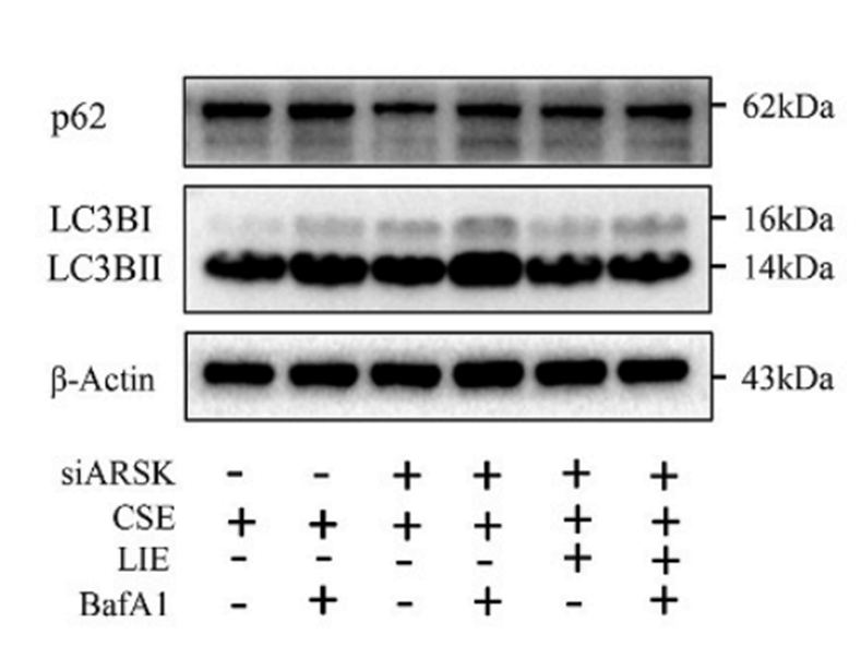
Liensinine (LIE) (40 μM) treatment effectively suppressed autophagy by Western blot.
Liensinine perchlorate purchased from MedChemExpress. Usage Cited in: Redox Biol. 2025 Oct:86:103793. [Abstract]
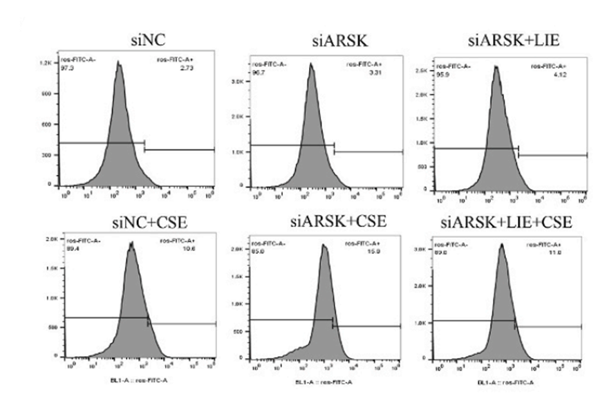
Liensinine (LIE) (40 μM) improved mitochondrial damage induced by ARSK knockdown, as evidenced by the reductions in intracellular and mitochondrial ROS levels.
Liensinine perchlorate purchased from MedChemExpress. Usage Cited in: Redox Biol. 2025 Oct:86:103793. [Abstract]
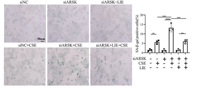
SA-β-gal staining results demonstrated that Liensinine (LIE) (40 μM) treatment obviously decreased the percentage of positive cells.
Liensinine perchlorate purchased from MedChemExpress. Usage Cited in: Redox Biol. 2025 Oct:86:103793. [Abstract]
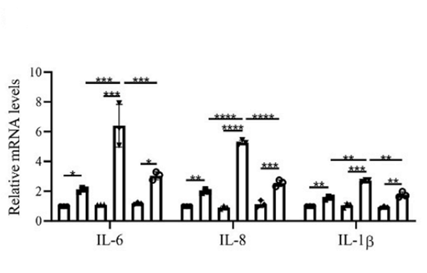
The expressions of IL-6, IL-8 and IL-1β were both relived in HBE cells by Liensinine (LIE) (40 μM).
-
Cell Death Dis
Crotonylated BEX2 interacts with NDP52 and enhances mitophagy to modulate chemotherapeutic agent-induced apoptosis in non-small-cell lung cancer cells. [Abstract]2023 Sep 30;14(9):645. PMID: 37777549 -
Int J Biol Sci
Tubule-mitophagic secretion of SerpinG1 reprograms macrophages to instruct anti-septic acute kidney injury efficacy of high-dose ascorbate mediated by NRF2 transactivation. [Abstract]2022 Aug 8;18(13):5168-5184. PMID: 35982894 -
Anim Nutr
Caffeic acid phenethyl ester ameliorates high-fat diet-induced muscle textural deterioration in grass carp (Ctenopharyngodon idellus) by modulating adipose-muscle crosstalk via myostatin-taz signaling. [Abstract]2026 Feb 6:25:160-174. PMID: 41938616 -
Chin Med
Targeting G-protein-coupled receptors and gut microbiota: Ge-Lian Qi-Shen decoction elevates GLP-1 to combat non-alcoholic fatty liver disease. [Abstract]2026 Jan 24;21(1):51. PMID: 41580820 -
J Ethnopharmacol
Integrating UHPLC-Q-TOF-MS/MS, network pharmacology, bioinformatics and experimental validation to uncover the anti-cancer mechanisms of TiaoPi AnChang decoction in colorectal cancer. [Abstract]2024 Nov 15:334:118576. PMID: 39002822 -
Life Sci
Auto- and paracrine rewiring of NIX-mediated mitophagy by insulin-like growth factor-binding protein 7 in septic AKI escalates inflammation-coupling tubular damage. [Abstract]2023 Jun 1:322:121653. PMID: 37011875 -
Int J Mol Sci
Liensinine Prevents Acute Myocardial Ischemic Injury via Inhibiting the Inflammation Response Mediated by the Wnt/β-Catenin Signaling Pathway. [Abstract]2025 May 10;26(10):4566. PMID: 40429711 -
Molecules
Molecular Mechanisms Underlying the Anti-Tumor Activity of Lotus-Derived Alkaloids in Breast Cancer. [Abstract]2026 Mar 12;31(6):947. PMID: 41900047 -
J Mol Med (Berl)
Liensinine can improve vascular remodeling in hypertension through the ferroptosis-related TLR4 inflammatory pathway. [Abstract]2025 Dec 26;104(1):13. PMID: 41452356 -
J Biol Chem
Activation of Nemo-like Kinase in Diamond Blackfan Anemia suppresses early erythropoiesis by preventing mitochondrial biogenesis. [Abstract]2024 Aug;300(8):107542. PMID: 38992436 -
Adipocyte
The combination of nuclear receptor NR1D1 and ULK1 promotes mitophagy in adipocytes to ameliorate obesity. [Abstract]2022 Dec;11(1):202-212. PMID: 35410572 -
Tissue Cell
Liensinine induces autophagy and apoptosis in hepatocellular carcinoma via reactive oxygen species-mediated inhibition of the PI3K/AKT/mTOR pathway. [Abstract]2026 Jun 12:103:103696. PMID: 42314534 -
Genes Nutr
Liensinine alleviates type 2 diabetes mellitus through modulating the pancreatic β cell function and gut microbiota. [Abstract]2025 Dec 24;20(1):29. PMID: 41444518 -
J Int Med Res
RNA sequencing-based evaluation of the mechanisms underlying the liensinine-mediated inhibition of hypopharyngeal cancer proliferation. [Abstract]2026 Feb;54(2):3000605261419604. PMID: 41698653 -
-
-
-
-
J Oncol
2022 Jul 1:2022:1533779. PMID: 35813859 -
Oxid Med Cell Longev
Liensinine Inhibits Osteosarcoma Growth by ROS-Mediated Suppression of the JAK2/STAT3 Signaling Pathway. [Abstract]2022 Jan 25:2022:8245614. PMID: 35116094


Solvent & Solubility
In Vitro:
DMSO : 100 mg/mL (Need ultrasonic; Hygroscopic DMSO has a significant impact on the solubility of product, please use newly opened DMSO)
In Vivo:
Select the appropriate dissolution method based on your experimental animal and administration route.
- For the following dissolution methods, please ensure to first prepare a clear stock solution using an In Vitro approach and then sequentially add co-solvents:
- To ensure reliable experimental results, the clarified stock solution can be appropriately stored based on storage conditions. As for the working solution for In Vivo experiments, it is recommended to prepare freshly and use it on the same day.
- The percentages shown for the solvents indicate their volumetric ratio in the final prepared solution. If precipitation or phase separation occurs during preparation, heat and/or sonication can be used to aid dissolution.
Add each solvent one by one: 10% DMSO 40% PEG300 5% Tween-80 45% Saline
Solubility: ≥ 2.08 mg/mL; Clear solution
This protocol yields a clear solution of ≥ 2.08 mg/mL (saturation unknown).
Taking 1 mL working solution as an example, add 100 μL DMSO stock solution (20.8 mg/mL) to 400 μL PEG300, and mix evenly; then add 50 μL Tween-80 and mix evenly; then add 450 μL Saline to adjust the volume to 1 mL.
Preparation of Saline: Dissolve 0.9 g sodium chloride in ddH₂O and dilute to 100 mL to obtain a clear Saline solution.
Add each solvent one by one: 10% DMSO 90% (20% SBE-β-CD in Saline)
Solubility: ≥ 2.08 mg/mL; Clear solution
This protocol yields a clear solution of ≥ 2.08 mg/mL (saturation unknown).
Taking 1 mL working solution as an example, add 100 μL DMSO stock solution (20.8 mg/mL) to 900 μL 20% SBE-β-CD in Saline, and mix evenly.
Preparation of 20% SBE-β-CD in Saline (4°C, storage for one week): 2 g SBE-β-CD powder is dissolved in 10 mL Saline, completely dissolve until clear.
In Vivo Dissolution Calculator
Please enter the basic information of animal experiments:
-
-
-
-
Recommended: Prepare an additional quantity of animals to account for potential losses during experiments.
Please enter your animal formula composition:
-
%DMSO +
Recommended: Keep the proportion of DMSO in working solution below 2% if your animal is weak.
-
%+
-
+%Tween-80 + +
-
%Saline +
The co-solvents required include: DMSO, . All of co-solvents are available by MedChemExpress (MCE). , Tween 80. All of co-solvents are available by MedChemExpress (MCE).
Working solution concentration: 0.22 mg/mL
Method for preparing stock solution: mg drug dissolved in μL DMSO. Stock solution concentration: mg/mL. * In solvent : -80°C, 6 months; -20°C, 1 month (sealed storage, away from moisture and light)
1. Take μL DMSO stock solution;
2. Add μL .
μL , mix evenly;
3. Then add μL Tween 80, mix evenly;
4. Then add μL
Please ensure that the stock solution in the first step is dissolved to a clear state, and add co-solvents in sequence. You can use ultrasonic heating (ultrasonic cleaner, recommended frequency 20-40 kHz), vortexing, etc. to assist dissolution.

Protocols
-
RNA extraction experimental
By lysing cells, releasing RNA, and removing impurities such as proteins and DNA, high-purity RNA products are finally obtained. The commonly used traditional method is the guanidine isothiocyanate/phenol/chloroform method (Trizol), which is suitable for a variety of animal materials including animal tissues, microorganisms, cultured cells, etc., and most plant materials.
-
Human pluripotent stem cell endothelial-cell differentiation
Human pluripotent stem cell endothelial differentiation is based on stepwise developmental patterning: early activation of WNT/GSK3β inhibition promotes mesodermal or vascular progenitor entry, followed by endothelial specification using VEGF-related signaling, BMP4, FGF2, Notch modulation, or cAMP depending on the published protocol. Endothelial differentiation is read out by acquisition of CD31, CD34, VE-cadherin/CD144, KDR/VEGFR2, vWF, Tie2, NOS3, acetylated LDL uptake, tube/network formation, barrier function, and in vivo vessel-forming capacity where tested.
-
Vascular/Branching Fractal Analysis
Vascular/branching fractal analysis quantifies the geometric complexity of vessel trees or vascular networks from segmented 2D images, commonly by converting vessels into binary and/or skeletonized maps and estimating fractal dimension using box-counting or related approaches. Fractal dimension is interpreted as an image-derived readout of vascular branching complexity, space filling, or density, and has been applied to retinal photographs, fluorescein angiography, OCT angiography, capillary perfusion maps, and in vitro Matrigel angiogenesis networks. The assay readout is generated from vessel-positive pixels after image preprocessing, vessel segmentation, binarization, and optional skeletonization; reported outputs include fractal dimension, vessel density, branchpoint density, endpoint density, vessel length density, tortuosity, and generation-based branching metrics when VESGEN-style analysis is used. The biological interpretation is limited to quantitative vascular patterning and s
-
Annexin V plus membrane-impermeant dye apoptosis staining
Annexin V-based apoptosis assays rely on the detection of phosphatidylserine (PS) externalization from the inner leaflet of the plasma membrane to the outer leaflet, an early biochemical hallmark of apoptosis. Fluorescently labeled Annexin V binds PS in a calcium-dependent manner, enabling identification of early apoptotic cells by flow cytometry or fluorescence microscopy. When combined with a membrane-impermeant DNA-binding dye (e. g. , propidium iodide), this approach allows discrimination between viable (Annexin V−/dye−), early apoptotic (Annexin V+/dye−), and late apoptotic or necrotic (Annexin V+/dye+) cell populations by assessing membrane integrity and PS exposure.
-
ROS/oxidative-stress fluorescent staining
ROS/oxidative-stress fluorescent staining uses cell-permeant fluorogenic probes that become fluorescent after oxidation inside cells or tissues; commonly used examples include DCFH-DA/DCFDA for broad cellular oxidant detection, DHE for superoxide-related signal detection, MitoSOX for mitochondrial superoxide-related signal detection, and CellROX probes for oxidative-stress-associated fluorescence readouts. The assay detects probe oxidation rather than a single ROS species unless the probe and analysis method have been chemically validated for that species. DCFH-DA enters cells, is deacetylated by intracellular esterases to DCFH, and produces fluorescent DCF after oxidation, so the readout is used as an operational measure of total cellular oxidative stress rather than a species-specific ROS measurement. DHE and MitoSOX can report superoxide-related oxidation, but red fluorescence alone can include non-specific ethidium-like oxidation products; HPLC or optimized spectral approaches are
-
Research Protocol for Inflammation-related Diseases
The NLRP3 inflammasome is a cytosolic innate immune signaling platform that integrates priming signals and danger-signal activation to promote caspase-1 activation, maturation of IL-1β and IL-18, and gasdermin D-mediated pyroptotic cell death. The core experimental logic is to determine whether inflammatory disease phenotypes are driven by increased NLRP3 expression, ASC-containing inflammasome assembly, caspase-1 cleavage, GSDMD cleavage, and extracellular release of IL-1β/IL-18 rather than by nonspecific cell injury alone. The pathway is strongly linked to inflammation-related disease phenotypes because monosodium urate crystals activate NALP3/NLRP3 inflammasome signaling in gout-like crystal inflammation, cholesterol crystals activate NLRP3 inflammasomes in atherogenesis models, and DSS-induced intestinal inflammation has been reported to involve NLRP3 inflammasome activity. However, experimental colitis studies also show context-dependent protective effects of NLRP3 inflammasome co
-
Liver Cancer Modeling
Liver cancer can be classified into primary liver cancer and secondary liver cancer. Secondary liver cancer is the metastatic liver cancer. Primary liver cancer includes hepatocellular carcinoma (HCC), intrahepatic cholangiocarcinoma (ICC) and fibrolamellar HCC, of which HCC is the most common form, accounting for approximately 90% of primary liver cancers[1]. HCC mouse models include chemical agent-induced models, transplanted tumor models, and genetic engineered models.
-
Alzheimer’s Disease Modeling
Alzheimer’s Disease (AD) is a neurodegenerative disorder characterized by a progressive decline in cognitive functions and loss of specific types of neurons and synapses. Alzheimer's symptoms can be simulated in mice by injecting drugs (such as Aβ) or genetically modified.
-
Apoptosis
Apoptosis, also called programmed cell death, is generally characterized by distinct morphological characteristics.
-
TUNEL staining for apoptotic DNA fragmentation
TUNEL staining detects DNA strand breaks by using terminal deoxynucleotidyl transferase to add labeled nucleotides to exposed 3′-OH DNA termini, generating either microscopic staining in fixed cells or tissue sections, or fluorescence/cytometric signal in cell suspensions. TUNEL positivity reflects DNA fragmentation but should not be interpreted alone as definitive apoptosis, because TUNEL can also label necrotic, autolytic, mechanically damaged, or DNA-repair-associated DNA breaks.
-
LPS-Induced Endotoxemia/Systemic Inflammation
Lipopolysaccharide (LPS)-induced endotoxemia is a widely used in vivo model of acute systemic inflammation in which LPS, a Gram-negative bacterial endotoxin, activates innate immune signaling primarily through TLR4, leading to rapid and transient induction of pro-inflammatory cytokines such as TNF-α, IL-6, and IL-1β in circulation and tissues. This cytokine surge is commonly used as a measurable readout of systemic inflammatory activation and immune dysregulation, and is typically assessed within hours after intraperitoneal LPS administration in mouse models of endotoxemia. The model captures key features of systemic inflammatory response syndrome, including cytokine release, immune cell activation, and downstream tissue responses, and has been used to evaluate anti-inflammatory interventions such as cytokine modulation, lipid mediators, and immune cell-targeting therapies.
-
Autophagy
Autophagy is a process in which eukaryotic cells use lysosomes to degrade their own cytoplasmic proteins and damaged organelles under the regulation of autophagy related gene (Atg). Microtubule-associated proteins light chain 3 (LC3) is recognized as autophagy marker, which transfers from cytoplasmic LC3 (LC3-I) to membrane type (LC3-II). LC3-II/I ratio could be detected by Western Blot and fluorescence microscopy.
-
Endothelial Tube Formation Assay
Endothelial tube formation assay evaluates the ability of endothelial cells to attach, migrate, align, and organize into capillary-like networks when cultured on gelled basement membrane extract or Matrigel; the readout is the morphology and quantity of tube-like networks, which reflects an in vitro endothelial morphogenesis step related to angiogenesis. Basement membrane extract/Matrigel provides laminin-rich extracellular matrix cues that support endothelial differentiation into capillary-like structures, but it can contain biologically active growth factors, so growth-factor-reduced matrix is preferred when testing defined angiogenic stimulators or inhibitors.
-
Apoptosis Solutions
Apoptosis is a regulated, generally non-lytic cell-death pathway that removes unwanted, damaged, infected, or abnormal cells through coordinated morphological changes, caspase activation, DNA fragmentation, and membrane remodeling. The intrinsic apoptosis pathway is controlled mainly by mitochondrial outer membrane permeabilization, BCL-2 family proteins, cytochrome c release, apoptosome formation, caspase-9 activation, and downstream executioner caspase-3/7 activation. The extrinsic apoptosis pathway is initiated by death receptors such as Fas, TNFR, and TRAIL receptors, which recruit adaptor proteins and activate caspase-8 before engaging executioner caspases or mitochondrial amplification through BID cleavage. Apoptosis is linked to many phenotypes, including cancer cell killing, tissue homeostasis, immune regulation, neurodegeneration, infection response, and treatment-induced cytotoxicity; unresolved questions include how apoptosis interacts with necroptosis, pyroptosis, ferroptos
-
Lysosome and acidic-vesicle live-cell staining
Lysosome and acidic-vesicle live-cell staining detects acidic intracellular compartments by using membrane-permeant acidotropic probes that accumulate in low-pH vesicles, including lysosomes, late endosomes, autolysosomes, and acidic phagosomes. LysoTracker staining is commonly used as an intensity-based readout of acidic lysosomal compartment abundance or enlargement, while acridine orange produces green fluorescence in less concentrated compartments and red fluorescence after concentration-dependent accumulation in acidic vesicular organelles. Loss or reduction of acridine-orange red signal can be used as a readout of lysosomal membrane permeabilization or reduced acidic-vesicle integrity. This protocol is designed for live cultured cells and can be adapted for fluorescence microscopy, high-content imaging, plate-reader readout, or flow cytometry when the selected literature supports the readout. Because these dyes report acidotropic accumulation rather than lysosome identity alone,
-
Macroautophagy Solutions
Macroautophagy is a conserved lysosome-dependent degradation pathway in which cytoplasmic material is sequestered into double-membrane autophagosomes and delivered to lysosomes for degradation and recycling. The pathway supports cellular homeostasis during nutrient limitation, organelle stress, protein-aggregate accumulation, infection, differentiation, and tissue remodeling by coupling cargo sequestration, autophagosome maturation, lysosomal fusion, and degradation of cargo-derived macromolecules. The core molecular sequence includes initiation by nutrient- and stress-regulated autophagy machinery, autophagosome nucleation, LC3/ATG8-family conjugation to autophagosomal membranes, cargo selection through receptors such as SQSTM1/p62, autophagosome-lysosome fusion, and lysosomal degradation. LC3 was identified as a mammalian homolog of yeast Atg8 that localizes to autophagosomal membranes after processing, and p62/SQSTM1 was shown to connect ubiquitinated cargo with autophagic degradati
-
Endothelial Cell Migration/Angiogenic Sprouting Assay
Endothelial cell migration and angiogenic sprouting assays are in vitro (and partially ex vivo-adapted) functional models that quantify the ability of endothelial cells to undergo coordinated migration, extracellular matrix invasion, and multicellular organization into capillary-like sprouts in response to pro-angiogenic stimuli such as VEGF, bFGF, or conditioned microenvironments. These assays are used to model early angiogenic events including tip-cell formation, directional migration, and lumen-like sprout extension, which collectively reflect angiogenic activation and vascular morphogenesis processes observed in vivo.


Purity & Documentation
-
Data Sheet (289 KB)
-
SDS (252 KB)
- English - EN (252 KB)
- Français - FR (252 KB)
- Deutsch - DE (252 KB)
- Norwegian - NO (252 KB)
- Español - ES (252 KB)
- Swedish - SV (252 KB)
- Italian - IT (252 KB)
- Korean - KR (252 KB)
- Portuguese - PT (252 KB)
-
Handling Instructions (2659 KB)
References
[1]. Jia F, et al. Liensinine Inhibits Osteosarcoma Growth by ROS-Mediated Suppression of the JAK2/STAT3 Signaling Pathway. Oxid Med Cell Longev. 2022;2022:8245614. Published 2022 Jan 25. [Content Brief]
[2]. Zhang W, et al. Liensinine pretreatment reduces inflammation, oxidative stress, apoptosis, and autophagy to alleviate sepsis acute kidney injury. Int Immunopharmacol. 2023;122:110563. [Content Brief]
[3]. Qiao W, et al. Liensinine ameliorates ischemia-reperfusion-induced brain injury by inhibiting autophagy via PI3K/AKT signaling. Funct Integr Genomics. 2023;23(2):140. Published 2023 Apr 28. [Content Brief]
[4]. Wu MC, et al. Liensinine and neferine exert neuroprotective effects via the autophagy pathway in transgenic Caenorhabditis elegans. BMC Complement Med Ther. 2023;23(1):386. Published 2023 Oct 27. [Content Brief]
[5]. Liu J, et al. Liensinine reshapes the immune microenvironment and enhances immunotherapy by reprogramming metabolism through the AMPK-HIF-1α axis in hepatocellular carcinoma. J Exp Clin Cancer Res. 2025;44(1):208. Published 2025 Jul 15. [Content Brief]

Calculators
Concentration (start) × Volume (start) = Concentration (final) × Volume (final)


Liensinine perchlorate Related Classifications
HY-N5014 Related Classifications
 Powered by Bioz