



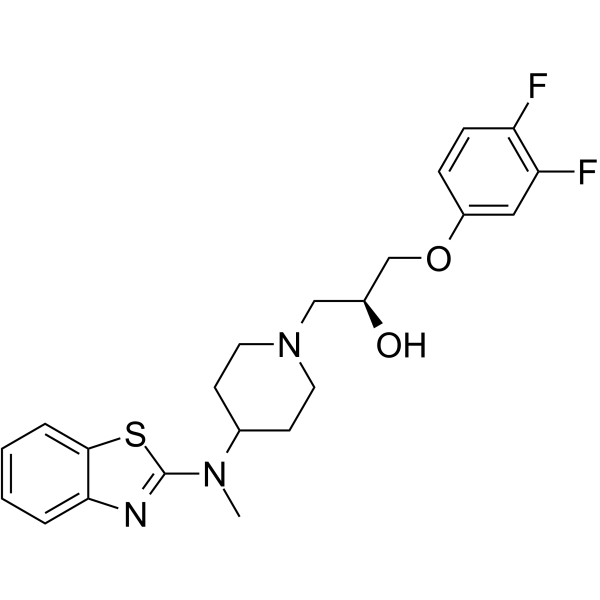
Lubeluzole
Lubeluzole is the S-isomer of benzothiazole derivative. Lubeluzole can inhibit glutamate release, glutamate-activated NO synthesis and block voltage-gated Sodium Channel and Calcium Channel. Lubeluzole exhibits anti-ischemic and neuroprotective effects. Lubeluzole also shows anti-bacterial and anti-diarrheal potential. Lubeluzole can inhibit cardiac sodium channel and prolong cardiac action potential. Lubeluzole can inhibit cancer cells proliferation and invasion and shows chemosensitizing effect. Lubeluzole can be used for the researches of cancer, infection, neurological and cardiovascular disease such as stroke, infectious diarrhea and ovarian.
商品は「研究用試薬」です。人や動物の医療用・臨床診断用・食品用の製品ではありません。
研究用途以外に使用した場合、当社は一切の責任を負いかねます。
- CAS 番号: 144665-07-6
- 分子式: C22H25F2N3O2S
- 分子量:433.51
-
保管条件:
Please store the product under the recommended conditions in the Certificate of Analysis.

To place orders, for customer services and technical support, please contact: MedChemExpress USA
Tel: 609-228-6898 E-mail: [email protected] [email protected]


-
 生物活性
生物活性
-
化学情報
- プロトコル
- 純度とドキュメンテーション
- 参考文献
-
 Help & FAQs


Calcium Channel アイソフォーム固有の製品をすべて表示
More

生物活性
製品説明
Cellular Effect
|
Cell Line

|
Type | Value | Description | References |
|---|---|---|---|---|
| A2780 | IC50 |
5.7 μM
Compound: (S)-1a, Lubeluzole
|
Cytotoxicity against human A2780 cells after 72 hrs by MTT assay
Cytotoxicity against human A2780 cells after 72 hrs by MTT assay
|
[PMID: 23886686] |
| A549 | IC50 |
14 μM
Compound: (S)-1a, Lubeluzole
|
Cytotoxicity against human A549 cells after 72 hrs by MTT assay
Cytotoxicity against human A549 cells after 72 hrs by MTT assay
|
[PMID: 23886686] |
| CHO | IC50 |
0.26 μM
Compound: Lubeluzole
|
In vitro inhibition of [14C]- guanidinium influx in Chinese hamster ovary (CHO) cells expressing rat brain sodium channel type IIA (CNaIIA-1)
In vitro inhibition of [14C]- guanidinium influx in Chinese hamster ovary (CHO) cells expressing rat brain sodium channel type IIA (CNaIIA-1)
|
[PMID: 9685245] |
| Sf9 | IC50 |
40 μM
Compound: (S)-1
|
Inhibition of full-length recombinant human CamKII expressed in insect Sf9 cells incubated for 30 mins in presence of 5 uM CaM/[gamma32P]ATP by scintillation counting analysis
Inhibition of full-length recombinant human CamKII expressed in insect Sf9 cells incubated for 30 mins in presence of 5 uM CaM/[gamma32P]ATP by scintillation counting analysis
|
[PMID: 27043269] |
体外実験
Lubeluzole (0.1-100 nM, 19 h) protects neurons from glutamate-induced excitotoxicity in primary mixed hippocampal cultures[1].
Lubeluzole (64-128 μg/mL) inhibits Yersinia enterocolitica DSM 4780 and Bacillus cereus DSM 4313[2].
Lubeluzole (25.6-204.8 μg/mL) exhibits synergistic antibacterial effects against Enterococcus faecalis ATCC 29212, Staphylococcus aureus ATCC 29213, Pseudomonas aeruginosa ATCC 27853, and Escherichia coli ATCC 25922 companied with Minocycline (HY-17412A)[2].
Lubeluzole (0.001-1 μM, 30 mins) inhibits acetylcholine-induced contraction in the isolated rat proximal colon model[2].
Lubeluzole (0.01-100 μM) inhibits cardiac sodium channels (INa) in isolated guinea-pig ventricular myocytes[3].
Lubeluzole (0.001-1 μM) lengthens action potential duration at 50% and 90% of repolarization in rabbit isolated Purkinje fibres[4].
Lubeluzole (0.005-100 μM, 72 h) exhibits anti-proliferative activity in A2780/DX3, A2780, A549, MDA-MB-231, and HepG2 cells, with IC50 values ranging from 4.7 to 29.6 μM[5].
Lubeluzole (0.5-50 μM, 72 h) synergistically induces apoptosis companied with Doxorubicin (HY-15142A) in A2780/DX3 and A2780 cells[5].
Lubeluzole (6.5-30.8 μM, 4 h) increases Doxorubicin accumulation in A2780/DX3 cells[5].
Lubeluzole (6.5-30.8 μM, 72 h) downregulates MDR1 expression in A2780/DX3 cells[5].
Lubeluzole (0.05-0.5 μM, 1 h) synergistically enhances Doxorubicin-induced oxidative stress damage in A2780/DX3 cells[5].
Lubeluzole (0.005-0.5 μM, 18 h) reduces intracellular NO levels in A2780/DX3 cells[5].
Lubeluzole (7.3623.6 μM, 16 h) inhibits cell invasion in MDA-MB-231 cells[5].
MedChemExpress (MCE) has not independently confirmed the accuracy of these methods. They are for reference only.
体内実験
Lubeluzole (0.63-2.5 mg/kg, half dose via i.p. and the other half dose via i.v. over 1 h, immediately after MCAO) reduces infarct volume in MCAO rats models[1].
Lubeluzole (2.5 mg/kg, half dose via i.p. and the other half dose via i.v. over 1 h, 3 h after MCAO) reduces infarct volume in MCAO rats models[1].
MedChemExpress (MCE) has not independently confirmed the accuracy of these methods. They are for reference only.
-
Animal Model:MCAO rats models[1]
-
Dosage:0.63, 1.25 and 2.5 mg/kg
-
Administration:Half dose via i.p. and the other half dose via i.v., over 1 h, immediately after MCAO
-
Result:Decreased the infarct volume to approximately 70%.
Did not affect physiological parameters, such as mean arterial blood pressure, blood glucose, arterial pH, pCO2 and pO2.


化学情報
-
CAS 番号 144665-07-6
-
分子量 433.51
-
分子式 C22H25F2N3O2S
-
SMILES
CN(C1CCN(CC1)C[C@H](O)COC2=CC(F)=C(C=C2)F)C3=NC4=CC=CC=C4S3
-
輸送条件
Room temperature in continental US; may vary elsewhere.
-
保管条件
Please store the product under the recommended conditions in the Certificate of Analysis.


純度とドキュメンテーション
参考文献
[1]. Culmsee C, et al. Lubeluzole protects hippocampal neurons from excitotoxicity in vitro and reduces brain damage caused by ischemia. Eur J Pharmacol. 1998 Jan 26;342(2-3):193-201. [Content Brief]
[2]. Cavalluzzi MM, et al. Lubeluzole: from anti-ischemic drug to preclinical antidiarrheal studies. Pharmacol Rep. 2021 Feb;73(1):172-184. [Content Brief]
[3]. Le Grand B, et al. Study of the interaction of lubeluzole with cardiac sodium channels. J Cardiovasc Pharmacol. 2003 Nov;42(5):581-7. [Content Brief]
[4]. Le Grand B, et al. Lubeluzole-induced prolongation of cardiac action potential in rabbit Purkinje fibres. Fundam Clin Pharmacol. 2000 Mar-Apr;14(2):159-62. [Content Brief]
[5]. Viale M, et al. Lubeluzole Repositioning as Chemosensitizing Agent on Multidrug-Resistant Human Ovarian A2780/DX3 Cancer Cells. Molecules. 2022 Nov 15;27(22):7870. [Content Brief]

Calculators
濃度 (開始) × 体積 (開始) = 濃度 (終了) × 体積 (終了)


Lubeluzole 関連分類
HY-105084 関連分類
 Powered by Bioz