



LY5

Based on 1 Customer Validation
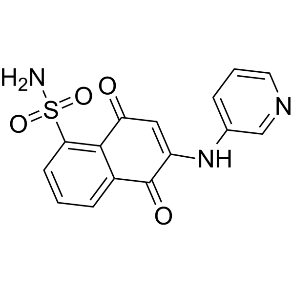
LY5 is a STAT3 inhibitor with an IC50 value of 0.5 μM. LY5 induces apoptosis and inhibits STAT3 phosphorylation. LY5 shows antitumor activity in vivo, it can be used for the research of cancer.
For research use only. We do not sell to patients.
- Purity : 97.02%
- CAS No.: 1436382-03-4
- Formula: C15H11N3O4S
- Molecular Weight:329.33
-
Storage:Powder -20°C, 3 years ; In solvent -80°C, 6 months , -20°C, 1 month

To place orders, for customer services and technical support, please contact: MedChemExpress USA
Tel: 609-228-6898 E-mail: [email protected] [email protected]


-
 Biological Activity
-
Chemical Information
-
Solvent & Solubility
- Protocols
- Purity & Documentation
- References
-
 Help & FAQs




Biological Activity
Description
IC50 & Target
IC50: 0.5 μM (STAT3)[1]
Cellular Effect
|
Cell Line

|
Type | Value | Description | References |
|---|---|---|---|---|
| SJRH30 | IC50 |
0.55 μM
Compound: 8, LY5
|
Cytotoxicity against human Rh30 cells assessed as cell viability by MTT assay
Cytotoxicity against human Rh30 cells assessed as cell viability by MTT assay
|
[PMID: 23651330] |
| U2OS | IC50 |
0.5 μM
Compound: 8, LY5
|
Cytotoxicity against human U2OS cells assessed as cell viability by MTT assay
Cytotoxicity against human U2OS cells assessed as cell viability by MTT assay
|
[PMID: 23651330] |
In Vitro
LY5 shows inhibition effects to U2OS, RH30 and RD2 cancer cells with IC50 values of 0.52, 0.55 and 1.39 μM, respectively[1]. LY5 (0.25-1 μM; 16 h) induces apoptosis and inhibits STAT3 phosphorylation in human sarcoma cancer cells[1]. LY5 (0.25-1 μM; 5 h) inhibits STAT3 phosphorylation induced by IL-6[1].
MedChemExpress (MCE) has not independently confirmed the accuracy of these methods. They are for reference only.
-
Cell Line:RH30 and EW8 cell lines
-
Concentration:0.25-1 μM
-
Incubation Time:16 hours
-
Result:Completely inhibited Tyr705 phosphorylation at 0.5 μM and dose-dependently decreased in formation of P-STAT3.
In Vivo
MedChemExpress (MCE) has not independently confirmed the accuracy of these methods. They are for reference only.
-
Animal Model:Nude mice with MDA-MB-231 cancer cells injection[1]
-
Dosage:5 mg/kg
-
Administration:Intraperitoneal injection; 5 mg/kg; once daily; for 21 days
-
Result:Suppressed tumor growth and significantly reduced the tumor sizes.
- Kinase activity and phosphorylation assays
- Western Blot
- Detection of Apoptosis
- TUNEL staining for apoptotic DNA fragmentation
- Annexin V plus membrane-impermeant dye apoptosis staining
- Apoptosis Solutions
- Protocol for Kinase activity and phosphorylation assays
- Subcutaneous Cell-Line-Derived Xenograft


Chemical Information
-
CAS No. 1436382-03-4
-
Appearance Solid
-
Molecular Weight 329.33
-
Formula C15H11N3O4S
-
Color Brown to red
-
SMILES
NS(=O)(C1=C2C(C(C(NC3=CC=CN=C3)=CC2=O)=O)=CC=C1)=O
-
Shipping
Room temperature in continental US; may vary elsewhere.
-
Storage
Powder -20°C 3 years In solvent -80°C 6 months -20°C 1 month


Solvent & Solubility
In Vitro:
DMSO : 6.25 mg/mL (18.98 mM; Need ultrasonic; Hygroscopic DMSO has a significant impact on the solubility of product, please use newly opened DMSO)
Please refer to the solubility information to select the appropriate solvent. Once prepared, please aliquot and store the solution to prevent product inactivation from repeated freeze-thaw cycles.
Storage method and period of stock solution: -80°C, 6 months; -20°C, 1 month. When stored at -80°C, please use it within 6 months. When stored at -20°C, please use it within 1 month.
Please refer to the solubility information to select the appropriate solvent. Once prepared, please aliquot and store the solution to prevent product inactivation from repeated freeze-thaw cycles.
Storage method and period of stock solution: -80°C, 6 months; -20°C, 1 month. When stored at -80°C, please use it within 6 months. When stored at -20°C, please use it within 1 month.
Concentration (start) × Volume (start) = Concentration (final) × Volume (final)


Purity & Documentation
-
Data Sheet (278 KB)
-
SDS (251 KB)
- English - EN (251 KB)
- Français - FR (251 KB)
- Deutsch - DE (251 KB)
- Norwegian - NO (251 KB)
- Español - ES (251 KB)
- Swedish - SV (251 KB)
- Italian - IT (251 KB)
- Korean - KR (251 KB)
- Portuguese - PT (251 KB)
-
Handling Instructions (2659 KB)
References


Complete Stock Solution Preparation Table
Please refer to the solubility information to select the appropriate solvent. Once prepared, please aliquot and store the solution to prevent product inactivation from repeated freeze-thaw cycles.
Storage method and period of stock solution: -80°C, 6 months; -20°C, 1 month. When stored at -80°C, please use it within 6 months. When stored at -20°C, please use it within 1 month.
| Optional Solvent | Concentration Solvent Mass | 1 mg | 5 mg | 10 mg | 25 mg |
|---|---|---|---|---|---|
| DMSO | 1 mM | 3.0365 mL | 15.1823 mL | 30.3647 mL | 75.9117 mL |
| 5 mM | 0.6073 mL | 3.0365 mL | 6.0729 mL | 15.1823 mL | |
| 10 mM | 0.3036 mL | 1.5182 mL | 3.0365 mL | 7.5912 mL | |
| 15 mM | 0.2024 mL | 1.0122 mL | 2.0243 mL | 5.0608 mL |


LY5 Related Classifications
HY-12442 Related Classifications
 Powered by Bioz