



Lycoramine hydrobromide
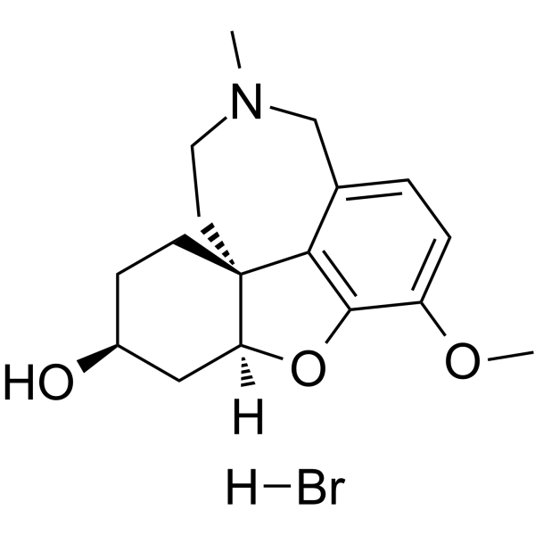
Lycoramine hydrobromide is a blood-brain barrier-penetrating Acetylcholinesterase inhibitor and Galanthamine (HY-76299) derivative. Lycoramine hydrobromide induces Amyloid-beta plaque clearance. Lycoramine hydrobromide induces reversal of cognitive decline and memory improvement. Lycoramine hydrobromide can be used for research on Alzheimer's disease and myasthenia gravis.
For research use only. We do not sell to patients.
- CAS No.: 89505-76-0
- Formula: C17H24BrNO3
- Molecular Weight:370.28
-
Storage:
Please store the product under the recommended conditions in the Certificate of Analysis.

To place orders, for customer services and technical support, please contact: MedChemExpress USA
Tel: 609-228-6898 E-mail: [email protected] [email protected]


-
 Biological Activity
-
Chemical Information
- Protocols
- Purity & Documentation
- References
-
 Help & FAQs




Biological Activity
Description
IC50 & Target
[3]|
AChE |
In Vitro
Lycoramine hydrobromide produces weaker AChE inhibition than Galanthamine (HY-76299) in the HPLC-IMER-MS bioanalytical system[1].
Lycoramine hydrobromide exhibits 42% higher acetylcholinesterase inhibitory activity than galantamine under in vitro conditions[3].
MedChemExpress (MCE) has not independently confirmed the accuracy of these methods. They are for reference only. Further protocols information, click here.
In Vivo
MedChemExpress (MCE) has not independently confirmed the accuracy of these methods. They are for reference only.
-
Animal Model:5xFAD transgenic mice (12 month-old, sex equally distributed across groups, over-expressing APP with K670N, M671L, V717I mutations and PS1 with M146L and L286V mutations)[3]
-
Dosage:4 mg/kg
-
Administration:i.p.; daily; 7 days
-
Result:Decreased escape latency in the acquisition phase of the Morris water maze test.
Increased time spent in the target quadrant during the probe phase.
Detected in all dissected brain regions (cortex, cerebellum, hippocampus, and remaining regions), confirming blood-brain barrier passage.
Caused a dramatic decrease in Aβ load, greater than galantamine.
Identified 2556 proteins in the cortex, with 145 proteins showing significant changes (fold change ≥ 1.3; q < 0.05).
Decreased cortical protein levels of clusterin (fc = 0.71, q = 0.00), apolipoprotein E (fc = 0.74, q = 0.009), microtubule-associated protein tau (fc = 0.49, q < 0.000), MAP1A (fc = 0.69, q < 0.000), MAP2 (fc = 0.44, q < 0.000), α-synuclein (fc = 0.49, q < 0.000), Tppp/25 (fc = 0.63, q < 0.000), GFAP (fc = 0.69, q = 0.004), Gja-1 (fc = 0.6, q = 0.007), AK-1 (fc = 0.67, q = 0.002), EHD3 (fc = 0.75, q = 0.019), and Synaptotagmin-5 (fc = 0.44, q = 0.006).
Increased cortical protein levels of GABRB2 (fc = 1.3, q = 0.011), Neurobeachin (fc = 1.3, q = 0.006), heat shock 70 kDa protein 1-like (fc = 1.35, q < 0.000), AT2B4/PMCA4 (fc = 1.81, q = 0.001), OSBP-1 (fc = 1.37, q = 0.018), and SRBS-1 (fc = 1.9, q = 0.004).
Increased cortical protein levels of Vimentin (fc = 1.43) and Gap junction alpha-1 protein (fc = 1.68).


Chemical Information
-
CAS No. 89505-76-0
-
Molecular Weight 370.28
-
Formula C17H24BrNO3
-
SMILES
O[C@H]1CC[C@@]23CCN(C)CC4=CC=C(OC)C(O[C@@]3([H])C1)=C24.[H]Br
-
Structure Classification
-
Initial Source
-
Shipping
Room temperature in continental US; may vary elsewhere.
-
Storage
Please store the product under the recommended conditions in the Certificate of Analysis.

Protocols
-
Protocol for Pharmacokinetic Study
Pharmacokinetic studies quantify how an organism handles a drug over time through absorption, distribution, metabolism, and excretion, and the core experimental readout is the concentration-time profile of parent drug and, when relevant, metabolites in biological matrices such as plasma, whole blood, urine, bile, or tissue. Pharmacokinetic analysis links dose, route, exposure, clearance, half-life, distribution, bioavailability, and systemic exposure to drug efficacy and toxicity hypotheses rather than measuring a signaling pathway directly. The literature links pharmacokinetics to drug-development phenotypes by showing that drug metabolism and pharmacokinetics influence compound progression, exposure-response interpretation, safety margins, dosing strategy, and failure risk during discovery and development. DMPK science contributes to compound optimization by integrating physicochemical properties, in vitro metabolism, transporter behavior, in vivo exposure, and pharmacodynamic contex
-
Amyloid: Congo Red Amyloid Staining
Congo red amyloid staining is a histochemical method used to detect extracellular amyloid deposits in tissue sections based on the affinity of Congo red dye for β-pleated sheet-rich protein aggregates. When bound to amyloid, Congo red produces characteristic apple-green birefringence under polarized light microscopy, which is widely regarded as a diagnostic feature of amyloid deposition in histopathology. The diagnostic principle relies on the combination of dye binding (congophilia) and optical anisotropy under polarized illumination, which distinguishes amyloid from most non-amyloid eosinophilic extracellular deposits in routine histological evaluation. Amyloid identification by Congo red staining remains a cornerstone in diagnostic pathology despite the availability of adjunct methods such as immunohistochemistry and mass spectrometry, particularly because of its ability to localize deposits directly within tissue architecture. The specificity of Congo red-positive deposits is incre
-
Alzheimer’s Disease Modeling
Alzheimer’s Disease (AD) is a neurodegenerative disorder characterized by a progressive decline in cognitive functions and loss of specific types of neurons and synapses. Alzheimer's symptoms can be simulated in mice by injecting drugs (such as Aβ) or genetically modified.


Purity & Documentation
References
[1].
Yuan Y, et al. Online acetylcholinesterase inhibition evaluation by high-performance liquid chromatography-mass spectrometry hyphenated with an immobilized enzyme reactor. J Chromatogr A. 2020 Jan 4;1609:460506.
[Content Brief]
[2]. IRWIN RL, et al. Cholinesterase inhibition by galanthamine and lycoramine. Biochem Pharmacol. 1960 May;3:147-8. [Content Brief]

Calculators
Concentration (start) × Volume (start) = Concentration (final) × Volume (final)


Lycoramine hydrobromide Related Classifications
HY-N6619 Related Classifications
 Powered by Bioz