



Mevalonic acid lithium salt
Based on 13 publication(s) in Google Scholar
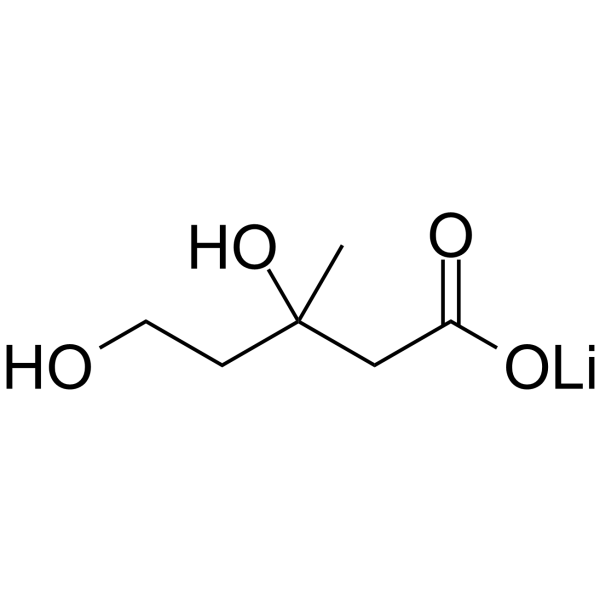
Mevalonic acid (MVA) lithium salt is a precursor substance of the mevalonate pathway, which is essential for cell growth and proliferation. Mevalonic acid lithium salt is effective in inhibiting Simvastatin (HY-17502)-induced decrease in C2C12 cell viability in vitro. Mevalonic acid lithium salt can be used in studies of myopathy and heart failure.
For research use only. We do not sell to patients.
- Purity : 98.0%
- CAS No.: 2618458-93-6
- Formula: C6H11LiO4
- Molecular Weight:154.09
-
Storage:
4°C, sealed storage, away from moisture
* In solvent : -80°C, 6 months; -20°C, 1 month (sealed storage, away from moisture)

To place orders, for customer services and technical support, please contact: MedChemExpress USA
Tel: 609-228-6898 E-mail: [email protected] [email protected]


-
 Biological Activity
-
Chemical Information
-
Solvent & Solubility
- Protocols
- Purity & Documentation
- References
-
 Help & FAQs


-
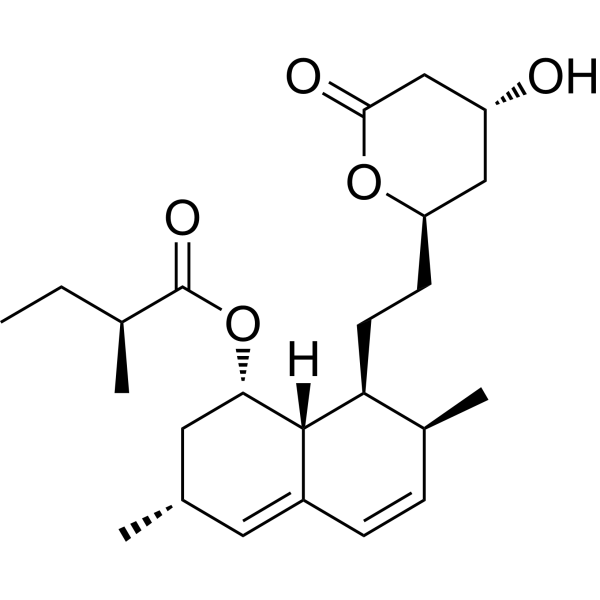
HY-N0504
LovastatinHMG-CoA Inhibitor -
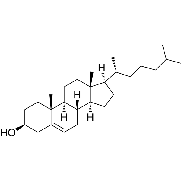
HY-N0322
Cholesterol (from animal)ERRα Agonist -
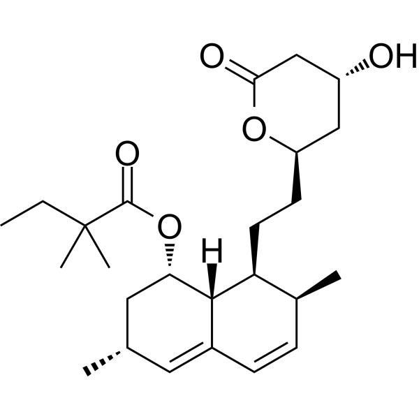
HY-17502
SimvastatinHMGCR Inhibitor -
HY-113134
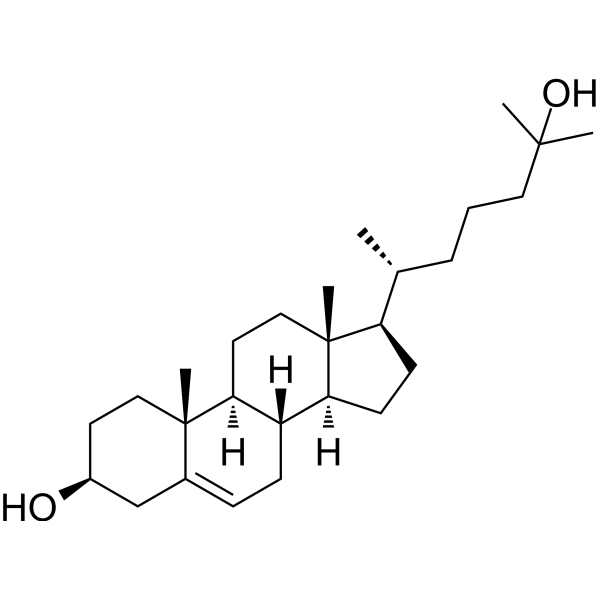
25-HydroxycholesterolCholesterol Metabolite -
HY-W011474
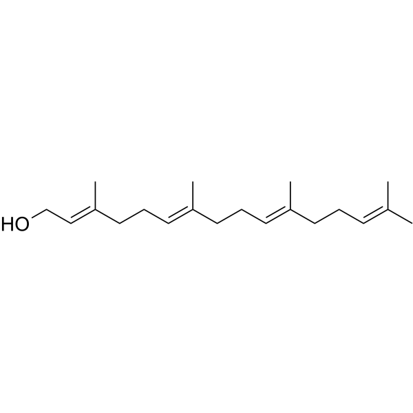
GeranylgeraniolVitamin K2 -

HY-13259
MG-132Proteasome Inhibitor -
HY-N0111
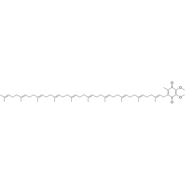
Coenzyme Q10Antioxidant -

HY-N6782
OligomycinH+-ATP-Synthase Inhibitor -

HY-100558
Bafilomycin A1V-ATPase Inhibitor -

HY-10071
Y-27632ROCK Inhibitor -

HY-10182
LaduviglusibGSK-3 Inhibitor -
HY-116767
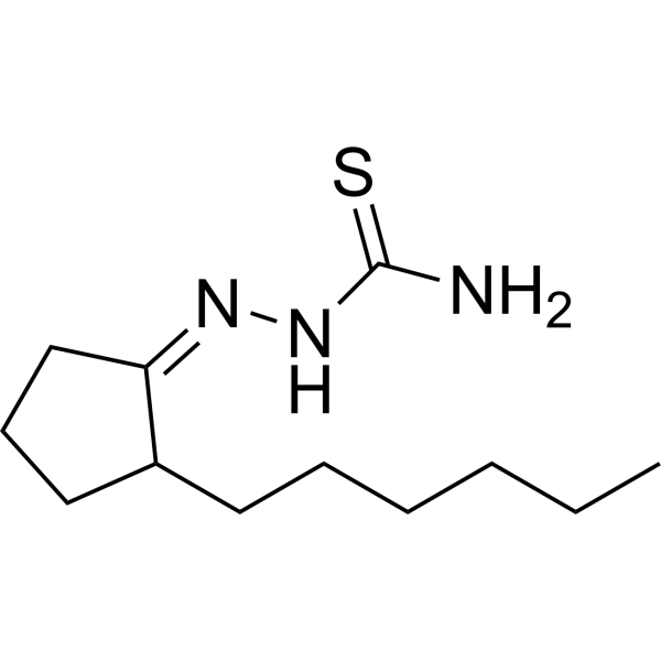
BLT-1SR-BI Inhibitor
Publications Citing Use of MedChemExpress (MCE) Mevalonic acid lithium salt
More- Nature. 2025 Jul;643(8070):192-200. [Abstract]
- Acta Pharm Sin B. 2023 Jun;13(6):2585-2600. [Abstract]
- Acta Pharmacol Sin. 2021 Nov;42(11):1875-1887. [Abstract]
- Dev Cell. 2026 Apr 29:S1534-5807(26)00150-4. [Abstract]
- Cell Biosci. 2021 Oct 9;11(1):179. [Abstract]
- Nat Prod Bioprospect. 2026 Jan 10;16(1):11. [Abstract]
- Biomedicines. 2022 Oct 5;10(10):2489. [Abstract]
- Biochim Biophys Acta Mol Cell Biol Lipids. 2022 Dec;1867(12):159217. [Abstract]
- Integr Cancer Ther. 2026 Jan-Dec:25:15347354261450966. [Abstract]
- Life Sci Alliance. 2022 Jan 4;5(3):e202101256. [Abstract]
- Drugs R D. 2023 Dec;23(4):439-451. [Abstract]
- Biochem Biophys Res Commun. 2020 Feb 19;522(4):862-868. [Abstract]
- Eur J Histochem. 2026 Jul 23;70(3).
 Customer Validation & Images

-
Bio/Physico-chemical Assay
-
Histological Imaging/Staining
-
In Vivo Efficacy Study
-
Cell Proliferation/Viability Assay
-
WB
All Endogenous Metabolite Isoforms
More

Biological Activity
Description
IC50 & Target
|
Human Endogenous Metabolite |
In Vitro
Mevalonic acid lithium salt (80, 90, 100, 110 μM; 72 h) shows prevention of simvastatin-induced loss of viability of C2C12myotube cells in vitro[1].
MedChemExpress (MCE) has not independently confirmed the accuracy of these methods. They are for reference only. Further protocols information, click here.
-
Cell Line:C2C12 cells (simvastatin-induced)
-
Concentration:80, 90, 100, 110 µM
-
Incubation Time:72 h
-
Result:Showed no decline in cell viability.
Clinical Trial
| NCT Number | Sponsor | Condition | Start Date |
Phase

|
|---|---|---|---|---|
| NCT01329991 | Plexxikon| | 2011-05 | PHASE1 |


Chemical Information
-
CAS No. 2618458-93-6
-
Appearance Solid
-
Molecular Weight 154.09
-
Formula C6H11LiO4
-
Color White to off-white
-
SMILES
O=C(O[Li])CC(C)(O)CCO
-
Synonyms
MVA lithium salt
-
Structure Classification
-
Initial Source
-
Shipping
Room temperature in continental US; may vary elsewhere.
-
Storage
4°C, sealed storage, away from moisture
* In solvent : -80°C, 6 months; -20°C, 1 month (sealed storage, away from moisture)


Publications (13)
-
Journal Impact Factor
-
Most Recent
-
Nature
2025 Jul;643(8070):192-200. PMID: 39695227 -
Acta Pharm Sin B
2023 Jun;13(6):2585-2600. PMID: 37425040 -
Acta Pharmacol Sin
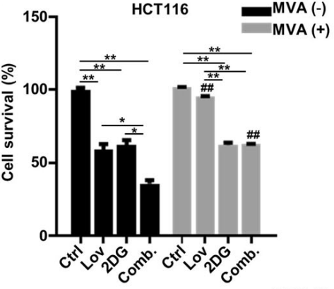
Autophagy inhibitors increase the susceptibility of KRAS-mutant human colorectal cancer cells to a combined treatment of 2-deoxy-D-glucose and lovastatin. [Abstract]2021 Nov;42(11):1875-1887. PMID: 33608672
Mevalonic acid lithium salt purchased from MedChemExpress. Usage Cited in: Acta Pharmacol Sin. 2021 Nov;42(11):1875-1887. [Abstract]
HCT116 and LoVo cells were cultured with lovastatin (15 μM) and 2DG (5 mM) alone or in combination for 72 h in either the presence or absence of 200 mM Mevalonate, and cell proliferation was determined. *P < 0.05, **P < 0.01. ##P < 0.01 vs corresponding Mevalonic acid lithium salt (MVA) (-) group.
-
Dev Cell
2026 Apr 29:S1534-5807(26)00150-4. PMID: 42061405 -
Cell Biosci
2021 Oct 9;11(1):179. PMID: 34627383
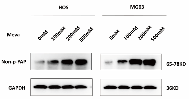
Mevalonic acid lithium salt purchased from MedChemExpress. Usage Cited in: Cell Biosci. 2021 Oct 9;11(1):179. [Abstract]
Western blotting was used to assess changes in the expression of unphosphorylated YAP in HOS and MG63 cells following a 6 h Mevalonic acid lithium salt (Meva) treatment.
-
Nat Prod Bioprospect
2026 Jan 10;16(1):11. PMID: 41518459
Mevalonic acid lithium salt purchased from MedChemExpress. Usage Cited in: Nat Prod Bioprospect. 2026 Jan 10;16(1):11. [Abstract]
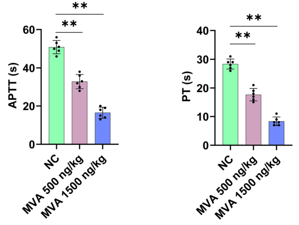
Mevalonic acid lithium salt (MVA, 500 or 1500 ng/kg; i.v.) significantly shortened activated partial thromboplastin time (APTT) and prothrombin time (PT) in C57BL/C mice.
Mevalonic acid lithium salt purchased from MedChemExpress. Usage Cited in: Nat Prod Bioprospect. 2026 Jan 10;16(1):11. [Abstract]
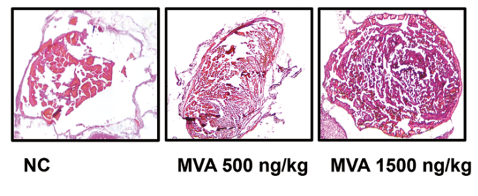
Mice were subjected to the inferior vena cava IVC stasis model for 5 h to evaluate venous thrombosis formation, with subsequent pathological assessment by hematoxylin and eosin (HE) staining. The results showed that Mevalonic acid lithium salt (MVA, 500 or 1500 ng/kg; i.v.) treatment markedly increased both thrombus weight.
Mevalonic acid lithium salt purchased from MedChemExpress. Usage Cited in: Nat Prod Bioprospect. 2026 Jan 10;16(1):11. [Abstract]
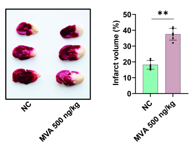
Mice were administered injections of either 500 ng/kg Mevalonic acid lithium salt (MVA). On 24 h after transient middle cerebral artery occlusion (tMCAO), typical images of coronal brain sections stained with 2,3,5-triphenyltetrazolium chloride (TTC). The results showed that MVA led to a significant increase in infarct volume.
-
Biomedicines
c-Myc Sustains Pancreatic Cancer Cell Survival and mutp53 Stability through the Mevalonate Pathway. [Abstract]2022 Oct 5;10(10):2489. PMID: 36289751 -
Biochim Biophys Acta Mol Cell Biol Lipids
PCSK9 deficiency results in a specific shedding of excess LDLR in female mice only: Role of hepatic cholesterol. [Abstract]2022 Dec;1867(12):159217. PMID: 35985474 -
Integr Cancer Ther
Scutellaria Barbata Water Extract Suppresses Breast Cancer Growth and Lung Metastasis by Inducing Ferroptosis. [Abstract]2026 Jan-Dec:25:15347354261450966. PMID: 42244109 -
Life Sci Alliance
Necrotic debris and STING exert therapeutically relevant effects on tumor cholesterol homeostasis. [Abstract]2022 Jan 4;5(3):e202101256. PMID: 34983824 -
Drugs R D
Simvastatin Preferentially Targets FLT3/ITD Acute Myeloid Leukemia by Inhibiting MEK/ERK and p38-MAPK Signaling Pathways. [Abstract]2023 Dec;23(4):439-451. PMID: 37847357 -
Biochem Biophys Res Commun
Combinatorial screening of a panel of FDA-approved drugs identifies several candidates with anti-Ebola activities. [Abstract]2020 Feb 19;522(4):862-868. PMID: 31806372 -


Solvent & Solubility
In Vitro:
H2O : 100 mg/mL (648.97 mM; Need ultrasonic)
DMSO : 50 mg/mL (324.49 mM; Need ultrasonic; Hygroscopic DMSO has a significant impact on the solubility of product, please use newly opened DMSO)
Please refer to the solubility information to select the appropriate solvent. Once prepared, please aliquot and store the solution to prevent product inactivation from repeated freeze-thaw cycles.
Storage method and period of stock solution: -80°C, 6 months; -20°C, 1 month (sealed storage, away from moisture). When stored at -80°C, please use it within 6 months. When stored at -20°C, please use it within 1 month.
* Note: If you choose water as the stock solution, please dilute it to the working solution, then filter and sterilize it with a 0.22 μm filter before use.
Please refer to the solubility information to select the appropriate solvent. Once prepared, please aliquot and store the solution to prevent product inactivation from repeated freeze-thaw cycles.
Storage method and period of stock solution: -80°C, 6 months; -20°C, 1 month (sealed storage, away from moisture). When stored at -80°C, please use it within 6 months. When stored at -20°C, please use it within 1 month.
* Note: If you choose water as the stock solution, please dilute it to the working solution, then filter and sterilize it with a 0.22 μm filter before use.
Concentration (start) × Volume (start) = Concentration (final) × Volume (final)
In Vivo:
Select the appropriate dissolution method based on your experimental animal and administration route.
- For the following dissolution methods, please ensure to first prepare a clear stock solution using an In Vitro approach and then sequentially add co-solvents:
- To ensure reliable experimental results, the clarified stock solution can be appropriately stored based on storage conditions. As for the working solution for In Vivo experiments, it is recommended to prepare freshly and use it on the same day.
- The percentages shown for the solvents indicate their volumetric ratio in the final prepared solution. If precipitation or phase separation occurs during preparation, heat and/or sonication can be used to aid dissolution.
Add each solvent one by one: 10% DMSO 40% PEG300 5% Tween-80 45% Saline
Solubility: ≥ 2.5 mg/mL (16.22 mM); Clear solution
This protocol yields a clear solution of ≥ 2.5 mg/mL (saturation unknown).
Taking 1 mL working solution as an example, add 100 μL DMSO stock solution (25.0 mg/mL) to 400 μL PEG300, and mix evenly; then add 50 μL Tween-80 and mix evenly; then add 450 μL Saline to adjust the volume to 1 mL.
Preparation of Saline: Dissolve 0.9 g sodium chloride in ddH₂O and dilute to 100 mL to obtain a clear Saline solution.
Add each solvent one by one: 10% DMSO 90% (20% SBE-β-CD in Saline)
Solubility: ≥ 2.5 mg/mL (16.22 mM); Clear solution
This protocol yields a clear solution of ≥ 2.5 mg/mL (saturation unknown).
Taking 1 mL working solution as an example, add 100 μL DMSO stock solution (25.0 mg/mL) to 900 μL 20% SBE-β-CD in Saline, and mix evenly.
Preparation of 20% SBE-β-CD in Saline (4°C, storage for one week): 2 g SBE-β-CD powder is dissolved in 10 mL Saline, completely dissolve until clear.
For the following dissolution methods, please prepare the working solution directly:
It is recommended to prepare fresh solutions and use them promptly within a short period of time.
The percentages shown for the solvents indicate their volumetric ratio in the final prepared solution. If precipitation or phase separation occurs during preparation, heat and/or sonication can be used to aid dissolution.
Add each solvent one by one: PBS
Solubility: 25 mg/mL (162.24 mM); Clear solution; Need ultrasonic
In Vivo Dissolution Calculator
Please enter the basic information of animal experiments:
-
-
-
-
Recommended: Prepare an additional quantity of animals to account for potential losses during experiments.
Working solution concentration: 0.22 mg/mL
This product has good water solubility, please refer to the measured solubility data in water/PBS/Saline for details.

Protocols
-
Cell Cytotoxicity Assay
Cytotoxicity assays are usually based on the assessment of cell membrane damage, which can also be indirectly detected by measuring cell viability. Detection methods include MTT assay, CKK-8 assay, LDH assay and ATP assay, etc.
-
Mammalian live/dead viability and cytotoxicity staining
Live/dead viability and cytotoxicity staining assays are based on the simultaneous detection of intracellular esterase activity in metabolically active (viable) cells and membrane integrity loss in non-viable cells. In commonly used dual-staining approaches, membrane-permeant fluorogenic substrates are converted by intracellular esterases into fluorescent products in live cells, while impermeant DNA-binding dyes selectively enter cells with compromised plasma membranes and label nucleic acids in dead or dying cells, enabling discrimination between viable and non-viable populations by fluorescence microscopy or flow cytometry.
-
Protocol for Cell Counting and Cell Density Analysis
Cell counting and cell-density analysis estimate the number of cells in a known volume or field area. Manual hemocytometer counting uses a chamber of defined geometry to convert counted cells into cells/mL, while automated counters and image-analysis workflows detect cell objects from optical, brightfield, fluorescence, impedance, or digital-image features. Trypan blue viability counting is based on dye exclusion: viable cells with intact membranes exclude dye, while non-viable cells with compromised membranes stain blue. The readout is total cell density, viable-cell density, dead-cell density, and percent viability. Cell density can also be estimated from microscopy images by counting objects per image area, from flow cytometry using calibrated volume or reference particles, or from in situ microscopy in bioreactors after calibration against reference methods such as hemocytometer or flow cytometry.
-
Somatic Cell Culture
A method of simulating the in vivo environment in vitro to maintain the cell growth, differentation and main functions.
-
CCK-8/WST-8 Cell Proliferation Assay
The CCK-8/WST-8 assay is based on the reduction of the water-soluble tetrazolium salt WST-8 to a water-soluble formazan product by cellular dehydrogenases in metabolically active cells, where the generated formazan amount is proportional to the number of living cells and is quantified by measuring absorbance in the visible range, providing a colorimetric readout for cell viability and proliferation assessment. This class of tetrazolium-based assays improves upon earlier MTT-based systems by producing a water-soluble formazan, eliminating the need for organic solubilization steps and enabling direct spectrophotometric measurement in culture medium.
-
Research Protocol for Cardiovascular Diseases
Cardiovascular disease can be modeled as maladaptive cardiac remodeling, where ischemic injury or pressure overload activates inflammatory signaling, fibroblast activation, extracellular-matrix deposition, cardiomyocyte hypertrophy, vascular remodeling, and progressive ventricular dysfunction. The TGF-β/SMAD axis is a central profibrotic pathway after myocardial injury and pressure overload, while innate immune and cytokine pathways regulate leukocyte recruitment, scar formation, and adverse remodeling. Key unresolved questions include which inflammatory signals are reparative versus harmful, when fibrosis is protective versus maladaptive, and whether pathway inhibition improves function without weakening necessary infarct healing or compensatory remodeling.
-
Cell Counting-Based Growth Curve Assay
Cell counting-based growth curve assays quantify cell proliferation by directly measuring changes in viable cell number over time using manual or automated counting methods such as hemocytometer-based counting or instrument-assisted cell enumeration, enabling construction of growth curves that reflect population expansion dynamics in response to culture conditions. A widely used approach is trypan blue exclusion with hemocytometer counting, where membrane-compromised (non-viable) cells take up the dye, allowing discrimination between viable and non-viable cells while simultaneously enabling total cell number quantification. Repeated sampling across time points allows estimation of proliferation rate, growth phases, and comparative growth kinetics between experimental conditions.
-
MTT Cell Proliferation Assay
The MTT assay is a colorimetric endpoint assay for estimating viable cell number, cell growth, cytotoxicity, or cell activation in cultured mammalian cells. Living cells reduce the yellow tetrazolium salt MTT into purple/blue formazan, while dead cells do not generate the same signal; the resulting color can be quantified with a multiwell spectrophotometer. MTT reduction is commonly interpreted as a readout of metabolic activity that often correlates with viable cell number, but it should not be treated as a direct cell-counting method unless the assay is optimized for the cell type and experimental condition. Studies show that MTT reduction can involve mitochondrial and non-mitochondrial reducing systems, and formazan may accumulate in intracellular lipid droplets rather than simply marking mitochondria.
-
Cell Viability Determination by MTT Colorimetric Assay
The following protocol uses the MTT colorimetric assay as a classic literature-established method for assessing cell viability/metabolic activity in cultured mammalian cells. MTT[3-(4,5-dimethylthiazol-2-yl)-2,5-diphenyltetrazolium bromide] is reduced by metabolically active cells to a colored formazan product; the amount of formazan is quantified spectrophotometrically and provides an indirect measure of metabolically active viable cells. Importantly, MTT reduction reflects cellular oxidoreductase/metabolic activity rather than an absolute direct count of living cells, so changes in cellular metabolism can alter the signal independently of cell number.


Purity & Documentation
-
Data Sheet (271 KB)
-
SDS (393 KB)
- English - EN (393 KB)
- Français - FR (393 KB)
- Deutsch - DE (393 KB)
- Norwegian - NO (393 KB)
- Español - ES (393 KB)
- Swedish - SV (393 KB)
- Italian - IT (393 KB)
- Korean - KR (393 KB)
- Portuguese - PT (393 KB)
-
Handling Instructions (2659 KB)
References
[1]. Moschetti A, et al. Coenzyme Q nanodisks counteract the effect of statins on C2C12 myotubes. Nanomedicine. 2021 Oct;37:102439. [Content Brief]
[2]. Soma MR, et al. Cholesterol and mevalonic acid modulation in cell metabolism and multiplication. Toxicol Lett. 1992 Dec;64-65 Spec No:1-15. [Content Brief]


Complete Stock Solution Preparation Table
Please refer to the solubility information to select the appropriate solvent. Once prepared, please aliquot and store the solution to prevent product inactivation from repeated freeze-thaw cycles.
Storage method and period of stock solution: -80°C, 6 months; -20°C, 1 month (sealed storage, away from moisture). When stored at -80°C, please use it within 6 months. When stored at -20°C, please use it within 1 month.
| Optional Solvent | Concentration Solvent Mass | 1 mg | 5 mg | 10 mg | 25 mg |
|---|---|---|---|---|---|
| DMSO / H2O | 1 mM | 6.4897 mL | 32.4486 mL | 64.8971 mL | 162.2428 mL |
| 5 mM | 1.2979 mL | 6.4897 mL | 12.9794 mL | 32.4486 mL | |
| 10 mM | 0.6490 mL | 3.2449 mL | 6.4897 mL | 16.2243 mL | |
| 15 mM | 0.4326 mL | 2.1632 mL | 4.3265 mL | 10.8162 mL | |
| 20 mM | 0.3245 mL | 1.6224 mL | 3.2449 mL | 8.1121 mL | |
| 25 mM | 0.2596 mL | 1.2979 mL | 2.5959 mL | 6.4897 mL | |
| 30 mM | 0.2163 mL | 1.0816 mL | 2.1632 mL | 5.4081 mL | |
| 40 mM | 0.1622 mL | 0.8112 mL | 1.6224 mL | 4.0561 mL | |
| 50 mM | 0.1298 mL | 0.6490 mL | 1.2979 mL | 3.2449 mL | |
| 60 mM | 0.1082 mL | 0.5408 mL | 1.0816 mL | 2.7040 mL | |
| 80 mM | 0.0811 mL | 0.4056 mL | 0.8112 mL | 2.0280 mL | |
| 100 mM | 0.0649 mL | 0.3245 mL | 0.6490 mL | 1.6224 mL |
* Note: If you choose water as the stock solution, please dilute it to the working solution, then filter and sterilize it with a 0.22 μm filter before use.


Mevalonic acid lithium salt Related Classifications
HY-113071A Related Classifications
 Powered by Bioz