



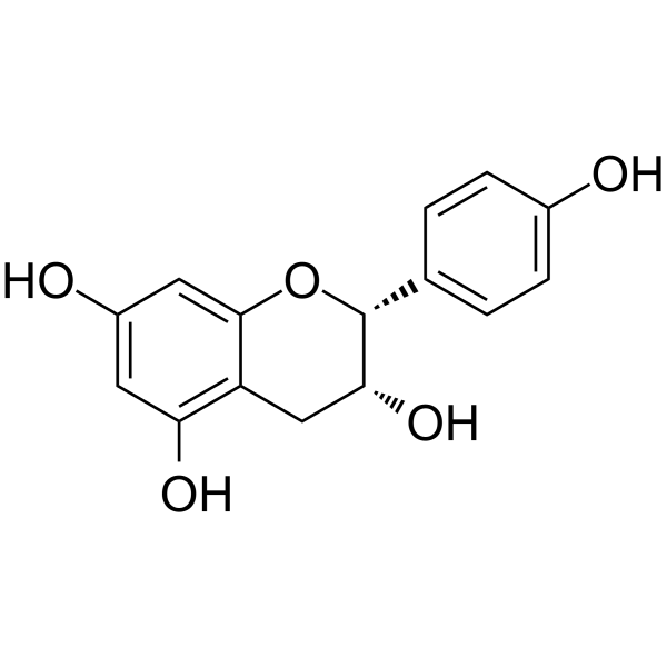
(-)-Epiafzelechin

Based on 1 Customer Validation
(−)-Epiafzelechin is a COX-1 inhibitor with an IC50 of approximately 15 μM against COX-1 activity. (−)-Epiafzelechin also exhibits DPPH free radical scavenging activity, with an EC50 of 20.9 μM. (−)-Epiafzelechin promotes osteoblast differentiation by increasing ALP activity, ECM collagen content and Runx2 expression, while reducing the growth of osteoclast precursors and RANKL-induced TRAP activity, thereby regulating the bone remodeling process. (−)-Epiafzelechin alleviates ovariectomy-induced bone loss. (−)-Epiafzelechin can be used in studies related to bone remodeling, postmenopausal osteoporosis, inflammatory responses and oxidative stress.
For research use only. We do not sell to patients.
- Purity : 99.87%
- CAS No.: 24808-04-6
- Formula: C15H14O5
- Molecular Weight:274.27
-
Storage:
4°C, sealed storage, away from moisture and light
* In solvent : -80°C, 6 months; -20°C, 1 month (sealed storage, away from moisture and light)

To place orders, for customer services and technical support, please contact: MedChemExpress USA
Tel: 609-228-6898 E-mail: [email protected] [email protected]


-
 Biological Activity
-
Chemical Information
-
Solvent & Solubility
- Protocols
- Purity & Documentation
- References
-
 Help & FAQs




Biological Activity
Description
IC50 & Target
[2]|
COX-1 15 μM (IC50) |
Cellular Effect
|
Cell Line

|
Type | Value | Description | References |
|---|---|---|---|---|
| H9 | EC50 |
70 μg/mL
Compound: 1
|
Inhibition of HIV-1 replication in H9 (human lymphoma) cells.
Inhibition of HIV-1 replication in H9 (human lymphoma) cells.
|
10.1016/0960-894X(96)00095-9 |
| H9 | IC50 |
100 μg/mL
Compound: 1
|
Inhibition of uninfected H9 lymphocytic cell growth
Inhibition of uninfected H9 lymphocytic cell growth
|
10.1016/0960-894X(96)00095-9 |
In Vitro
(-)-Epiafzelechin (0.1 nM-1 μM) stimulates proliferation, alkaline phosphatase activity, collagen production, and Runx2 mRNA expression in mouse preosteoblastic cells MC3T3-E1, but does not promote mineralization; instead, it inhibits mineralization at higher concentrations[1].
(-)-Epiafzelechin (12-45 μM) potently inhibits COX-1 activity in ram seminal vesicle microsomes, with an IC50 value of 15 μM[2].
(-)-Epiafzelechin promotes the proliferation, differentiation and bone nodule mineralization of rat osteoblasts[3].
(-)-Epiafzelechin (10 nM-1 μM) promotes the differentiation of mouse preosteoblast MC3T3-E1 by increasing alkaline phosphatase activity, extracellular matrix collagen content, and osteoblast-specific gene expression, without altering calcium deposition[5].
(-)-Epiafzelechin (10 nM-1 μM) inhibits RANKL-induced osteoclast maturation and function in RAW264.7 osteoclast precursor cells by reducing cell viability, the number of tartrate-resistant acid phosphatase (TRAP)-positive multinucleated cells, and TRAP activity[5].
(−)-Epiafzelechin (10−7-10−6 M; 5 d) reduces the number of TRAP-positive multinucleated osteoclasts and decreases TRAP activity in RANKL (100 ng/mL)-induced RAW264.7 cells[1].
(−)-Epiafzelechin (10−14 M) enhances ERα-mediated ERE luciferase activity, but does not significantly increase ERβ-mediated ERE luciferase activity within the tested concentration range[5].
MedChemExpress (MCE) has not independently confirmed the accuracy of these methods. They are for reference only. Further protocols information, click here.
Parmacokinetics
In Vivo
(-)-Epiafzelechin (100 mg/kg; p.o.; single administration) reduces carrageenan-induced paw swelling in mice, with a significant decrease in swelling observed at 1 h, 4 h, and 6 h after carrageenan challenge[2].
MedChemExpress (MCE) has not independently confirmed the accuracy of these methods. They are for reference only.
-
Animal Model:C57BL/6J (mature, 3-4 months old, ovariectomized)[1]
-
Dosage:500 µg/kg
-
Administration:p.o.; daily; 6 weeks
-
Result:Reduced OVX-induced body weight gain.
Reduced urinary calcium excretion to levels matching sham-operated mice.
Reduced serum osteocalcin levels and urinary deoxypyridinoline levels in OVX mice.
Increased trabecular bone mineral density at the proximal tibia by 53% and at lumbar vertebra L4 by 17.5% in OVX mice.
Increased bone volume/total volume to 19.3%, trabecular thickness to 0.050 mm, and connectivity density to 159.4 mm-3, while decreasing trabecular separation to 0.210 mm and structural model index to 1.85 at the proximal tibia in OVX mice.
Increased bone volume/total volume to 25.2%, trabecular number to 4.42 mm-1, and connectivity density to 134.0 mm-3, while decreasing trabecular separation to 0.169 mm and structural model index to 0.82 at lumbar vertebra L4 in OVX mice.
Did not increase uterine index in OVX mice.
-
Animal Model:ICR mice (male, 25 g)[2]
-
Dosage:100 mg/kg
-
Administration:p.o.; single dose
-
Result:Reduced paw edema to 16.6% at 1 h post-carrageenin injection.
Reduced paw edema to 28.6% at 2 h post-carrageenin injection.
Reduced paw edema to 43.1% at 4 h post-carrageenin injection.
Reduced paw edema to 42.5% at 6 h post-carrageenin injection.


Chemical Information
-
CAS No. 24808-04-6
-
Appearance Solid
-
Molecular Weight 274.27
-
Formula C15H14O5
-
Color White to off-white
-
SMILES
O[C@H]1[C@@H](C2=CC=C(O)C=C2)OC3=CC(O)=CC(O)=C3C1
-
Structure Classification
-
Initial Source
-
Shipping
Room temperature in continental US; may vary elsewhere.
-
Storage
4°C, sealed storage, away from moisture and light
* In solvent : -80°C, 6 months; -20°C, 1 month (sealed storage, away from moisture and light)


Solvent & Solubility
In Vitro:
DMSO : 50 mg/mL (182.30 mM; Need ultrasonic; Hygroscopic DMSO has a significant impact on the solubility of product, please use newly opened DMSO)
Please refer to the solubility information to select the appropriate solvent. Once prepared, please aliquot and store the solution to prevent product inactivation from repeated freeze-thaw cycles.
Storage method and period of stock solution: -80°C, 6 months; -20°C, 1 month (sealed storage, away from moisture and light). When stored at -80°C, please use it within 6 months. When stored at -20°C, please use it within 1 month.
Please refer to the solubility information to select the appropriate solvent. Once prepared, please aliquot and store the solution to prevent product inactivation from repeated freeze-thaw cycles.
Storage method and period of stock solution: -80°C, 6 months; -20°C, 1 month (sealed storage, away from moisture and light). When stored at -80°C, please use it within 6 months. When stored at -20°C, please use it within 1 month.
Concentration (start) × Volume (start) = Concentration (final) × Volume (final)
In Vivo:
Select the appropriate dissolution method based on your experimental animal and administration route.
- For the following dissolution methods, please ensure to first prepare a clear stock solution using an In Vitro approach and then sequentially add co-solvents:
- To ensure reliable experimental results, the clarified stock solution can be appropriately stored based on storage conditions. As for the working solution for In Vivo experiments, it is recommended to prepare freshly and use it on the same day.
- The percentages shown for the solvents indicate their volumetric ratio in the final prepared solution. If precipitation or phase separation occurs during preparation, heat and/or sonication can be used to aid dissolution.
Add each solvent one by one: 10% DMSO 40% PEG300 5% Tween-80 45% Saline
Solubility: ≥ 1.25 mg/mL (4.56 mM); Clear solution
This protocol yields a clear solution of ≥ 1.25 mg/mL (saturation unknown).
Taking 1 mL working solution as an example, add 100 μL DMSO stock solution (12.5 mg/mL) to 400 μL PEG300, and mix evenly; then add 50 μL Tween-80 and mix evenly; then add 450 μL Saline to adjust the volume to 1 mL.
Preparation of Saline: Dissolve 0.9 g sodium chloride in ddH₂O and dilute to 100 mL to obtain a clear Saline solution.
Add each solvent one by one: 10% DMSO 90% (20% SBE-β-CD in Saline)
Solubility: ≥ 1.25 mg/mL (4.56 mM); Clear solution
This protocol yields a clear solution of ≥ 1.25 mg/mL (saturation unknown).
Taking 1 mL working solution as an example, add 100 μL DMSO stock solution (12.5 mg/mL) to 900 μL 20% SBE-β-CD in Saline, and mix evenly.
Preparation of 20% SBE-β-CD in Saline (4°C, storage for one week): 2 g SBE-β-CD powder is dissolved in 10 mL Saline, completely dissolve until clear.
In Vivo Dissolution Calculator
Please enter the basic information of animal experiments:
-
-
-
-
Recommended: Prepare an additional quantity of animals to account for potential losses during experiments.
Please enter your animal formula composition:
-
%DMSO +
Recommended: Keep the proportion of DMSO in working solution below 2% if your animal is weak.
-
%+
-
+%Tween-80 + +
-
%Saline +
The co-solvents required include: DMSO, . All of co-solvents are available by MedChemExpress (MCE). , Tween 80. All of co-solvents are available by MedChemExpress (MCE).
Working solution concentration: 0.22 mg/mL
Method for preparing stock solution: mg drug dissolved in μL DMSO. Stock solution concentration: mg/mL. * In solvent : -80°C, 6 months; -20°C, 1 month (sealed storage, away from moisture and light)
1. Take μL DMSO stock solution;
2. Add μL .
μL , mix evenly;
3. Then add μL Tween 80, mix evenly;
4. Then add μL
Please ensure that the stock solution in the first step is dissolved to a clear state, and add co-solvents in sequence. You can use ultrasonic heating (ultrasonic cleaner, recommended frequency 20-40 kHz), vortexing, etc. to assist dissolution.

Protocols
-
Fibrosis/Collagen Morphometry
Fibrosis and collagen morphometry is based on the quantitative visualization of fibrillar collagen deposition in tissue sections using histochemical stains such as Sirius Red (Picrosirius Red) or Masson's trichrome, followed by image-based or polarization-enhanced analysis to estimate collagen proportional area as a surrogate of extracellular matrix accumulation during fibrotic remodeling. Sirius Red combined with polarized light microscopy enhances detection of collagen fibers due to birefringence properties, enabling more specific visualization of collagen type I and III fibrils compared to conventional bright-field histology, while whole-section or region-restricted digital morphometry reduces field-selection bias in fibrosis assessment. Alternative quantitative approaches include second harmonic generation (SHG) and two-photon excited fluorescence microscopy, which enable label-free detection of fibrillar collagen and have been validated against histological staining and biochemica
-
Collagen: Sirius Red Staining
Sirius Red or picrosirius red staining is a histochemical method for visualizing collagen-rich extracellular matrix in tissue sections, and collagen fibers are detected as red-stained structures under bright-field microscopy with enhanced birefringence under polarized light. Picrosirius red is useful for assessing total collagen organization, distribution, and fibrosis burden, but polarized color should not be interpreted as a definitive collagen type I versus type III readout because color is affected by fiber orientation, thickness, and packing.
-
RNA extraction experimental
By lysing cells, releasing RNA, and removing impurities such as proteins and DNA, high-purity RNA products are finally obtained. The commonly used traditional method is the guanidine isothiocyanate/phenol/chloroform method (Trizol), which is suitable for a variety of animal materials including animal tissues, microorganisms, cultured cells, etc., and most plant materials.
-
Cell Cytotoxicity Assay
Cytotoxicity assays are usually based on the assessment of cell membrane damage, which can also be indirectly detected by measuring cell viability. Detection methods include MTT assay, CKK-8 assay, LDH assay and ATP assay, etc.
-
Mesenchymal stromal/stem cell osteogenic differentiation
Mesenchymal stromal/stem cells can be induced toward an osteoblast-like lineage in vitro by culture in osteogenic medium containing dexamethasone, ascorbic acid or ascorbate-2-phosphate, and β-glycerophosphate; the differentiation process is commonly evaluated by alkaline phosphatase activity, osteogenic marker expression, collagenous matrix formation, and calcium-rich matrix mineralization. The main readouts are alkaline phosphatase activity as an early osteogenic marker and Alizarin Red S staining as a calcium-deposit readout for mineralized extracellular matrix; Alizarin Red S can be inspected microscopically or extracted and measured colorimetrically at 405 nm.
-
ROS/oxidative-stress fluorescent staining
ROS/oxidative-stress fluorescent staining uses cell-permeant fluorogenic probes that become fluorescent after oxidation inside cells or tissues; commonly used examples include DCFH-DA/DCFDA for broad cellular oxidant detection, DHE for superoxide-related signal detection, MitoSOX for mitochondrial superoxide-related signal detection, and CellROX probes for oxidative-stress-associated fluorescence readouts. The assay detects probe oxidation rather than a single ROS species unless the probe and analysis method have been chemically validated for that species. DCFH-DA enters cells, is deacetylated by intracellular esterases to DCFH, and produces fluorescent DCF after oxidation, so the readout is used as an operational measure of total cellular oxidative stress rather than a species-specific ROS measurement. DHE and MitoSOX can report superoxide-related oxidation, but red fluorescence alone can include non-specific ethidium-like oxidation products; HPLC or optimized spectral approaches are
-
Osteoclast differentiation from monocyte/macrophage precursors
Osteoclast differentiation is an in vitro induction assay in which monocyte/macrophage-lineage precursors are exposed to macrophage colony-stimulating factor (M-CSF) and receptor activator of NF-κB ligand (RANKL), generating multinucleated osteoclasts that are commonly identified by tartrate-resistant acid phosphatase (TRAP) staining and functionally confirmed by resorption pits on dentin, bone, or mineralized substrates. M-CSF supports survival and expansion of osteoclast precursors, while RANKL binding to RANK drives osteoclast commitment, fusion, maturation, and resorptive function; osteoprotegerin inhibits this pathway by binding RANKL and preventing RANK activation. The main readouts are the number of TRAP-positive multinucleated cells, formation of F-actin rings, and resorbed surface area; TRAP-positive multinucleated cells indicate osteoclast differentiation, whereas pit formation on dentin, bone, or mineralized coating indicates functional bone-resorbing activity.


Purity & Documentation
-
Data Sheet (288 KB)
-
SDS (252 KB)
- English - EN (252 KB)
- Français - FR (252 KB)
- Deutsch - DE (252 KB)
- Norwegian - NO (252 KB)
- Español - ES (252 KB)
- Swedish - SV (252 KB)
- Italian - IT (252 KB)
- Korean - KR (252 KB)
- Portuguese - PT (252 KB)
-
Handling Instructions (2659 KB)
References
[1]. Wong KC, et al. (-)-Epiafzelechin Protects against Ovariectomy-induced Bone Loss in Adult Mice and Modulate Osteoblastic and Osteoclastic Functions In Vitro. Nutrients. 2017 May 22;9(5):530. [Content Brief]
[2]. Min KR, et al. (-)-Epiafzelechin: cyclooxygenase-1 inhibitor and anti-inflammatory agent from aerial parts of Celastrus orbiculatus. Planta medica. 1999 Jun;65(5):460-2. [Content Brief]
[4]. Hori K, et al. Antioxidant phenolic compounds from the rhizomes of Astilbe rivularis. Natural product research. 2018 Feb;32(4):453-456. [Content Brief]


Complete Stock Solution Preparation Table
Please refer to the solubility information to select the appropriate solvent. Once prepared, please aliquot and store the solution to prevent product inactivation from repeated freeze-thaw cycles.
Storage method and period of stock solution: -80°C, 6 months; -20°C, 1 month (sealed storage, away from moisture and light). When stored at -80°C, please use it within 6 months. When stored at -20°C, please use it within 1 month.
| Optional Solvent | Concentration Solvent Mass | 1 mg | 5 mg | 10 mg | 25 mg |
|---|---|---|---|---|---|
| DMSO | 1 mM | 3.6460 mL | 18.2302 mL | 36.4604 mL | 91.1511 mL |
| 5 mM | 0.7292 mL | 3.6460 mL | 7.2921 mL | 18.2302 mL | |
| 10 mM | 0.3646 mL | 1.8230 mL | 3.6460 mL | 9.1151 mL | |
| 15 mM | 0.2431 mL | 1.2153 mL | 2.4307 mL | 6.0767 mL | |
| 20 mM | 0.1823 mL | 0.9115 mL | 1.8230 mL | 4.5576 mL | |
| 25 mM | 0.1458 mL | 0.7292 mL | 1.4584 mL | 3.6460 mL | |
| 30 mM | 0.1215 mL | 0.6077 mL | 1.2153 mL | 3.0384 mL | |
| 40 mM | 0.0912 mL | 0.4558 mL | 0.9115 mL | 2.2788 mL | |
| 50 mM | 0.0729 mL | 0.3646 mL | 0.7292 mL | 1.8230 mL | |
| 60 mM | 0.0608 mL | 0.3038 mL | 0.6077 mL | 1.5192 mL | |
| 80 mM | 0.0456 mL | 0.2279 mL | 0.4558 mL | 1.1394 mL | |
| 100 mM | 0.0365 mL | 0.1823 mL | 0.3646 mL | 0.9115 mL |


(-)-Epiafzelechin Related Classifications
HY-N0943 Related Classifications
 Powered by Bioz