



MY-943

Based on 1 Customer Validation
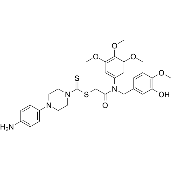
MY-943 is a potent tubulin polymerization and LSD1 inhibitor with anticancer activity. MY-943 induces G2/M phase arrest and apoptosis, and inhibits cell migration. MY-943 can be used for gastric cancer research.
For research use only. We do not sell to patients.
- Purity : 98.01%
- CAS No.: 3043676-46-3
- Formula: C30H36N4O6S2
- Molecular Weight:612.76
-
Storage:Powder -20°C, 3 years , 4°C, 2 years ; In solvent -80°C, 6 months , -20°C, 1 month

To place orders, for customer services and technical support, please contact: MedChemExpress USA
Tel: 609-228-6898 E-mail: [email protected] [email protected]


-
 Biological Activity
-
Chemical Information
-
Solvent & Solubility
- Protocols
- Purity & Documentation
- References
-
 Help & FAQs




Biological Activity
Description
Cellular Effect
|
Cell Line

|
Type | Value | Description | References |
|---|---|---|---|---|
| A549 | IC50 |
253.9 nM
Compound: I-25; MY-943
|
Antiproliferative activity against human A549 cells assessed as inhibition of cell growth incubated for 48 hrs by MTT assay
Antiproliferative activity against human A549 cells assessed as inhibition of cell growth incubated for 48 hrs by MTT assay
|
[PMID: 36940611] |
| DU-145 | IC50 |
49.9 nM
Compound: I-25; MY-943
|
Antiproliferative activity against human DU-145 cells assessed as inhibition of cell growth incubated for 48 hrs by MTT assay
Antiproliferative activity against human DU-145 cells assessed as inhibition of cell growth incubated for 48 hrs by MTT assay
|
[PMID: 36940611] |
| ECa-109 cell line | IC50 |
114.4 nM
Compound: I-25; MY-943
|
Antiproliferative activity against human EC109 cells assessed as inhibition of cell growth incubated for 48 hrs by MTT assay
Antiproliferative activity against human EC109 cells assessed as inhibition of cell growth incubated for 48 hrs by MTT assay
|
[PMID: 36940611] |
| HCT-116 | IC50 |
0.044 μM
Compound: I-25; MY-943
|
Antiproliferative activity against human HCT-116 cells assessed as inhibition of cell growth incubated for 48 hrs by MTT assay
Antiproliferative activity against human HCT-116 cells assessed as inhibition of cell growth incubated for 48 hrs by MTT assay
|
[PMID: 36940611] |
| HGC-27 | IC50 |
24.5 nM
Compound: I-25; MY-943
|
Antiproliferative activity against human HGC-27 cells assessed as inhibition of cell growth incubated for 48 hrs by MTT assay
Antiproliferative activity against human HGC-27 cells assessed as inhibition of cell growth incubated for 48 hrs by MTT assay
|
[PMID: 36940611] |
| Huh-7 | IC50 |
47.9 nM
Compound: I-25; MY-943
|
Antiproliferative activity against human Huh-7 cells assessed as inhibition of cell growth incubated for 48 hrs by MTT assay
Antiproliferative activity against human Huh-7 cells assessed as inhibition of cell growth incubated for 48 hrs by MTT assay
|
[PMID: 36940611] |
| KYSE-450 | IC50 |
0.03 μM
Compound: I-25; MY-943
|
Antiproliferative activity against human KYSE-450 cells assessed as inhibition of cell growth incubated for 48 hrs by MTT assay
Antiproliferative activity against human KYSE-450 cells assessed as inhibition of cell growth incubated for 48 hrs by MTT assay
|
[PMID: 36940611] |
| MCF7 | IC50 |
34.4 nM
Compound: I-25; MY-943
|
Antiproliferative activity against human MCF7 cells assessed as inhibition of cell growth incubated for 48 hrs by MTT assay
Antiproliferative activity against human MCF7 cells assessed as inhibition of cell growth incubated for 48 hrs by MTT assay
|
[PMID: 36940611] |
| MGC-803 | IC50 |
0.017 μM
Compound: I-25; MY-943
|
Antiproliferative activity against human MGC-803 cells assessed as inhibition of cell growth incubated for 48 hrs by MTT assay
Antiproliferative activity against human MGC-803 cells assessed as inhibition of cell growth incubated for 48 hrs by MTT assay
|
[PMID: 36940611] |
| NCI-H520 | IC50 |
22.5 nM
Compound: I-25; MY-943
|
Antiproliferative activity against human NCI-H520 cells assessed as inhibition of cell growth incubated for 48 hrs by MTT assay
Antiproliferative activity against human NCI-H520 cells assessed as inhibition of cell growth incubated for 48 hrs by MTT assay
|
[PMID: 36940611] |
| PC-3 | IC50 |
44.6 nM
Compound: I-25; MY-943
|
Antiproliferative activity against human PC-3 cells assessed as inhibition of cell growth incubated for 48 hrs by MTT assay
Antiproliferative activity against human PC-3 cells assessed as inhibition of cell growth incubated for 48 hrs by MTT assay
|
[PMID: 36940611] |
| SGC-7901 | IC50 |
19.5 nM
Compound: I-25; MY-943
|
Antiproliferative activity against human SGC-7901 cells assessed as inhibition of cell growth incubated for 48 hrs by MTT assay
Antiproliferative activity against human SGC-7901 cells assessed as inhibition of cell growth incubated for 48 hrs by MTT assay
|
[PMID: 36940611] |
| SMMC-7721 | IC50 |
27 nM
Compound: I-25; MY-943
|
Antiproliferative activity against human SMMC-7721 cells assessed as inhibition of cell growth incubated for 48 hrs by MTT assay
Antiproliferative activity against human SMMC-7721 cells assessed as inhibition of cell growth incubated for 48 hrs by MTT assay
|
[PMID: 36940611] |
| TE-1 | IC50 |
31.4 nM
Compound: I-25; MY-943
|
Antiproliferative activity against human TE-1 cells assessed as inhibition of cell growth incubated for 48 hrs by MTT assay
Antiproliferative activity against human TE-1 cells assessed as inhibition of cell growth incubated for 48 hrs by MTT assay
|
[PMID: 36940611] |
In Vitro
MY-943 exhibits the anti-proliferative activities against three kinds of cancer cells with IC50 values of 0.019 μM for MGC-803, 0.044 μM for HCT-116 and 0.030 μM for KYSE450[1].
MY-943 (10, 20, 30 nM; 20, 40, 48, 60 h) dose-dependently and time-dependently inhibits the cell viability of MGC-803 and SGC-7901 cells[1].
MY-943 (1, 5, 10 μm; 48 h) dose-dependently weakens the alkylation of β-tubulin in the presence of EBI, and prevents the formation of β-tubulin:EBI adduct band in MGC-803 and SGC-7901 cells[1].
MY-943 (10, 20, 30 nM; 8, 16, 24 nM; 48 h) concentration-dependently inhibits tubulin polymerization in MGC-803 and SGC-7901 cells[1].
MY-943 (10, 20, 30 nM; 8, 16, 24 nM; 48 h) dose-dependently induces cell apoptosis[1].
MY-943 dose-dependently down-regulates the expression levels of Bcl-2 and Mcl-1 (anti-apoptotic proteins), and dose-dependently increases the expression levels of cleaved Caspase-3 and Caspase-7[1].
MY-943 (10, 20, 30 nM; 8, 16, 24 nM; 48 h) dose-dependently down-regulates the expression levels of Weel, CyclinB1 and CDC2, and dose-dependently increases the expression levels of p-Histone H3, H3K4me1 and H3K4me2[1].
MY-943 (10, 20, 30 nM; 8, 16, 24 nM; 48 h) effectively and dose-dependently induces G2/M phase arrest[1].
MY-943 (10, 20, 30 nM; 8, 16, 24 nM; 48 h) significantly inhibits the migration ability of gastric cancer cells MGC-803 and SGC-7901[1].
MedChemExpress (MCE) has not independently confirmed the accuracy of these methods. They are for reference only.
-
Cell Line:MGC-803 and SGC-7901 cells
-
Concentration:10, 20, 30 nM
-
Incubation Time:20, 40, 48, 60 h
-
Result:Dose-dependently inhibited the cell viability of MGC-803 and SGC-7901 cells (10, 20, 30 nM; 48 h). Time-dependently inhibited the cell viability of MGC-803 and SGC-7901 cells (10, 20, 30 nM; 20, 40, 60 h).
-
Cell Line:MGC-803 and SGC-7901 cells
-
Concentration:1, 5, 10 μM
-
Incubation Time:48 h
-
Result:Dose-dependently weakened the alkylation of β-tubulin in the presence of EBI, and prevented the formation of β-tubulin:EBI adduct band in MGC-803 and SGC-7901 cells.
-
Cell Line:MGC-803 and SGC-7901 cells
-
Concentration:10, 20, 30 nM for SGC-7901 cells; 8, 16, 24 nM for MGC-803 cells
-
Incubation Time:48 h
-
Result:Concentration-dependently inhibited tubulin polymerization in MGC-803 and SGC-7901 cells, thereby destroying the microtubule network.
-
Cell Line:MGC-803 and SGC-7901 cells
-
Concentration:10, 20, 30 nM for SGC-7901 cells; 8, 16, 24 nM for MGC-803 cells
-
Incubation Time:48 h
-
Result:A concentration-dependently made cell nuclei brighten, shrink, and vary in size, and when the concentration was 24 nM for MGC-803 cells or 30 nM for SGC-7901 cells, the nucleus appeared broken, showing the morphological characteristics of apoptotic cells and the proportion of dead cells increased.
-
Cell Line:MGC-803 and SGC-7901 cells
-
Concentration:10, 20, 30 nM for SGC-7901 cells; 8, 16, 24 nM for MGC-803 cells
-
Incubation Time:48 h
-
Result:Effectively and dose-dependently induced G2/M phase arrest. After the treatment with 24 nmol/L (MGC-803 cells) or 30 nmol/L (SGC-7901 cells), the percentages of G2/M phase in MGC-803 and SGC-7901 cells were 60% and 74%. While the percentages of G2/M in untreated groups were 33% (MGC-803 cells) and 32% (SGC-7901 cells), respectively.
-
Cell Line:MGC-803 and SGC-7901 cells
-
Concentration:10, 20, 30 nM for SGC-7901 cells; 8, 16, 24 nM for MGC-803 cells
-
Incubation Time:48 h
-
Result:Down-regulated the expression levels of Bcl-2 and Mcl-1 (anti-apoptotic proteins) in a dose-dependent manner, significantly increased the protein levels of cleaved Caspase-3 and Caspase-7.
-
Cell Line:MGC-803 and SGC-7901 cells
-
Concentration:10, 20, 30 nM for SGC-7901 cells; 8, 16, 24 nM for MGC-803 cells
-
Incubation Time:48 h
-
Result:Down-regulated the expression levels of the proliferation related proteins Weel and CDC2 in dose-dependent manners, thus leading to the decrease of cdc2 phosphorylation (thr161). While decreased the expression level of CyclinB1 (a G2 phase related protein), and obviously increased the expression level of p-Histone H3 (a M phase marker protein) in a dose-dependent manner.
-
Cell Line:MGC-803 and SGC-7901 cells
-
Concentration:10, 20, 30 nM for SGC-7901 cells; 8, 16, 24 nM for MGC-803 cells
-
Incubation Time:48 h
-
Result:With the increase of concentrations, increased the expression levels of H3K4me1 and H3K4me2, indicating that inhibited cellular activity of LSD1 in MGC-803 and SGC-7901 cells.
-
Cell Line:MGC-803 and SGC-7901 cells
-
Concentration:10, 20, 30 nM for SGC-7901 cells; 8, 16, 24 nM for MGC-803 cells
-
Incubation Time:48 h
-
Result:Exhibited significant inhibitory effects on the migratory ability of gastric cancer cells MGC-803 and SGC-7901 cells.
In Vivo
MedChemExpress (MCE) has not independently confirmed the accuracy of these methods. They are for reference only.
-
Animal Model:BALB/c-nu nude mice[1]
-
Dosage:25 mg/kg
-
Administration:25 mg/kg/day; i.p.; 21 days
-
Result:Significantly inhibited the growth of gastric cancer and greatly reduced the weight and volume of the tumor tissues.
- Scratch/Wound-Healing Migration Assay
- Annexin V plus membrane-impermeant dye apoptosis staining
- Cell migration
- Neural Crest/Neuronal Cell Migration Explant Assay
- Protocol for Cell Cycle
- Detection of Apoptosis
- TUNEL staining for apoptotic DNA fragmentation
- Flow cytometric DNA-content cell-cycle staining
- Zigmond Chamber Chemotaxis Assay
- Under-Agarose Cell Migration Assay
- Transwell/Boyden Chamber Migration Assay
- Apoptosis Solutions
- 3D Collagen/Hydrogel Matrix Migration Assay
- Cell-Exclusion Zone Migration Assay
- Dunn/Insall Chamber Chemotaxis Assay


Chemical Information
-
CAS No. 3043676-46-3
-
Appearance Solid
-
Molecular Weight 612.76
-
Formula C30H36N4O6S2
-
Color Light yellow to yellow
-
SMILES
COC1=CC(N(C(CSC(N2CCN(CC2)C3=CC=C(C=C3)N)=S)=O)CC4=CC(O)=C(C=C4)OC)=CC(OC)=C1OC
-
Shipping
Room temperature in continental US; may vary elsewhere.
-
Storage
Powder -20°C 3 years 4°C 2 years In solvent -80°C 6 months -20°C 1 month


Solvent & Solubility
In Vitro:
DMSO : 100 mg/mL (163.20 mM; Need ultrasonic; Hygroscopic DMSO has a significant impact on the solubility of product, please use newly opened DMSO)
Please refer to the solubility information to select the appropriate solvent. Once prepared, please aliquot and store the solution to prevent product inactivation from repeated freeze-thaw cycles.
Storage method and period of stock solution: -80°C, 6 months; -20°C, 1 month. When stored at -80°C, please use it within 6 months. When stored at -20°C, please use it within 1 month.
Please refer to the solubility information to select the appropriate solvent. Once prepared, please aliquot and store the solution to prevent product inactivation from repeated freeze-thaw cycles.
Storage method and period of stock solution: -80°C, 6 months; -20°C, 1 month. When stored at -80°C, please use it within 6 months. When stored at -20°C, please use it within 1 month.
Concentration (start) × Volume (start) = Concentration (final) × Volume (final)
In Vivo:
Select the appropriate dissolution method based on your experimental animal and administration route.
- For the following dissolution methods, please ensure to first prepare a clear stock solution using an In Vitro approach and then sequentially add co-solvents:
- To ensure reliable experimental results, the clarified stock solution can be appropriately stored based on storage conditions. As for the working solution for In Vivo experiments, it is recommended to prepare freshly and use it on the same day.
- The percentages shown for the solvents indicate their volumetric ratio in the final prepared solution. If precipitation or phase separation occurs during preparation, heat and/or sonication can be used to aid dissolution.
Add each solvent one by one: 10% DMSO 40% PEG300 5% Tween-80 45% Saline
Solubility: 2.5 mg/mL (4.08 mM); Clear solution; Need ultrasonic
This protocol yields a clear solution of 2.5 mg/mL.
Taking 1 mL working solution as an example, add 100 μL DMSO stock solution (25.0 mg/mL) to 400 μL PEG300, and mix evenly; then add 50 μL Tween-80 and mix evenly; then add 450 μL Saline to adjust the volume to 1 mL.
Preparation of Saline: Dissolve 0.9 g sodium chloride in ddH₂O and dilute to 100 mL to obtain a clear Saline solution.
Add each solvent one by one: 10% DMSO 90% (20% SBE-β-CD in Saline)
Solubility: 2.5 mg/mL (4.08 mM); Suspended solution; Need ultrasonic
This protocol yields a suspended solution of 2.5 mg/mL. Suspended solution can be used for oral and intraperitoneal injection.
Taking 1 mL working solution as an example, add 100 μL DMSO stock solution (25.0 mg/mL) to 900 μL 20% SBE-β-CD in Saline, and mix evenly.
Preparation of 20% SBE-β-CD in Saline (4°C, storage for one week): 2 g SBE-β-CD powder is dissolved in 10 mL Saline, completely dissolve until clear.
In Vivo Dissolution Calculator
Please enter the basic information of animal experiments:
-
-
-
-
Recommended: Prepare an additional quantity of animals to account for potential losses during experiments.
Please enter your animal formula composition:
-
%DMSO +
Recommended: Keep the proportion of DMSO in working solution below 2% if your animal is weak.
-
%+
-
+%Tween-80 + +
-
%Saline +
The co-solvents required include: DMSO, . All of co-solvents are available by MedChemExpress (MCE). , Tween 80. All of co-solvents are available by MedChemExpress (MCE).
Working solution concentration: 0.22 mg/mL
Method for preparing stock solution: mg drug dissolved in μL DMSO. Stock solution concentration: mg/mL.
1. Take μL DMSO stock solution;
2. Add μL .
μL , mix evenly;
3. Then add μL Tween 80, mix evenly;
4. Then add μL
Please ensure that the stock solution in the first step is dissolved to a clear state, and add co-solvents in sequence. You can use ultrasonic heating (ultrasonic cleaner, recommended frequency 20-40 kHz), vortexing, etc. to assist dissolution.


Purity & Documentation
-
Data Sheet (281 KB)
-
SDS (252 KB)
- English - EN (252 KB)
- Français - FR (252 KB)
- Deutsch - DE (252 KB)
- Norwegian - NO (252 KB)
- Español - ES (252 KB)
- Swedish - SV (252 KB)
- Italian - IT (252 KB)
- Korean - KR (252 KB)
- Portuguese - PT (252 KB)
-
Handling Instructions (2659 KB)
References


Complete Stock Solution Preparation Table
Please refer to the solubility information to select the appropriate solvent. Once prepared, please aliquot and store the solution to prevent product inactivation from repeated freeze-thaw cycles.
Storage method and period of stock solution: -80°C, 6 months; -20°C, 1 month. When stored at -80°C, please use it within 6 months. When stored at -20°C, please use it within 1 month.
| Optional Solvent | Concentration Solvent Mass | 1 mg | 5 mg | 10 mg | 25 mg |
|---|---|---|---|---|---|
| DMSO | 1 mM | 1.6320 mL | 8.1598 mL | 16.3196 mL | 40.7990 mL |
| 5 mM | 0.3264 mL | 1.6320 mL | 3.2639 mL | 8.1598 mL | |
| 10 mM | 0.1632 mL | 0.8160 mL | 1.6320 mL | 4.0799 mL | |
| 15 mM | 0.1088 mL | 0.5440 mL | 1.0880 mL | 2.7199 mL | |
| 20 mM | 0.0816 mL | 0.4080 mL | 0.8160 mL | 2.0400 mL | |
| 25 mM | 0.0653 mL | 0.3264 mL | 0.6528 mL | 1.6320 mL | |
| 30 mM | 0.0544 mL | 0.2720 mL | 0.5440 mL | 1.3600 mL | |
| 40 mM | 0.0408 mL | 0.2040 mL | 0.4080 mL | 1.0200 mL | |
| 50 mM | 0.0326 mL | 0.1632 mL | 0.3264 mL | 0.8160 mL | |
| 60 mM | 0.0272 mL | 0.1360 mL | 0.2720 mL | 0.6800 mL | |
| 80 mM | 0.0204 mL | 0.1020 mL | 0.2040 mL | 0.5100 mL | |
| 100 mM | 0.0163 mL | 0.0816 mL | 0.1632 mL | 0.4080 mL |


MY-943 Related Classifications
HY-149249 Related Classifications
 Powered by Bioz