



PF-3758309 hydrochloride
Based on 12 publication(s) in Google Scholar
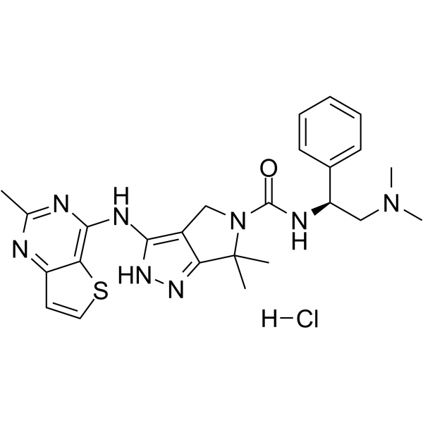
PF-3758309 (PF-03758309) hydrochloride is a potent, orally available, and reversible ATP-competitive inhibitor of PAK4 (Kd= 2.7 nM; Ki=18.7 nM). PF-3758309 hydrochloride has the expected cellular functions of a PAK4 inhibitor: inhibition of anchorage-independent growth, induction of apoptosis, cytoskeletal remodeling, and inhibition of proliferation.
For research use only. We do not sell to patients.
- CAS No.: 1279034-84-2
- Formula: C25H31ClN8OS
- Molecular Weight:527.08
-
Storage:
Please store the product under the recommended conditions in the Certificate of Analysis.

To place orders, for customer services and technical support, please contact: MedChemExpress USA
Tel: 609-228-6898 E-mail: [email protected] [email protected]


-
 Biological Activity
-
Chemical Information
- Protocols
- Purity & Documentation
- References
-
 Help & FAQs


Publications Citing Use of MedChemExpress (MCE) PF-3758309 hydrochloride
More- Science. 2017 Dec 1;358(6367):eaan4368. [Abstract]
- Nat Commun. 2026 Feb 12;17(1):1214. [Abstract]
- Sci Transl Med. 2018 Jul 18;10(450):eaaq1093. [Abstract]
- Br J Pharmacol. 2026 Jul 24.
- Int J Mol Sci. 2024 Sep 21;25(18):10138. [Abstract]
- Biochem J. 2022 Oct 14;479(19):2131-2151. [Abstract]
- Exp Cell Res. 2020 Oct 15;395(2):112187. [Abstract]
- Anticancer Drugs. 2024 Jan 1;35(1):46-54. [Abstract]
- bioRxiv. 2026 May 29.
- bioRxiv. 2025 April 26.
- bioRxiv. 2024 Oct 22:2024.10.22.619411. [Abstract]
- Necmettin Erbakan University. 2022 Jun.
 Customer Validation & Images

-
Cell Proliferation/Viability Assay

-
Others

-
Cell Imaging/Staining


Biological Activity
Description
IC50 & Target
[1]|
PAK4 18.7 nM (Ki) |
PAK1 13.7 nM (Ki) |
PAK5 18.1 nM (Ki) |
PAK6 17.1 nM (Ki) |
PAK2 190 nM (IC50) |
PAK3 99 nM (IC50) |
PAK4 2.7 nM (Kd) |
In Vitro
PF-3758309 hydrochloride has similar enzymatic potency against the kinase domains of the other group B PAKs (PAK5, Ki=18.1 nM; PAK6, Ki=17.1 nM) and group A PAK1 (Ki=13.7 nM), but is less active against the other two group A PAKs (PAK2, IC50=190 nM; PAK3, IC50=99 nM)[1].
In cells, PF-3758309 hydrochloride inhibits phosphorylation of the PAK4 substrate GEF-H1 (IC50=1.3 nM) and anchorage-independent growth of a panel of tumor cell lines (IC50=4.7 nM)[1].
PF-3758309 hydrochloride also inhibits endogenous pGEF-H1 accumulation in HCT116 cells. PF-3758309 potently inhibits cellular proliferation (IC50=20 nM) and anchorage-independent growth (IC50=27 nM) of A549 cells[1].
MedChemExpress (MCE) has not independently confirmed the accuracy of these methods. They are for reference only. Further protocols information, click here.
In Vivo
MedChemExpress (MCE) has not independently confirmed the accuracy of these methods. They are for reference only.
-
Animal Model:Female nu/nu, CRL breed 6–8 weeks old mice (bearing HCT116 and A549 tumors)[1]
-
Dosage:7.5-30 mg/kg
-
Administration:Oral administration; twice daily for 9-18 days
-
Result:Significant tumor growth inhibition (TGI) in HCT116 and A549 models.


Chemical Information
-
CAS No. 1279034-84-2
-
Molecular Weight 527.08
-
Formula C25H31ClN8OS
-
SMILES
O=C(N1C(C)(C)C2=NNC(NC3=C4C(C=CS4)=NC(C)=N3)=C2C1)N[C@@H](C5=CC=CC=C5)CN(C)C.[H]Cl
-
Synonyms
PF-03758309 hydrochloride
-
Shipping
Room temperature in continental US; may vary elsewhere.
-
Storage
Please store the product under the recommended conditions in the Certificate of Analysis.


Publications (12)
-
Journal Impact Factor
-
Most Recent
-
Science
2017 Dec 1;358(6367):eaan4368. PMID: 29191878 -
Nat Commun
Human iPSC-based Modeling of Pulmonary Fibrosis Reveals p300/CBP Inhibition Suppresses Alveolar Transitional Cell State. [Abstract]2026 Feb 12;17(1):1214. PMID: 41680175 -
Sci Transl Med
PP2A inhibition is a druggable MEK inhibitor resistance mechanism in KRAS-mutant lung cancer cells. [Abstract]2018 Jul 18;10(450):eaaq1093. PMID: 30021885 -
-
Int J Mol Sci
2024 Sep 21;25(18):10138. PMID: 39337621
PF-3758309 hydrochloride purchased from MedChemExpress. Usage Cited in: Int J Mol Sci. 2024 Sep 21;25(18):10138. [Abstract]
NAMPT activity in response to PAK4 inhibitors (KPT-9274, PF-3758309, GNE2861, and LCH7749944), group I (PAK1-3) inhibitors (IPA-3 and FRAX486), phloretin (a natural compound unrelated to kinase inhibitors), MEK inhibitors (BI-847325 and PD0325901), the solvent control (DMSO), and FX866, the positive control.
-
Biochem J
Up-regulation of the PI3K/AKT and RHO/RAC/PAK signalling pathways in CHK1 inhibitor resistant Eµ-Myc lymphoma cells. [Abstract]2022 Oct 14;479(19):2131-2151. PMID: 36240067 -
Exp Cell Res
PAK5 promotes the cell stemness ability by phosphorylating SOX2 in lung squamous cell carcinomas. [Abstract]2020 Oct 15;395(2):112187. PMID: 32721391 -
Anticancer Drugs
2024 Jan 1;35(1):46-54. PMID: 37449977 -
-
PF-3758309 hydrochloride purchased from MedChemExpress. Usage Cited in: bioRxiv. 2025 April 26.
Viability of CUTO32 and LC-2/Ad cells upon co-treatment with pralsetinib and the PAK inhibitor PF-3758309 (CUTO32: 1 μM; LC2/Ad: 0.1 μM) for 3 days .
-
bioRxiv
Network analysis of α-synuclein pathology progression reveals p21-activated kinases as regulators of vulnerability. [Abstract]2024 Oct 22:2024.10.22.619411. PMID: 39484617
PF-3758309 hydrochloride purchased from MedChemExpress. Usage Cited in: bioRxiv. 2024 Oct 22:2024.10.22.619411. [Abstract]
Representative images of pS129 α-synuclein in vehicle treated neurons compared to neurons treated with the highest dose of PF-3758309 or GNE2861.
-

Protocols
-
Apoptosis
Apoptosis, also called programmed cell death, is generally characterized by distinct morphological characteristics.
-
TUNEL staining for apoptotic DNA fragmentation
TUNEL staining detects DNA strand breaks by using terminal deoxynucleotidyl transferase to add labeled nucleotides to exposed 3′-OH DNA termini, generating either microscopic staining in fixed cells or tissue sections, or fluorescence/cytometric signal in cell suspensions. TUNEL positivity reflects DNA fragmentation but should not be interpreted alone as definitive apoptosis, because TUNEL can also label necrotic, autolytic, mechanically damaged, or DNA-repair-associated DNA breaks.
-
Annexin V plus membrane-impermeant dye apoptosis staining
Annexin V-based apoptosis assays rely on the detection of phosphatidylserine (PS) externalization from the inner leaflet of the plasma membrane to the outer leaflet, an early biochemical hallmark of apoptosis. Fluorescently labeled Annexin V binds PS in a calcium-dependent manner, enabling identification of early apoptotic cells by flow cytometry or fluorescence microscopy. When combined with a membrane-impermeant DNA-binding dye (e. g. , propidium iodide), this approach allows discrimination between viable (Annexin V−/dye−), early apoptotic (Annexin V+/dye−), and late apoptotic or necrotic (Annexin V+/dye+) cell populations by assessing membrane integrity and PS exposure.
-
Apoptosis Solutions
Apoptosis is a regulated, generally non-lytic cell-death pathway that removes unwanted, damaged, infected, or abnormal cells through coordinated morphological changes, caspase activation, DNA fragmentation, and membrane remodeling. The intrinsic apoptosis pathway is controlled mainly by mitochondrial outer membrane permeabilization, BCL-2 family proteins, cytochrome c release, apoptosome formation, caspase-9 activation, and downstream executioner caspase-3/7 activation. The extrinsic apoptosis pathway is initiated by death receptors such as Fas, TNFR, and TRAIL receptors, which recruit adaptor proteins and activate caspase-8 before engaging executioner caspases or mitochondrial amplification through BID cleavage. Apoptosis is linked to many phenotypes, including cancer cell killing, tissue homeostasis, immune regulation, neurodegeneration, infection response, and treatment-induced cytotoxicity; unresolved questions include how apoptosis interacts with necroptosis, pyroptosis, ferroptos
-
Soft Agar Colony Formation Assay
Soft agar colony formation assay measures anchorage-independent growth, in which transformed or tumorigenic cells proliferate as colonies in a semisolid agar matrix while many non-transformed adherent cells fail to proliferate without attachment; classic studies showed that growth in semisolid medium correlates with tumorigenicity in nude mice, and later protocol papers describe the method as a stringent in vitro assay for malignant transformation. The readout is the number, size, morphology, or signal intensity of colonies formed within agar after incubation; published formats include manual colony counting after staining, 96-well or 384-well quantitative formats, DNA-binding dye detection, MTT/tetrazolium-based detection, digital image analysis, and PCR-based marker detection from soft agar cultures.
-
Phalloidin F-actin cytoskeleton staining
Phalloidin F-actin staining detects polymerized filamentous actin in fixed and permeabilized specimens by using fluorescent phalloidin or phalloidin-derived phallotoxins that bind actin filaments and generate a fluorescence microscopy readout corresponding to F-actin organization, including stress fibers, cortical actin, filament bundles, and tissue-specific actin networks. Phalloidin stabilizes F-actin by reducing actin subunit dissociation from filament ends, and fluorescent phallotoxins were established as tools for visualizing actin-containing structures in eukaryotic cells.
-
Cell Viability Determination by MTT Colorimetric Assay
The following protocol uses the MTT colorimetric assay as a classic literature-established method for assessing cell viability/metabolic activity in cultured mammalian cells. MTT[3-(4,5-dimethylthiazol-2-yl)-2,5-diphenyltetrazolium bromide] is reduced by metabolically active cells to a colored formazan product; the amount of formazan is quantified spectrophotometrically and provides an indirect measure of metabolically active viable cells. Importantly, MTT reduction reflects cellular oxidoreductase/metabolic activity rather than an absolute direct count of living cells, so changes in cellular metabolism can alter the signal independently of cell number.


Purity & Documentation
References
[1]. Murray, Brion W., et al. Small-molecule p21-activated kinase inhibitor PF3758309 is a potent inhibitor of oncogenic signaling and tumor growth. Proceedings of the National Academy of Sciences of the United States of America (2010), 107(20), 9446-9451, S94. [Content Brief]
[2]. Zhao ZS, et al. Do PAKs make good drug targets? F1000 Biol Rep. 2010 Sep 23;2:70. [Content Brief]
[3]. Ryu BJ, et al. PF-3758309, p21-activated kinase 4 inhibitor, suppresses migration and invasion of A549 human lung cancer cells via regulation of CREB, NF-κB, and β-catenin signalings. Mol Cell Biochem. 2014 Apr;389(1-2):69-77. [Content Brief]

Calculators
Concentration (start) × Volume (start) = Concentration (final) × Volume (final)


PF-3758309 hydrochloride Related Classifications
HY-13007A Related Classifications
 Powered by Bioz