



(Rac)-Hesperetin

Based on 1 Customer Validation
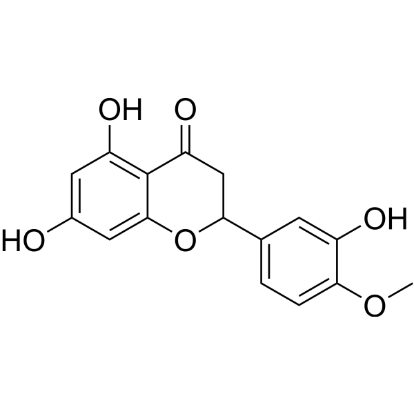
(Rac)-Hesperetin is the racemate of Hesperetin (HY-N0168), an orally active multi-target inhibitor. (Rac)-Hesperetin exhibits significant anti-tumor and anti-inflammatory activities by blocking the TGF-β1-mediated Fyn/RhoA signaling axis and the TLR4-MyD88-NF-κB inflammatory pathway. (Rac)-Hesperetin inhibits the formation of actin stress fibers and the migration and invasion of cancer cells, and is suitable for triple-negative breast cancer research. In inflammation models, (Rac)-Hesperetin effectively alleviates lung injury by reducing the release of pro-inflammatory mediators and regulating the activity of oxidative stress enzymes, and is suitable for acute lung injury research. (Rac)-Hesperetin also interferes with the entry and early replication processes of channel catfish virus, inhibits viral gene expression and progeny virus production, thereby protecting cells from virus-induced cytopathic effects.
For research use only. We do not sell to patients.
- Purity : 99.33%
- CAS No.: 69097-99-0
- Formula: C16H14O6
- Molecular Weight:302.28
-
Storage:Powder -20°C, 3 years , 4°C, 2 years ; In solvent -80°C, 6 months , -20°C, 1 month

To place orders, for customer services and technical support, please contact: MedChemExpress USA
Tel: 609-228-6898 E-mail: [email protected] [email protected]


-
 Biological Activity
-
Chemical Information
-
Solvent & Solubility
- Protocols
- Purity & Documentation
- References
-
 Help & FAQs


-
HY-W011641
(±)-NaringeninAnti-inflammatory Agents -
HY-K2014
PEI Transfection Reagent -
HY-10256
Adezmapimodp38 MAPK Inhibitor -

HY-N0168AR
(Rac)-Hesperetin (Standard)Racemate of Hesperetin -
HY-10578
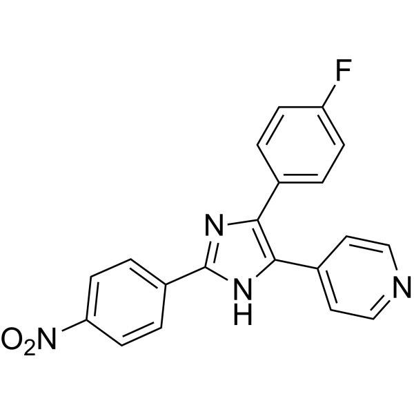
PD 169316p38 MAP Kinase Inhibitor -
HY-N7128
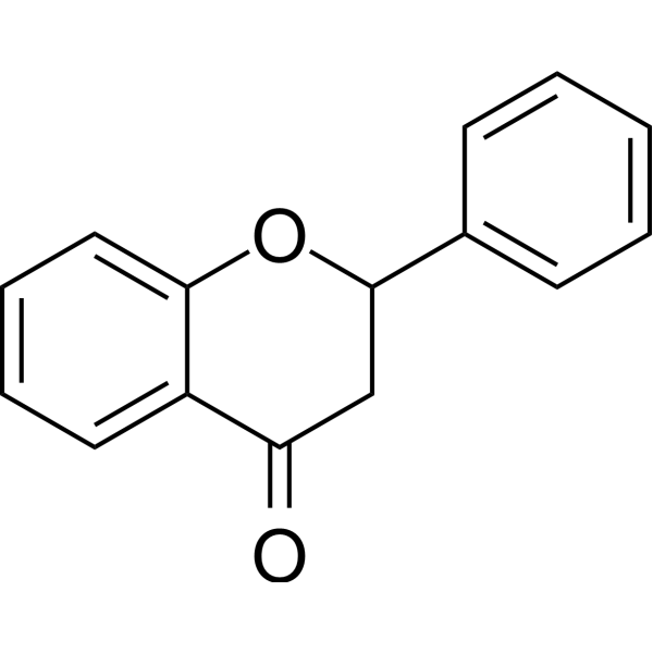
FlavanoneFlavones -

HY-10256R
Adezmapimod (Standard)p38 MAPK Inhibitor -
HY-N0674A
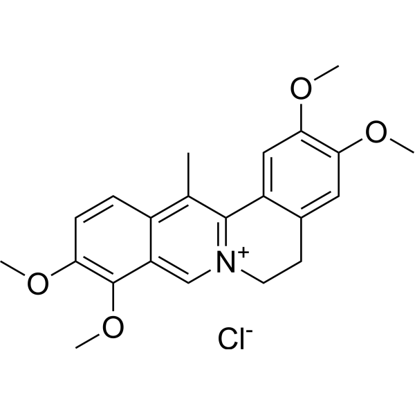
Dehydrocorydaline chloridep38 MAPK Activator -

HY-10578R
PD 169316 (Standard)Standard of PD 169316 -
HY-W011641R
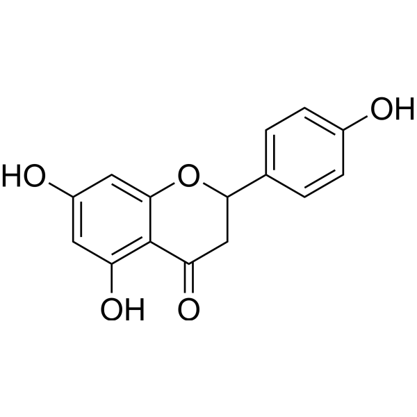
(±)-Naringenin (Standard) -
HY-14305A
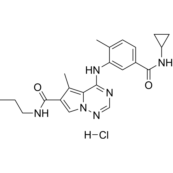
BMS-582949 hydrochloridep38α MAPK Inhibitor -

HY-N7128R
Flavanone (Standard)Flavones


Biological Activity
Description
In Vitro
Hesperetin (25, 50, 100 μM; 0, 24, 48, 72 h) significantly inhibits the migratory capacity of TGF-β1-induced human triple-negative breast cancer MDA-MB-231 cells in a dose-dependent manner[1].
Hesperetin (500 μg/mL; 36 h) significantly inhibits the expression of ORF39 and ORF59 proteins of CCV, completely alleviates the cytopathic effect on CCO cells induced by CCV infection, and exerts a cytoprotective effect[2].
MedChemExpress (MCE) has not independently confirmed the accuracy of these methods. They are for reference only. Further protocols information, click here.
-
Cell Line:Human triple-negative breast cancer MDA-MB-231 cells
-
Concentration:25, 50, 100 μM
-
Incubation Time:24, 48, 72 h
-
Result:Significantly inhibited TGF-β1-induced migration of MDA-MB-231 cells in a dose-dependent manner at 24 h, 48 h and 72 h after treatment.
In Vivo
MedChemExpress (MCE) has not independently confirmed the accuracy of these methods. They are for reference only.
-
Animal Model:C57 male mice (8 weeks old, 18-22 g) with LPS-induced acute lung injury[3]
-
Dosage:Hesperetin: 10 mg/kg, 20 mg/kg , 30 mg/kg ;
Positive control: 2 mg/kg Dexamethasone (oral gavage) -
Administration:oral gavage
-
Result:At 1 h after LPS intratracheal instillation, significantly suppressed the levels of interleukin-6 and tumor necrosis factor-α in bronchoalveolar lavage fluid, reduced the number of inflammatory cells in bronchoalveolar lavage fluid, ameliorated LPS-induced histopathologic changes of lung tissue and decreased lung inflammatory scores.
Reduced the lung wet/dry weight ratio, lowered the activities of myeloperoxidase and lactate dehydrogenase in lung tissue, enhanced superoxide dismutase activity, and downregulated the protein expression of Toll-like receptor 4 and myeloid differentiation factor 88 in lung tissue.
Suppressed the phosphorylation of nuclear factor-κB p65, IκB-α and TAK1, and inhibited the activation of nuclear factor-κB pathway in lung tissue, with the 30 mg/kg dosage showing the most significant therapeutic effect.


Chemical Information
-
CAS No. 69097-99-0
-
Appearance Solid
-
Molecular Weight 302.28
-
Formula C16H14O6
-
Color White to yellow
-
SMILES
O=C1CC(C2=CC=C(OC)C(O)=C2)OC3=CC(O)=CC(O)=C13
-
Structure Classification
-
Initial Source
-
Shipping
Room temperature in continental US; may vary elsewhere.
-
Storage
Powder -20°C 3 years 4°C 2 years In solvent -80°C 6 months -20°C 1 month


Solvent & Solubility
In Vitro:
DMSO : 116.67 mg/mL (385.97 mM; Need ultrasonic; Hygroscopic DMSO has a significant impact on the solubility of product, please use newly opened DMSO)
Please refer to the solubility information to select the appropriate solvent. Once prepared, please aliquot and store the solution to prevent product inactivation from repeated freeze-thaw cycles.
Storage method and period of stock solution: -80°C, 6 months; -20°C, 1 month. When stored at -80°C, please use it within 6 months. When stored at -20°C, please use it within 1 month.
Please refer to the solubility information to select the appropriate solvent. Once prepared, please aliquot and store the solution to prevent product inactivation from repeated freeze-thaw cycles.
Storage method and period of stock solution: -80°C, 6 months; -20°C, 1 month. When stored at -80°C, please use it within 6 months. When stored at -20°C, please use it within 1 month.
Concentration (start) × Volume (start) = Concentration (final) × Volume (final)
In Vivo:
Select the appropriate dissolution method based on your experimental animal and administration route.
- For the following dissolution methods, please ensure to first prepare a clear stock solution using an In Vitro approach and then sequentially add co-solvents:
- To ensure reliable experimental results, the clarified stock solution can be appropriately stored based on storage conditions. As for the working solution for In Vivo experiments, it is recommended to prepare freshly and use it on the same day.
- The percentages shown for the solvents indicate their volumetric ratio in the final prepared solution. If precipitation or phase separation occurs during preparation, heat and/or sonication can be used to aid dissolution.
Add each solvent one by one: 10% DMSO 90% (20% SBE-β-CD in Saline)
Solubility: ≥ 2.08 mg/mL (6.88 mM); Clear solution
This protocol yields a clear solution of ≥ 2.08 mg/mL (saturation unknown).
Taking 1 mL working solution as an example, add 100 μL DMSO stock solution (20.8 mg/mL) to 900 μL 20% SBE-β-CD in Saline, and mix evenly.
Preparation of 20% SBE-β-CD in Saline (4°C, storage for one week): 2 g SBE-β-CD powder is dissolved in 10 mL Saline, completely dissolve until clear.
In Vivo Dissolution Calculator
Please enter the basic information of animal experiments:
-
-
-
-
Recommended: Prepare an additional quantity of animals to account for potential losses during experiments.
Please enter your animal formula composition:
-
%DMSO +
Recommended: Keep the proportion of DMSO in working solution below 2% if your animal is weak.
-
%+
-
+%Tween-80 + +
-
%Saline +
The co-solvents required include: DMSO, . All of co-solvents are available by MedChemExpress (MCE). , Tween 80. All of co-solvents are available by MedChemExpress (MCE).
Working solution concentration: 0.22 mg/mL
Method for preparing stock solution: mg drug dissolved in μL DMSO. Stock solution concentration: mg/mL.
1. Take μL DMSO stock solution;
2. Add μL .
μL , mix evenly;
3. Then add μL Tween 80, mix evenly;
4. Then add μL
Please ensure that the stock solution in the first step is dissolved to a clear state, and add co-solvents in sequence. You can use ultrasonic heating (ultrasonic cleaner, recommended frequency 20-40 kHz), vortexing, etc. to assist dissolution.

Protocols
-
RT-PCR
Reverse transcription technology uses RNA as a template to synthesize DNA. RT-PCR is simple, specific and sensitive, and can be used to detect gene expression levels and expression differences in cells; detect RNA virus content; clone cDNA sequences of specific genes.
-
RNA extraction experimental
By lysing cells, releasing RNA, and removing impurities such as proteins and DNA, high-purity RNA products are finally obtained. The commonly used traditional method is the guanidine isothiocyanate/phenol/chloroform method (Trizol), which is suitable for a variety of animal materials including animal tissues, microorganisms, cultured cells, etc., and most plant materials.
-
Cell migration
Cell migration is a method that plays an important role in wound healing, cell differentiation, embryonic development, etc.
-
ROS/oxidative-stress fluorescent staining
ROS/oxidative-stress fluorescent staining uses cell-permeant fluorogenic probes that become fluorescent after oxidation inside cells or tissues; commonly used examples include DCFH-DA/DCFDA for broad cellular oxidant detection, DHE for superoxide-related signal detection, MitoSOX for mitochondrial superoxide-related signal detection, and CellROX probes for oxidative-stress-associated fluorescence readouts. The assay detects probe oxidation rather than a single ROS species unless the probe and analysis method have been chemically validated for that species. DCFH-DA enters cells, is deacetylated by intracellular esterases to DCFH, and produces fluorescent DCF after oxidation, so the readout is used as an operational measure of total cellular oxidative stress rather than a species-specific ROS measurement. DHE and MitoSOX can report superoxide-related oxidation, but red fluorescence alone can include non-specific ethidium-like oxidation products; HPLC or optimized spectral approaches are
-
Cell invasion
Cell invasion is the ability of cells to migrate from one area to another via the extracellular matrix. Cell invasion is the response of normal and cancer cells to chemical and mechanical stimuli. Before migrating to a new region, the extracellular matrix is degraded by proteases within the cell. Cell invasion often occurs during wound repair, vascularization and inflammation, abnormal tissue invasion, and tumor cell metastasis.
-
Research Protocol for Inflammation-related Diseases
The NLRP3 inflammasome is a cytosolic innate immune signaling platform that integrates priming signals and danger-signal activation to promote caspase-1 activation, maturation of IL-1β and IL-18, and gasdermin D-mediated pyroptotic cell death. The core experimental logic is to determine whether inflammatory disease phenotypes are driven by increased NLRP3 expression, ASC-containing inflammasome assembly, caspase-1 cleavage, GSDMD cleavage, and extracellular release of IL-1β/IL-18 rather than by nonspecific cell injury alone. The pathway is strongly linked to inflammation-related disease phenotypes because monosodium urate crystals activate NALP3/NLRP3 inflammasome signaling in gout-like crystal inflammation, cholesterol crystals activate NLRP3 inflammasomes in atherogenesis models, and DSS-induced intestinal inflammation has been reported to involve NLRP3 inflammasome activity. However, experimental colitis studies also show context-dependent protective effects of NLRP3 inflammasome co
-
Inhalation Toxicity Study
Inhalation toxicity studies expose rodents to a controlled aerosol, vapor, gas, or smoke atmosphere and assess respiratory and systemic toxicity using exposure-atmosphere characterization, clinical observations, body and organ weights, bronchoalveolar lavage fluid, histopathology, blood chemistry, hematology, and, when included, molecular endpoints such as transcriptomics, proteomics, lipidomics, or tissue burden analysis. The primary biological readouts are airway irritation, pulmonary inflammation, cytotoxicity, altered surfactant or lipid homeostasis, impaired particle clearance, and tissue remodeling, reflected by BALF cell differentials, BALF protein, LDH, phosphatase activities, cytokines, lung weight, microscopic respiratory-tract lesions, and retained lung burden.
-
Breast Cancer Modeling
Breast cancer is a heterogeneous cancer, and it has been distinguished into four subtypes: luminal A, luminal B, HER2-positive and basal-like. Molecular mutations, epigenetic alterations, hormone exposure and immune microenvironment are related to the progression of breast cancer.
-
Real Time qPCR (Q-PCR)
Real-time quantitative PCR (qPCR) quantifies an amplifiable nucleic-acid target by monitoring fluorescence during PCR cycling rather than measuring product only after amplification. The increase in fluorescence tracks accumulation of PCR product, and the quantification cycle (Cq; historically also Ct/CP) is related to the initial amount of target: samples containing more starting target generally reach the defined fluorescence threshold in fewer cycles.


Purity & Documentation
-
Data Sheet (279 KB)
-
SDS (393 KB)
- English - EN (393 KB)
- Français - FR (393 KB)
- Deutsch - DE (393 KB)
- Norwegian - NO (393 KB)
- Español - ES (393 KB)
- Swedish - SV (393 KB)
- Italian - IT (393 KB)
- Korean - KR (393 KB)
- Portuguese - PT (393 KB)
-
Handling Instructions (2659 KB)
References
[1]. Lu Q, et al. Hesperetin Inhibits TGF-β1-Induced Migration and Invasion of Triple Negative Breast Cancer MDA-MB-231 Cells via Suppressing Fyn/Paxillin/RhoA Pathway. Integr Cancer Ther. 2022;21:15347354221086900. [Content Brief]
[3]. Wang N, et al. Hesperetin ameliorates lipopolysaccharide-induced acute lung injury in mice through regulating the TLR4-MyD88-NF-κB signaling pathway. Arch Pharm Res. 2019;42(12):1063-1070. [Content Brief]


Complete Stock Solution Preparation Table
Please refer to the solubility information to select the appropriate solvent. Once prepared, please aliquot and store the solution to prevent product inactivation from repeated freeze-thaw cycles.
Storage method and period of stock solution: -80°C, 6 months; -20°C, 1 month. When stored at -80°C, please use it within 6 months. When stored at -20°C, please use it within 1 month.
| Optional Solvent | Concentration Solvent Mass | 1 mg | 5 mg | 10 mg | 25 mg |
|---|---|---|---|---|---|
| DMSO | 1 mM | 3.3082 mL | 16.5410 mL | 33.0819 mL | 82.7048 mL |
| 5 mM | 0.6616 mL | 3.3082 mL | 6.6164 mL | 16.5410 mL | |
| 10 mM | 0.3308 mL | 1.6541 mL | 3.3082 mL | 8.2705 mL | |
| 15 mM | 0.2205 mL | 1.1027 mL | 2.2055 mL | 5.5137 mL | |
| 20 mM | 0.1654 mL | 0.8270 mL | 1.6541 mL | 4.1352 mL | |
| 25 mM | 0.1323 mL | 0.6616 mL | 1.3233 mL | 3.3082 mL | |
| 30 mM | 0.1103 mL | 0.5514 mL | 1.1027 mL | 2.7568 mL | |
| 40 mM | 0.0827 mL | 0.4135 mL | 0.8270 mL | 2.0676 mL | |
| 50 mM | 0.0662 mL | 0.3308 mL | 0.6616 mL | 1.6541 mL | |
| 60 mM | 0.0551 mL | 0.2757 mL | 0.5514 mL | 1.3784 mL | |
| 80 mM | 0.0414 mL | 0.2068 mL | 0.4135 mL | 1.0338 mL | |
| 100 mM | 0.0331 mL | 0.1654 mL | 0.3308 mL | 0.8270 mL |


(Rac)-Hesperetin Related Classifications
HY-N0168A Related Classifications
 Powered by Bioz