



Ro 90-7501
Based on 3 publication(s) in Google Scholar
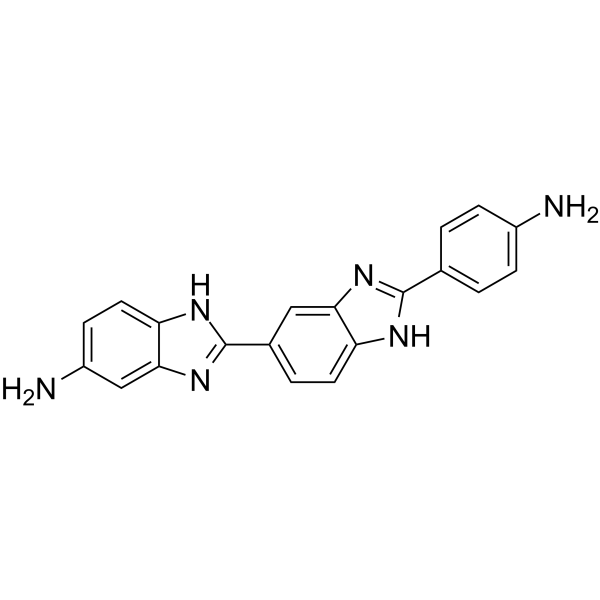
Ro 90-7501 is an amyloid β42 (Aβ42) fibril assembly inhibitor that reduces Aβ42-induced cytotoxicity (EC50 of 2 μM). Ro 90-7501 inhibits ATM phosphorylation and DNA repair. RO 90-7501 selectively enhances toll-like receptor 3 (TLR3) and RIG-I-like receptor (RLR) ligand-induced IFN-β gene expression and antiviral response. Ro 90-7501 also inhibits protein phosphatase 5 (PP5) in a TPR-dependent manner. Ro 90-7501 has significant radiosensitizing effects on cervical cancer cells.
For research use only. We do not sell to patients.
- Purity : 98.50%
- CAS No.: 293762-45-5
- Formula: C20H16N6
- Molecular Weight:340.38
-
Storage:Powder -20°C, 3 years , 4°C, 2 years ; In solvent -80°C, 2 years , -20°C, 1 year

To place orders, for customer services and technical support, please contact: MedChemExpress USA
Tel: 609-228-6898 E-mail: [email protected] [email protected]


-
 Biological Activity
-
Chemical Information
-
Solvent & Solubility
- Protocols
- Purity & Documentation
- References
-
 Help & FAQs


-
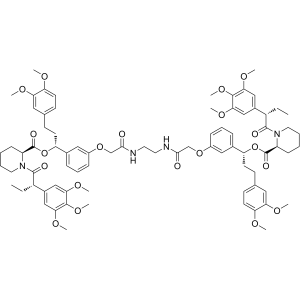
HY-16046
RimiducidDimerization Inducer -
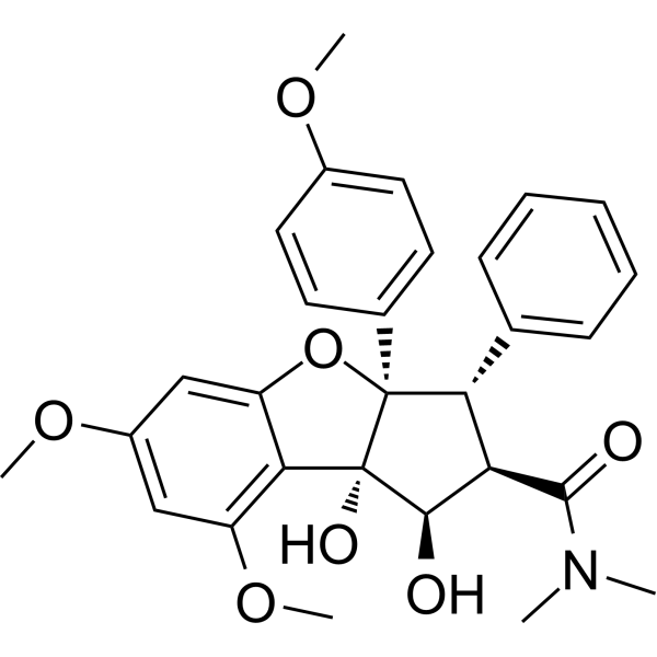
HY-19356
RocaglamideNF-κB Inhibitor -
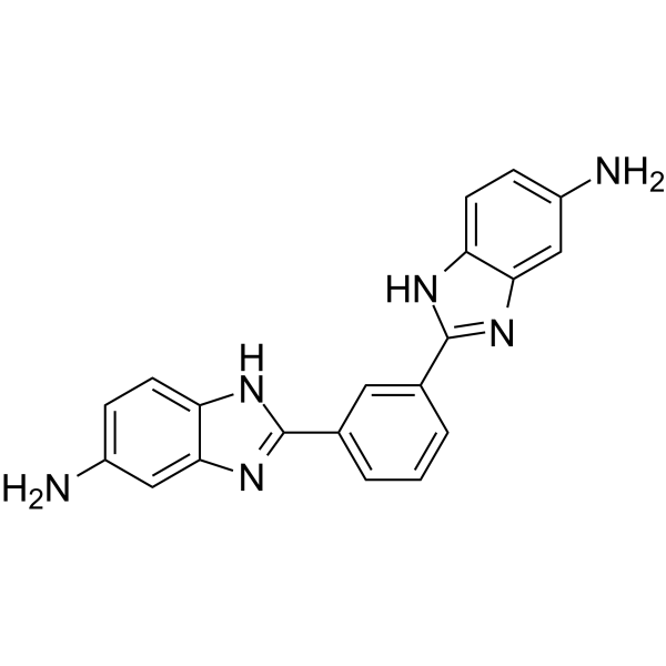
HY-138647
TGP-377/421MiR-377/421 Inhibitor -
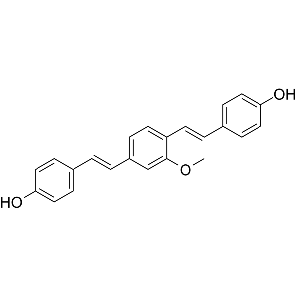
HY-103240
Methoxy-X04Fluorescent Dye -
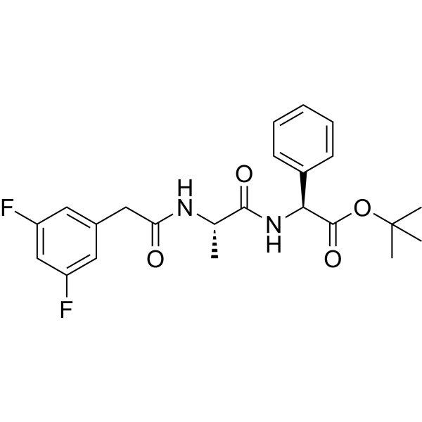
HY-13027
DAPTγ-secretase Inhibitor -
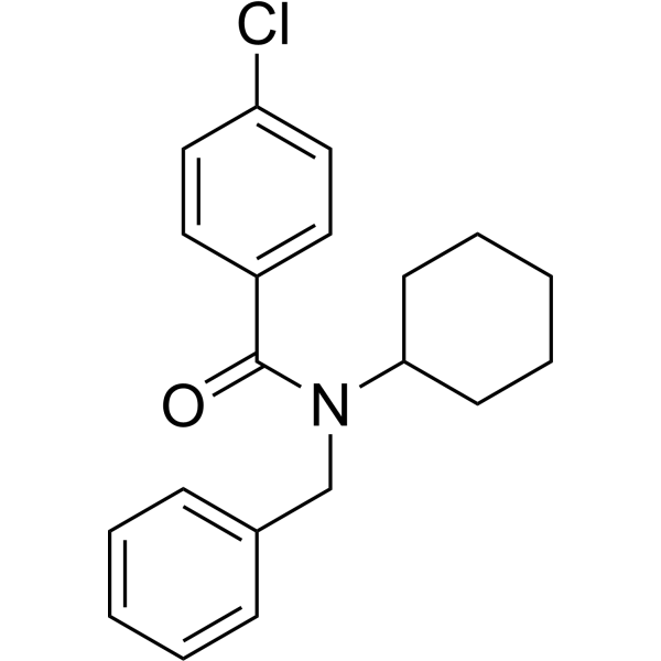
HY-19370
FPS-ZM1Amyloid-β Inhibitor -

HY-P1061
ColivelinNeuroprotectant/STAT3 Activator -

HY-P1363A
β-Amyloid (1-42), humanAmyloid β Peptide -

HY-P1363B
β-Amyloid (1-42), human, HFIP-treatedβ-Amyloid (1-42) Peptide -
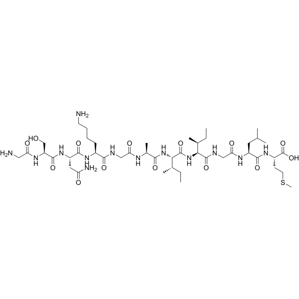
HY-P0128
β-Amyloid (25-35)Amyloid β Peptide -
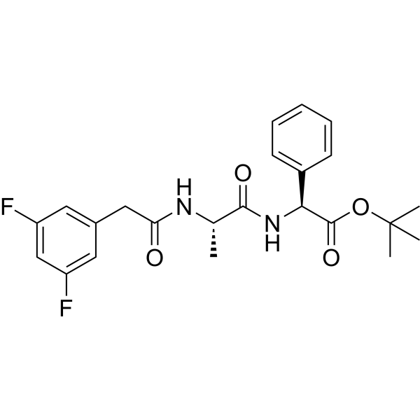
HY-13027G
DAPT (GMP)γ-Secretase Inhibitor -

HY-103241R
Ro 90-7501 (Standard)Reference Standard
Publications Citing Use of MedChemExpress (MCE) Ro 90-7501
More Customer Validation & Images

-
Cell Proliferation/Viability Assay

-
IF

-
Cell Migration/Invasion Assay

-
Apoptosis Analysis

-
WB


Biological Activity
Description
|
Amyloid β42 |
ATM |
Protein phosphatase 5 |
Apoptosis |
Cellular Effect
|
Cell Line

|
Type | Value | Description | References |
|---|---|---|---|---|
| Huh-7 | CC50 |
>100 μM
Compound: GNF-Pf-5510
|
NOVARTIS: Cytotoxicity against human hepatocellular carcinoma cell line (Huh7)
NOVARTIS: Cytotoxicity against human hepatocellular carcinoma cell line (Huh7)
|
[PMID: 18579783] |
In Vitro
Ro 90-7501 significantly enhances radiosensitivity compared with control HeLa and ME-180 cells. Ro 90-7501 significantly increases apoptosis and impaired cell cycle after irradiation. Ro 90-7501 suppresses the phosphorylation of ATM and its downstream proteins, such as H2AX, Chk1, and Chk2, after irradiation[1].
RO 90-7501, itself affects neither IFN-β nor NFκB promoter activity, but significantly enhances poly I:C-induced IFN-β promoter activation and inhibits the activation of NFκB in a dose-dependent manner. Treatment of cells with RO 90-7501 significantly enhances the antiviral activity of poly I:C[2].
MedChemExpress (MCE) has not independently confirmed the accuracy of these methods. They are for reference only. Further protocols information, click here.
In Vivo
MedChemExpress (MCE) has not independently confirmed the accuracy of these methods. They are for reference only.
-
Animal Model:Female BALB/c nude mice (8-week-old) with HeLa cells under irradiation[1]
-
Dosage:5 μg/g
-
Administration:Intraperitoneal injection; daily; for 21 days
-
Result:Tumor growth was significantly delayed in the combination group. Tumor volume was also significantly decreased in the irradiation group.


Chemical Information
-
CAS No. 293762-45-5
-
Appearance Solid
-
Molecular Weight 340.38
-
Formula C20H16N6
-
Color Brown to breen
-
SMILES
NC1=CC=C2C(N=C(C3=CC=C4C(N=C(C5=CC=C(N)C=C5)N4)=C3)N2)=C1
-
Shipping
Room temperature in continental US; may vary elsewhere.
-
Storage
Powder -20°C 3 years 4°C 2 years In solvent -80°C 2 years -20°C 1 year


Publications (3)
-
Journal Impact Factor
-
Most Recent
-
Int Immunopharmacol
Ro 90-7501 suppresses colon cancer progression by inducing immunogenic cell death and promoting RIG-1-mediated autophagy. [Abstract]2024 Nov 15;143(Pt 3):113631. PMID: 39549547
Ro 90-7501 purchased from MedChemExpress. Usage Cited in: Int Immunopharmacol. 2024 Nov 15;143(Pt 3):113631. [Abstract]
The effect of Ro 90-7501 (0, 10, 50, 100, 150, 200 μM) on the viability of HCT8 and SW480 cells.
Ro 90-7501 purchased from MedChemExpress. Usage Cited in: Int Immunopharmacol. 2024 Nov 15;143(Pt 3):113631. [Abstract]
The impact of Ro 90-7501 (0, 10, 100, 200 μM) on the proliferation of HCT8 and SW480 cells.
Ro 90-7501 purchased from MedChemExpress. Usage Cited in: Int Immunopharmacol. 2024 Nov 15;143(Pt 3):113631. [Abstract]
The influence of Ro 90-7501 (0, 10, 100, 200 μM) on the migration of HCT8 and SW480 cells.
Ro 90-7501 purchased from MedChemExpress. Usage Cited in: Int Immunopharmacol. 2024 Nov 15;143(Pt 3):113631. [Abstract]
Ro 90-7501(0, 10, 100, 200 μM) promotes apoptosis in HCT8 and SW480 cells.
Ro 90-7501 purchased from MedChemExpress. Usage Cited in: Int Immunopharmacol. 2024 Nov 15;143(Pt 3):113631. [Abstract]
The effect of Ro 90-7501 (0, 10, 100, 200 μM) on the expression of apoptosis-associated proteins.

Ro 90-7501 purchased from MedChemExpress. Usage Cited in: Int Immunopharmacol. 2024 Nov 15;143(Pt 3):113631. [Abstract]
IHC staining to detect the expression of Ki-67 in implanted tumors in mice treated with Ro 90-7501 (10 mg/kg, i.p.).

Ro 90-7501 purchased from MedChemExpress. Usage Cited in: Int Immunopharmacol. 2024 Nov 15;143(Pt 3):113631. [Abstract]
The effect of Ro 90-7501 (10 mg/kg, i.p.) on the volume and weight of implanted tumors in mice.

Ro 90-7501 purchased from MedChemExpress. Usage Cited in: Int Immunopharmacol. 2024 Nov 15;143(Pt 3):113631. [Abstract]
H&E staining to observe morphological changes in the heart, liver, spleen, lung, and kidney of mice treated with Ro 90-7501 (10 mg/kg, i.p.).
-
Int Immunopharmacol
Inhibitors of APE1 redox and ATM synergistically sensitize osteosarcoma cells to ionizing radiation by inducing ferroptosis. [Abstract]2024 Sep 30:139:112672. PMID: 39032469 -
Int J Biochem Cell Biol
Modified 5-aminolevulinic acid photodynamic therapy (M-PDT) inhibits cutaneous squamous cell carcinoma cell proliferation via targeting PP2A/PP5-mediated MAPK signaling pathway. [Abstract]2021 Aug:137:106036. PMID: 34217813


Solvent & Solubility
In Vitro:
DMSO : 41.67 mg/mL (122.42 mM; Need ultrasonic; Hygroscopic DMSO has a significant impact on the solubility of product, please use newly opened DMSO)
Please refer to the solubility information to select the appropriate solvent. Once prepared, please aliquot and store the solution to prevent product inactivation from repeated freeze-thaw cycles.
Storage method and period of stock solution: -80°C, 2 years; -20°C, 1 year. When stored at -80°C, please use it within 2 years. When stored at -20°C, please use it within 1 year.
Please refer to the solubility information to select the appropriate solvent. Once prepared, please aliquot and store the solution to prevent product inactivation from repeated freeze-thaw cycles.
Storage method and period of stock solution: -80°C, 2 years; -20°C, 1 year. When stored at -80°C, please use it within 2 years. When stored at -20°C, please use it within 1 year.
Concentration (start) × Volume (start) = Concentration (final) × Volume (final)
In Vivo:
Select the appropriate dissolution method based on your experimental animal and administration route.
- For the following dissolution methods, please ensure to first prepare a clear stock solution using an In Vitro approach and then sequentially add co-solvents:
- To ensure reliable experimental results, the clarified stock solution can be appropriately stored based on storage conditions. As for the working solution for In Vivo experiments, it is recommended to prepare freshly and use it on the same day.
- The percentages shown for the solvents indicate their volumetric ratio in the final prepared solution. If precipitation or phase separation occurs during preparation, heat and/or sonication can be used to aid dissolution.
Add each solvent one by one: 10% DMSO 40% PEG300 5% Tween-80 45% Saline
Solubility: 2.08 mg/mL (6.11 mM); Suspended solution; Need ultrasonic
This protocol yields a suspended solution of 2.08 mg/mL. Suspended solution can be used for oral and intraperitoneal injection.
Taking 1 mL working solution as an example, add 100 μL DMSO stock solution (20.8 mg/mL) to 400 μL PEG300, and mix evenly; then add 50 μL Tween-80 and mix evenly; then add 450 μL Saline to adjust the volume to 1 mL.
Preparation of Saline: Dissolve 0.9 g sodium chloride in ddH₂O and dilute to 100 mL to obtain a clear Saline solution.
Add each solvent one by one: 10% DMSO 90% (20% SBE-β-CD in Saline)
Solubility: ≥ 2.08 mg/mL (6.11 mM); Clear solution
This protocol yields a clear solution of ≥ 2.08 mg/mL (saturation unknown).
Taking 1 mL working solution as an example, add 100 μL DMSO stock solution (20.8 mg/mL) to 900 μL 20% SBE-β-CD in Saline, and mix evenly.
Preparation of 20% SBE-β-CD in Saline (4°C, storage for one week): 2 g SBE-β-CD powder is dissolved in 10 mL Saline, completely dissolve until clear.
In Vivo Dissolution Calculator
Please enter the basic information of animal experiments:
-
-
-
-
Recommended: Prepare an additional quantity of animals to account for potential losses during experiments.
Please enter your animal formula composition:
-
%DMSO +
Recommended: Keep the proportion of DMSO in working solution below 2% if your animal is weak.
-
%+
-
+%Tween-80 + +
-
%Saline +
The co-solvents required include: DMSO, . All of co-solvents are available by MedChemExpress (MCE). , Tween 80. All of co-solvents are available by MedChemExpress (MCE).
Working solution concentration: 0.22 mg/mL
Method for preparing stock solution: mg drug dissolved in μL DMSO. Stock solution concentration: mg/mL.
1. Take μL DMSO stock solution;
2. Add μL .
μL , mix evenly;
3. Then add μL Tween 80, mix evenly;
4. Then add μL
Please ensure that the stock solution in the first step is dissolved to a clear state, and add co-solvents in sequence. You can use ultrasonic heating (ultrasonic cleaner, recommended frequency 20-40 kHz), vortexing, etc. to assist dissolution.

Protocols
-
Kinase activity and phosphorylation assays
Kinase activity assays measure the ability of kinases to transfer phosphate groups from ATP to specific substrates, while phosphorylation assays detect the presence and levels of phosphorylated proteins. Common methods include radiolabeled ATP incorporation (e. g. ,), ADP release detection via bioluminescence (e. g. ,[3]), enzyme-linked immunosorbent assays (ELISA) for phospho-specific epitopes (e. g. ,[6]), and microtiter-based formats for high-throughput screening (e. g. ,[8]). The ADP-Glo assay quantifies kinase activity by measuring ADP produced during phosphorylation using a luciferase-based system. Radiometric assays involve autoradiography or scintillation counting after incorporation of 32P-labeled ATP into substrate proteins. ELISA-based approaches rely on phospho-specific antibodies to detect activated kinases in cell lysates or purified samples.
-
RT-PCR
Reverse transcription technology uses RNA as a template to synthesize DNA. RT-PCR is simple, specific and sensitive, and can be used to detect gene expression levels and expression differences in cells; detect RNA virus content; clone cDNA sequences of specific genes.
-
Western Blot
Western blotting (WB) is a commonly used experimental method in molecular biology, biochemistry, and immunogenetics for identifying and quantifying target proteins. It combines gel electrophoresis with immunoassay, enabling researchers to analyze protein expression, post-translational modifications, and molecular weight.
-
RNA extraction experimental
By lysing cells, releasing RNA, and removing impurities such as proteins and DNA, high-purity RNA products are finally obtained. The commonly used traditional method is the guanidine isothiocyanate/phenol/chloroform method (Trizol), which is suitable for a variety of animal materials including animal tissues, microorganisms, cultured cells, etc., and most plant materials.
-
Research Protocol for Infectious Diseases
Infectious-disease experiments test how pathogens interact with host barriers, innate immune receptors, inflammatory signaling, pathogen replication, and tissue injury; pattern-recognition receptors such as TLRs, RIG-I-like receptors, NOD-like receptors, and inflammasomes detect microbial molecules and activate NF-κB, interferon, and cytokine responses. The central hypothesis is that infection severity reflects the balance between pathogen burden and host response: protective inflammation restricts pathogen growth, whereas excessive or mislocalized inflammation contributes to tissue damage and disease phenotype. Unresolved questions include which host pathways are protective versus pathogenic, why some infection models fail to translate to human disease, and which combined readouts best predict clinically relevant infection outcomes.
-
Cell Cytotoxicity Assay
Cytotoxicity assays are usually based on the assessment of cell membrane damage, which can also be indirectly detected by measuring cell viability. Detection methods include MTT assay, CKK-8 assay, LDH assay and ATP assay, etc.
-
Mammalian live/dead viability and cytotoxicity staining
Live/dead viability and cytotoxicity staining assays are based on the simultaneous detection of intracellular esterase activity in metabolically active (viable) cells and membrane integrity loss in non-viable cells. In commonly used dual-staining approaches, membrane-permeant fluorogenic substrates are converted by intracellular esterases into fluorescent products in live cells, while impermeant DNA-binding dyes selectively enter cells with compromised plasma membranes and label nucleic acids in dead or dying cells, enabling discrimination between viable and non-viable populations by fluorescence microscopy or flow cytometry.
-
Protocol for Kinase activity and phosphorylation assays
Kinase activity assays measure transfer of phosphate from ATP to a protein or peptide substrate, generating phosphorylated substrate, ADP, or incorporated radiolabeled phosphate as the readout; phosphorylation assays measure site-specific phosphorylation in cells or tissues as a proxy for kinase-pathway activation, inhibition, or substrate regulation. Phosphorylation can be detected by phospho-specific Western blot, immunoprecipitation kinase assay, phospho-immunofluorescence, phospho-flow cytometry, luminescent ADP detection, radiolabeled ATP incorporation, or reporter-based pathway assays, and these readouts can be applied to cancer cells, primary neurons, mouse tumors, organoids, inflammatory macrophages, ferroptosis studies, and mitophagy studies when the kinase target is biologically relevant.
-
Amyloid: Congo Red Amyloid Staining
Congo red amyloid staining is a histochemical method used to detect extracellular amyloid deposits in tissue sections based on the affinity of Congo red dye for β-pleated sheet-rich protein aggregates. When bound to amyloid, Congo red produces characteristic apple-green birefringence under polarized light microscopy, which is widely regarded as a diagnostic feature of amyloid deposition in histopathology. The diagnostic principle relies on the combination of dye binding (congophilia) and optical anisotropy under polarized illumination, which distinguishes amyloid from most non-amyloid eosinophilic extracellular deposits in routine histological evaluation. Amyloid identification by Congo red staining remains a cornerstone in diagnostic pathology despite the availability of adjunct methods such as immunohistochemistry and mass spectrometry, particularly because of its ability to localize deposits directly within tissue architecture. The specificity of Congo red-positive deposits is incre
-
Real Time qPCR (Q-PCR)
Real-time quantitative PCR (qPCR) quantifies an amplifiable nucleic-acid target by monitoring fluorescence during PCR cycling rather than measuring product only after amplification. The increase in fluorescence tracks accumulation of PCR product, and the quantification cycle (Cq; historically also Ct/CP) is related to the initial amount of target: samples containing more starting target generally reach the defined fluorescence threshold in fewer cycles.
-
Cell Viability Determination by MTT Colorimetric Assay
The following protocol uses the MTT colorimetric assay as a classic literature-established method for assessing cell viability/metabolic activity in cultured mammalian cells. MTT[3-(4,5-dimethylthiazol-2-yl)-2,5-diphenyltetrazolium bromide] is reduced by metabolically active cells to a colored formazan product; the amount of formazan is quantified spectrophotometrically and provides an indirect measure of metabolically active viable cells. Importantly, MTT reduction reflects cellular oxidoreductase/metabolic activity rather than an absolute direct count of living cells, so changes in cellular metabolism can alter the signal independently of cell number.


Purity & Documentation
-
Data Sheet (274 KB)
-
SDS (394 KB)
- English - EN (394 KB)
- Français - FR (394 KB)
- Deutsch - DE (394 KB)
- Norwegian - NO (394 KB)
- Español - ES (394 KB)
- Swedish - SV (394 KB)
- Italian - IT (394 KB)
- Korean - KR (394 KB)
- Portuguese - PT (394 KB)
-
Handling Instructions (2659 KB)
References
[1]. Bohrmann B, et al. Self-assembly of beta-amyloid 42 is retarded by small molecular ligands at the stage of structural intermediates. J Struct Biol. 2000 Jun;130(2-3):232-46. [Content Brief]
[2]. Tamari K, et al. Ro 90-7501 Is a Novel Radiosensitizer for Cervical Cancer Cells that Inhibits ATM Phosphorylation. Anticancer Res. 2019 Sep;39(9):4805-4810. [Content Brief]
[3]. Guo F, et al. RO 90-7501 enhances TLR3 and RLR agonist induced antiviral response. PLoS One. 2012;7(10):e42583. [Content Brief]
[4]. Hong TJ, et al. Ro 90-7501 inhibits PP5 through a novel, TPR-dependent mechanism. Biochem Biophys Res Commun. 2017 Jan 8;482(2):215-220. [Content Brief]


Complete Stock Solution Preparation Table
Please refer to the solubility information to select the appropriate solvent. Once prepared, please aliquot and store the solution to prevent product inactivation from repeated freeze-thaw cycles.
Storage method and period of stock solution: -80°C, 2 years; -20°C, 1 year. When stored at -80°C, please use it within 2 years. When stored at -20°C, please use it within 1 year.
| Optional Solvent | Concentration Solvent Mass | 1 mg | 5 mg | 10 mg | 25 mg |
|---|---|---|---|---|---|
| DMSO | 1 mM | 2.9379 mL | 14.6895 mL | 29.3789 mL | 73.4473 mL |
| 5 mM | 0.5876 mL | 2.9379 mL | 5.8758 mL | 14.6895 mL | |
| 10 mM | 0.2938 mL | 1.4689 mL | 2.9379 mL | 7.3447 mL | |
| 15 mM | 0.1959 mL | 0.9793 mL | 1.9586 mL | 4.8965 mL | |
| 20 mM | 0.1469 mL | 0.7345 mL | 1.4689 mL | 3.6724 mL | |
| 25 mM | 0.1175 mL | 0.5876 mL | 1.1752 mL | 2.9379 mL | |
| 30 mM | 0.0979 mL | 0.4896 mL | 0.9793 mL | 2.4482 mL | |
| 40 mM | 0.0734 mL | 0.3672 mL | 0.7345 mL | 1.8362 mL | |
| 50 mM | 0.0588 mL | 0.2938 mL | 0.5876 mL | 1.4689 mL | |
| 60 mM | 0.0490 mL | 0.2448 mL | 0.4896 mL | 1.2241 mL | |
| 80 mM | 0.0367 mL | 0.1836 mL | 0.3672 mL | 0.9181 mL | |
| 100 mM | 0.0294 mL | 0.1469 mL | 0.2938 mL | 0.7345 mL |


Ro 90-7501 Related Classifications
HY-103241 Related Classifications
 Powered by Bioz