



ROCK-IN-4
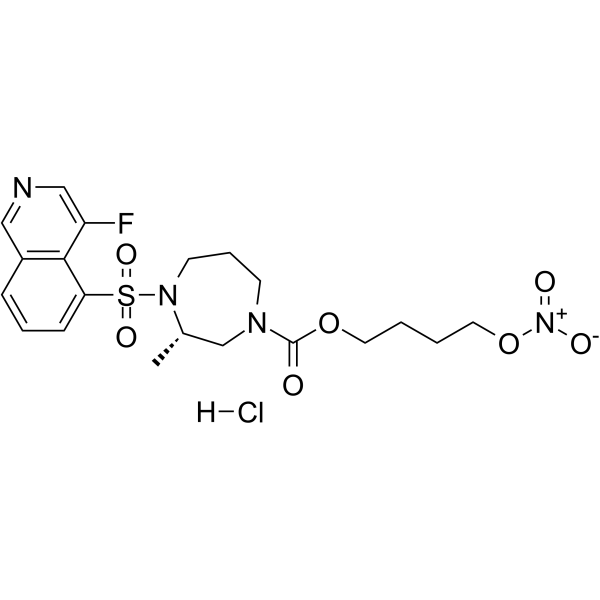
ROCK-IN-4 is a potent ROCK inhibitor maintaining NO releasing ability. ROCK-IN-4 reversibly depolymerizes F-actin, and suppresses mitochondrial respiration in human trabecular meshwork (HTM) cells. ROCK-IN-4 can be used for glaucoma or ocular hypertension research.
For research use only. We do not sell to patients.
- CAS No.: 2488395-07-7
- Formula: C20H26ClFN4O7S
- Molecular Weight:520.96
-
Storage:
Please store the product under the recommended conditions in the Certificate of Analysis.

To place orders, for customer services and technical support, please contact: MedChemExpress USA
Tel: 609-228-6898 E-mail: [email protected] [email protected]


-
 Biological Activity
-
Chemical Information
- Protocols
- Purity & Documentation
- References
-
 Help & FAQs




Biological Activity
Description
In Vitro
ROCK-IN-4 (RNO-6) (10 μM; 1 h) decreases p-MLC level and induces reversible F-actin depolymerization[1].
ROCK-IN-4 (10 μM; 24 h) involves in cGMP activation, increases cGMP concentration in human trabecular meshwork (HTM) cells[1].
ROCK-IN-4 (10 μM; 1 h) suppresses mitochondrial respiration by releasing NO and remarkably decreases the basal respiration, maximal respiration, and ATP production[1].
MedChemExpress (MCE) has not independently confirmed the accuracy of these methods. They are for reference only.
-
Cell Line:Human trabecular meshwork (HTM) cells
-
Concentration:10 μM
-
Incubation Time:1 hour
-
Result:Inhibited the phosphorylation of MLC, reduced the level of p-MLC.
-
Cell Line:Human trabecular meshwork (HTM) cells
-
Concentration:10 μM
-
Incubation Time:1 hour
-
Result:Exhibited conspicuous F-actin depolymerization, and after recovery for 4 h, recovered F-actin to the polymerization morphology, indicating a reversible depolymerization effect without permanent damage to cells.
In Vivo
ROCK-IN-4 (0.579% w/v for 25 μL; o.p. in left eye; single dose) shows low hyperemic effect and eye irritation in New Zealand White rabbits[1].
MedChemExpress (MCE) has not independently confirmed the accuracy of these methods. They are for reference only.
-
Animal Model:Chronic Ocular Hypertension Mouse Model induced by Microbead[1]
-
Dosage:0.26% (w/v), 10 μL
-
Administration:Ophthalmic drop; singel dose; administration in right eye 7 days after modeling, and measured IOP prior to and at 1, 2, 3, 4, and 6 h after instillation
-
Result:Reduced IOP (mmHg) to 3.68 ± 0.5 mmHg (19.9%) and 1.36 ± 0.6 mmHg (7.4%) at 1 and 4 h after instillation, respectively.
-
Animal Model:New Zealand White rabbits[1]
-
Dosage:0.579% (w/v); 25 μL
-
Administration:Ophthalmic drop; singel dose; administration in left eye before and at 1, 2, and 4 h after the first instillation
-
Result:Showed low side effects in conjunctival hyperemia.


Chemical Information
-
CAS No. 2488395-07-7
-
Molecular Weight 520.96
-
Formula C20H26ClFN4O7S
-
SMILES
C[C@H]1CN(C(OCCCCO[N+]([O-])=O)=O)CCCN1S(=O)(C2=CC=CC3=C2C(F)=CN=C3)=O.[H]Cl
-
Shipping
Room temperature in continental US; may vary elsewhere.
-
Storage
Please store the product under the recommended conditions in the Certificate of Analysis.


Purity & Documentation
References

Calculators
Concentration (start) × Volume (start) = Concentration (final) × Volume (final)


ROCK-IN-4 Related Classifications
HY-151189 Related Classifications
 Powered by Bioz