



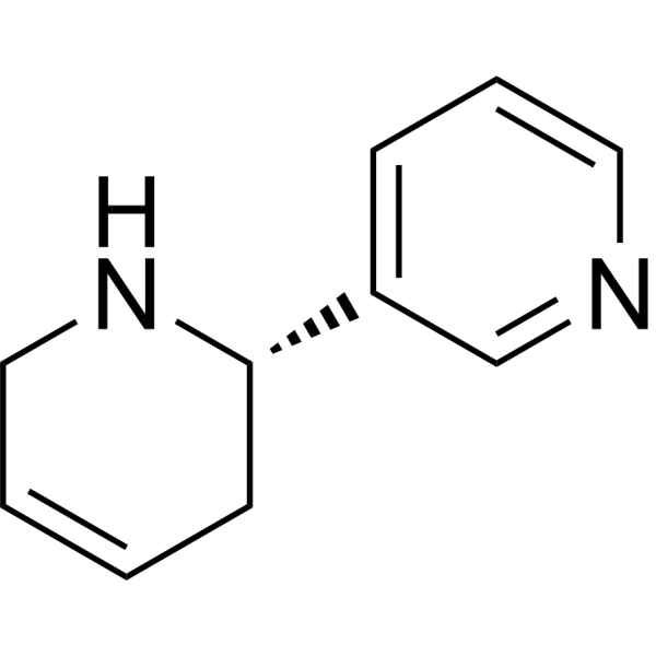
(S)-(-)-Anatabine
Based on 1 publication(s) in Google Scholar
(S)-(-)-Anatabine is an NFκB/BACE-1 inhibitor with blood-brain barrier penetration. (S)-(-)-Anatabine inhibits NFκB activation via phosphorylation of its p65 subunit. (S)-(-)-Anatabine inhibits BACE-1 transcription and reduces BACE-1 protein levels. (S)-(-)-Anatabine lowers production of Aβ1-40 and Aβ1-42 by reducing β-cleavage of amyloid precursor protein without affecting α-cleavage. (S)-(-)-Anatabine can be used for the research of Alzheimer's disease.
For research use only. We do not sell to patients.
- CAS No.: 581-49-7
- Formula: C10H12N2
- Molecular Weight:160.22
-
Storage:
Please store the product under the recommended conditions in the Certificate of Analysis.

To place orders, for customer services and technical support, please contact: MedChemExpress USA
Tel: 609-228-6898 E-mail: [email protected] [email protected]


-
 Biological Activity
-
Chemical Information
- Protocols
- Purity & Documentation
- References
-
 Help & FAQs


Publications Citing Use of MedChemExpress (MCE) (S)-(-)-Anatabine
More

Biological Activity
Description
IC50 & Target
[1]|
NF-κB |
BACE1 |
In Vitro
(S)-(-)-Anatabine (24 h) dose-dependently reduces Aβ1-40 and Aβ1-42 production with an IC50 of 640 μg/mL[1].
(S)-(-)-Anatabine (200-800 μg/mL; 3 h) dose-dependently inhibits TNFα-induced NFκB activation in HEK293 NFκB luciferase reporter cells[1].
(S)-(-)-Anatabine (600 μg/mL; 24 h) inhibits basal p65 NFκB phosphorylation in 7W CHO cells overexpressing human APP, and inhibits TNFα-induced p65 NFκB phosphorylation in HEK293 and SHSY-5Y cells[1].
(S)-(-)-Anatabine (500-1000 μg/mL; 30 min) fully inhibits TNFα-induced BACE-1 transcription in human neuronal-like SHSY-5Y cells[1].
(S)-(-)-Anatabine (5-1000 μg/mL; 24 h) dose-dependently reduces BACE-1 protein levels in human neuronal-like SHSY-5Y cells[1].
MedChemExpress (MCE) has not independently confirmed the accuracy of these methods. They are for reference only. Further protocols information, click here.
-
Cell Line:7W CHO cells overexpressing human APP, HEK293 cells, human neuronal-like SHSY-5Y cells
-
Concentration:600 μg/mL
-
Incubation Time:24 h
-
Result:Significantly reduced basal p65 NFκB phosphorylation relative to controls in 7W CHO cells.
Significantly reduced TNFα-induced p65 NFκB phosphorylation relative to TNFα-only treatment in HEK293 and SHSY-5Y cells.
-
Cell Line:human neuronal-like SHSY-5Y cells
-
Concentration:500 μg/mL; 1000 μg/mL
-
Incubation Time:30 min
-
Result:Fully prevented the TNFα-induced increase in BACE-1 mRNA levels at both 500 μg/mL and 1000 μg/mL.
Showed no significant difference in BACE-1 mRNA levels between control cells and cells treated with TNFα + anatabine.
-
Cell Line:human neuronal-like SHSY-5Y cells
-
Concentration:5 μg/mL; 20 μg/mL; 50 μg/mL; 100 μg/mL; 200 μg/mL; 800 μg/mL; 1000 μg/mL
-
Incubation Time:24 h
-
Result:Reduced BACE-1 protein levels dose-dependently.
Caused statistically significant reductions in BACE-1 protein levels relative to controls at all doses higher than 50 μg/mL.
Parmacokinetics
| Species | Dose | Route | AUC0-∞ (Brain) | Cmax (Brain) | AUC0-∞ (Plasma) | Cmax |
|---|---|---|---|---|---|---|
| Mice[1] | 2 mg/kg | i.v. | 172384 min·ng/mL | 5194.4 ng/mL | 49968 min·ng/mL | 1501.7 ng/mL |
In Vivo
and the maximal concentration (Cmax) achieving approximately 3.5 times greater in the brain of B6/SJL F1 mice than in the plasma[1].
MedChemExpress (MCE) has not independently confirmed the accuracy of these methods. They are for reference only.
-
Animal Model:B6/SJL F1 mice (43 week-old)[1]
-
Dosage:2 mg/kg
-
Administration:i.p.; single dose
-
Result:Exhibited AUC of 172384.9 ng·min/mL and 49968 ng·min/mL in the brain and plasma, respectively.
Exhibited Cmax of 5194.4 ng/mL and 1501.7 ng/mL in the brain and plasma, respectively.


Chemical Information
-
CAS No. 581-49-7
-
Molecular Weight 160.22
-
Formula C10H12N2
-
SMILES
[C@@H]1(C2=CC=CN=C2)CC=CCN1
-
Shipping
Room temperature in continental US; may vary elsewhere.
-
Storage
Please store the product under the recommended conditions in the Certificate of Analysis.


Publications (1)
-
Journal Impact Factor
-
Most Recent
-
Cardiovasc Toxicol
Chronic Anatabine Administration Attenuates Cardiovascular Activity by Targeting NF-κB/NLRP3/Caspase-1-Dependent Pyroptosis and Oxidative Stress in Paraventricular Nucleus of Hypertensive Rat. [Abstract]2025 Sep;25(9):1352-1368. PMID: 40690148

Protocols
-
Kinase activity and phosphorylation assays
Kinase activity assays measure the ability of kinases to transfer phosphate groups from ATP to specific substrates, while phosphorylation assays detect the presence and levels of phosphorylated proteins. Common methods include radiolabeled ATP incorporation (e. g. ,), ADP release detection via bioluminescence (e. g. ,[3]), enzyme-linked immunosorbent assays (ELISA) for phospho-specific epitopes (e. g. ,[6]), and microtiter-based formats for high-throughput screening (e. g. ,[8]). The ADP-Glo assay quantifies kinase activity by measuring ADP produced during phosphorylation using a luciferase-based system. Radiometric assays involve autoradiography or scintillation counting after incorporation of 32P-labeled ATP into substrate proteins. ELISA-based approaches rely on phospho-specific antibodies to detect activated kinases in cell lysates or purified samples.
-
Western Blot
Western blotting (WB) is a commonly used experimental method in molecular biology, biochemistry, and immunogenetics for identifying and quantifying target proteins. It combines gel electrophoresis with immunoassay, enabling researchers to analyze protein expression, post-translational modifications, and molecular weight.
-
Protocol for Kinase activity and phosphorylation assays
Kinase activity assays measure transfer of phosphate from ATP to a protein or peptide substrate, generating phosphorylated substrate, ADP, or incorporated radiolabeled phosphate as the readout; phosphorylation assays measure site-specific phosphorylation in cells or tissues as a proxy for kinase-pathway activation, inhibition, or substrate regulation. Phosphorylation can be detected by phospho-specific Western blot, immunoprecipitation kinase assay, phospho-immunofluorescence, phospho-flow cytometry, luminescent ADP detection, radiolabeled ATP incorporation, or reporter-based pathway assays, and these readouts can be applied to cancer cells, primary neurons, mouse tumors, organoids, inflammatory macrophages, ferroptosis studies, and mitophagy studies when the kinase target is biologically relevant.
-
Amyloid: Congo Red Amyloid Staining
Congo red amyloid staining is a histochemical method used to detect extracellular amyloid deposits in tissue sections based on the affinity of Congo red dye for β-pleated sheet-rich protein aggregates. When bound to amyloid, Congo red produces characteristic apple-green birefringence under polarized light microscopy, which is widely regarded as a diagnostic feature of amyloid deposition in histopathology. The diagnostic principle relies on the combination of dye binding (congophilia) and optical anisotropy under polarized illumination, which distinguishes amyloid from most non-amyloid eosinophilic extracellular deposits in routine histological evaluation. Amyloid identification by Congo red staining remains a cornerstone in diagnostic pathology despite the availability of adjunct methods such as immunohistochemistry and mass spectrometry, particularly because of its ability to localize deposits directly within tissue architecture. The specificity of Congo red-positive deposits is incre
-
Alzheimer’s Disease Modeling
Alzheimer’s Disease (AD) is a neurodegenerative disorder characterized by a progressive decline in cognitive functions and loss of specific types of neurons and synapses. Alzheimer's symptoms can be simulated in mice by injecting drugs (such as Aβ) or genetically modified.


Purity & Documentation
References

Calculators
Concentration (start) × Volume (start) = Concentration (final) × Volume (final)


(S)-(-)-Anatabine Related Classifications
HY-126047 Related Classifications
 Powered by Bioz