



Apcin-A

Based on 1 Customer Validation
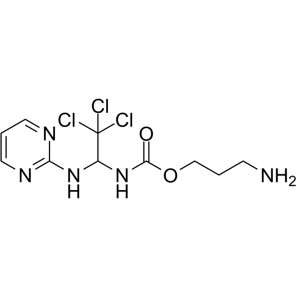
Apcin-A is a small molecule inhibitor that selectively targets the cell division cycle protein Cdc20 and is a derivative of Apcin (HY-110287). Apcin-A competitively binds to the D-box binding pocket of Cdc20 and inhibits substrate ubiquitination mediated by the anaphase promoting complex APC/C-Cdc20. Apcin-A also blocks the binding of Cdc20 to substrates (such as securin and cyclin B1), inhibiting anaphase initiation and cell cycle exit. Apcin-A can promote or prolong mitotic slippage in coordination with p31comet under conditions of high spindle assembly checkpoint (SAC) activity. Apcin-A can be used to develop anti-mitotic drugs and overcome tumor chemotherapy resistance. Apcin-A can be used to synthesize PROTAC CP5V (HY-130257)[1][2][3].
For research use only. We do not sell to patients.
- Purity : 98.0%
- CAS No.: 1683617-62-0
- Formula: C10H14Cl3N5O2
- Molecular Weight:342.61
-
Storage:Powder -20°C, 3 years , 4°C, 2 years ; In solvent -80°C, 6 months , -20°C, 1 month

To place orders, for customer services and technical support, please contact: MedChemExpress USA
Tel: 609-228-6898 E-mail: [email protected] [email protected]


-
 Biological Activity
-
Chemical Information
-
Solvent & Solubility
- Protocols
- Purity & Documentation
- References
-
 Help & FAQs


-
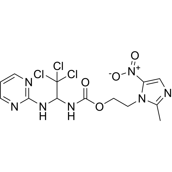
HY-110287
ApcinAPC/C Inhibitor -
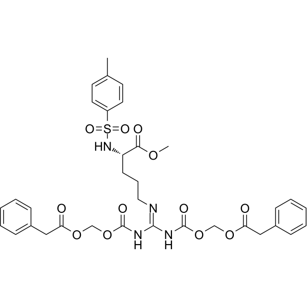
HY-124955
proTAMEAPC Inhibitor -

HY-P7109
EGF Protein, Human -
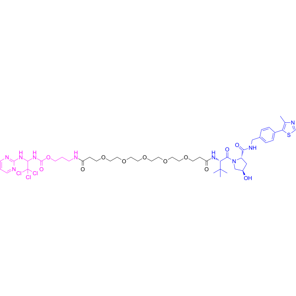
HY-130257
CP5VCdc20 PROTAC Degrader -
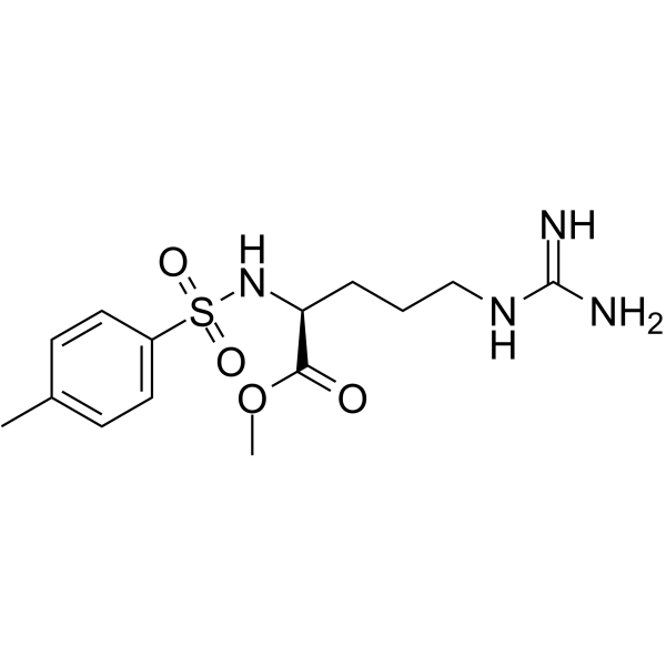
HY-13255
TAMEAPC Inhibitor -
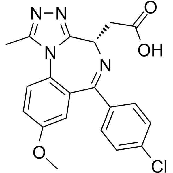
HY-107443
I-BET762 carboxylic acidBRD4 Inhibitor -
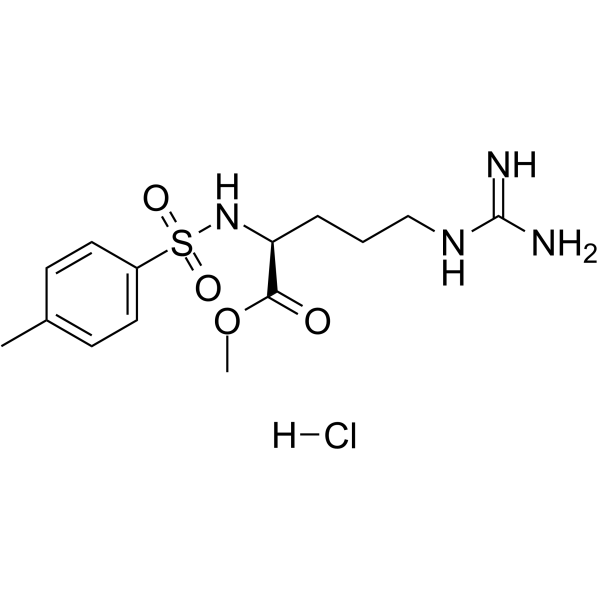
HY-13255A
TAME hydrochlorideAPC/C Inhibitor -
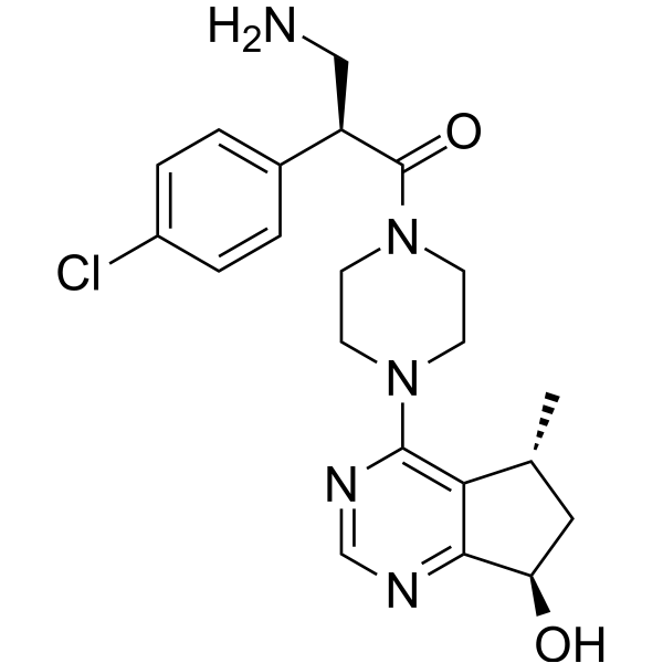
HY-130988
Ipatasertib-NH2Ligand For Target Protein -
HY-13030
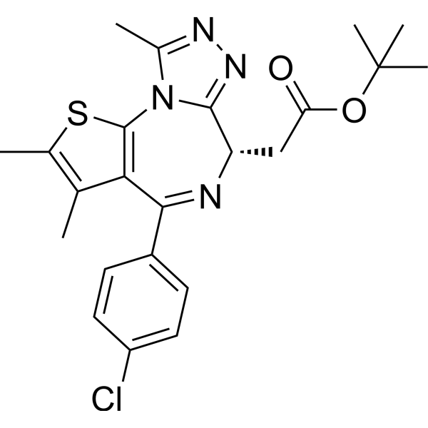
(+)-JQ-1BET Inhibitor -
HY-111606
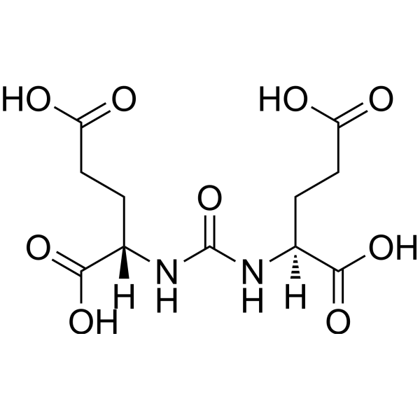
DUPALigands for Target Protein for PROTAC -
HY-107451
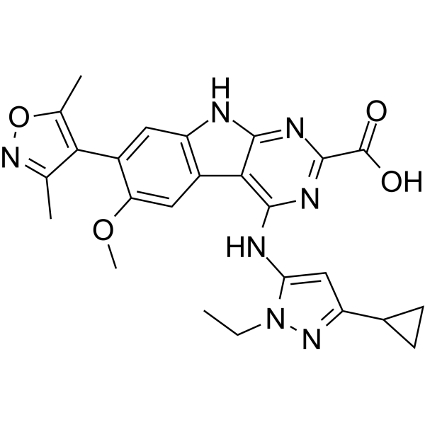
PROTAC BET-binding moiety 1Intermediate -
HY-P1918
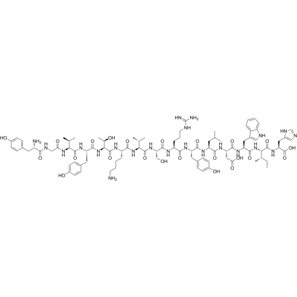
Activated Protein C (390-404), humanPolypeptide


Biological Activity
Description
IC50 & Target
|
Cdc20 |
In Vitro
Apcin-A (25 μM; 10 h) significantly reduces nocodazole (HY-13520)-induced mitotic fraction in HCT116 and HeLa cells, accelerates cyclin B1 degradation and CDC27 phosphorylation, and promotes mitotic slippage[1].
Apcin-A (500 μM; 5 min) synergizes with p31comet in a recombinant system to reduce MCC binding to APC/C and relieve MCC inhibition of APC/C[1].
Apcin-A (200 μM; 10 h) inhibits APC/C-mediated ubiquitination and degradation of securin and cyclin B1 in Xenopus egg extracts[2].
MedChemExpress (MCE) has not independently confirmed the accuracy of these methods. They are for reference only.


Chemical Information
-
CAS No. 1683617-62-0
-
Appearance Solid
-
Molecular Weight 342.61
-
Formula C10H14Cl3N5O2
-
Color Off-white to yellow
-
SMILES
O=C(OCCCN)NC(NC1=NC=CC=N1)C(Cl)(Cl)Cl
-
Shipping
Room temperature in continental US; may vary elsewhere.
-
Storage
Powder -20°C 3 years 4°C 2 years In solvent -80°C 6 months -20°C 1 month


Solvent & Solubility
In Vitro:
DMSO : 100 mg/mL (291.88 mM; Need ultrasonic; Hygroscopic DMSO has a significant impact on the solubility of product, please use newly opened DMSO)
Please refer to the solubility information to select the appropriate solvent. Once prepared, please aliquot and store the solution to prevent product inactivation from repeated freeze-thaw cycles.
Storage method and period of stock solution: -80°C, 6 months; -20°C, 1 month. When stored at -80°C, please use it within 6 months. When stored at -20°C, please use it within 1 month.
Please refer to the solubility information to select the appropriate solvent. Once prepared, please aliquot and store the solution to prevent product inactivation from repeated freeze-thaw cycles.
Storage method and period of stock solution: -80°C, 6 months; -20°C, 1 month. When stored at -80°C, please use it within 6 months. When stored at -20°C, please use it within 1 month.
Concentration (start) × Volume (start) = Concentration (final) × Volume (final)
In Vivo:
Select the appropriate dissolution method based on your experimental animal and administration route.
- For the following dissolution methods, please ensure to first prepare a clear stock solution using an In Vitro approach and then sequentially add co-solvents:
- To ensure reliable experimental results, the clarified stock solution can be appropriately stored based on storage conditions. As for the working solution for In Vivo experiments, it is recommended to prepare freshly and use it on the same day.
- The percentages shown for the solvents indicate their volumetric ratio in the final prepared solution. If precipitation or phase separation occurs during preparation, heat and/or sonication can be used to aid dissolution.
Add each solvent one by one: 10% DMSO 40% PEG300 5% Tween-80 45% Saline
Solubility: 2.5 mg/mL (7.30 mM); Suspended solution; Need ultrasonic
This protocol yields a suspended solution of 2.5 mg/mL. Suspended solution can be used for oral and intraperitoneal injection.
Taking 1 mL working solution as an example, add 100 μL DMSO stock solution (25.0 mg/mL) to 400 μL PEG300, and mix evenly; then add 50 μL Tween-80 and mix evenly; then add 450 μL Saline to adjust the volume to 1 mL.
Preparation of Saline: Dissolve 0.9 g sodium chloride in ddH₂O and dilute to 100 mL to obtain a clear Saline solution.
Add each solvent one by one: 10% DMSO 90% (20% SBE-β-CD in Saline)
Solubility: 2.5 mg/mL (7.30 mM); Suspended solution; Need ultrasonic
This protocol yields a suspended solution of 2.5 mg/mL. Suspended solution can be used for oral and intraperitoneal injection.
Taking 1 mL working solution as an example, add 100 μL DMSO stock solution (25.0 mg/mL) to 900 μL 20% SBE-β-CD in Saline, and mix evenly.
Preparation of 20% SBE-β-CD in Saline (4°C, storage for one week): 2 g SBE-β-CD powder is dissolved in 10 mL Saline, completely dissolve until clear.
In Vivo Dissolution Calculator
Please enter the basic information of animal experiments:
-
-
-
-
Recommended: Prepare an additional quantity of animals to account for potential losses during experiments.
Please enter your animal formula composition:
-
%DMSO +
Recommended: Keep the proportion of DMSO in working solution below 2% if your animal is weak.
-
%+
-
+%Tween-80 + +
-
%Saline +
The co-solvents required include: DMSO, . All of co-solvents are available by MedChemExpress (MCE). , Tween 80. All of co-solvents are available by MedChemExpress (MCE).
Working solution concentration: 0.22 mg/mL
Method for preparing stock solution: mg drug dissolved in μL DMSO. Stock solution concentration: mg/mL.
1. Take μL DMSO stock solution;
2. Add μL .
μL , mix evenly;
3. Then add μL Tween 80, mix evenly;
4. Then add μL
Please ensure that the stock solution in the first step is dissolved to a clear state, and add co-solvents in sequence. You can use ultrasonic heating (ultrasonic cleaner, recommended frequency 20-40 kHz), vortexing, etc. to assist dissolution.


Purity & Documentation
-
Data Sheet (272 KB)
-
SDS (251 KB)
- English - EN (251 KB)
- Français - FR (251 KB)
- Deutsch - DE (251 KB)
- Norwegian - NO (251 KB)
- Español - ES (251 KB)
- Swedish - SV (251 KB)
- Italian - IT (251 KB)
- Korean - KR (251 KB)
- Portuguese - PT (251 KB)
-
Handling Instructions (2659 KB)
References
[1]. Richeson KV, et al. Paradoxical mitotic exit induced by a small molecule inhibitor of APC/CCdc20. Nat Chem Biol. 2020 May;16(5):546-555. [Content Brief]
[2]. Sackton KL, et al. Synergistic blockade of mitotic exit by two chemical inhibitors of the APC/C. Nature. 2014 Oct 30;514(7524):646-9. [Content Brief]
[3]. Chi JJ, et al. A novel strategy to block mitotic progression for targeted therapy. EBioMedicine. 2019 Oct 25. pii: S2352-3964(19)30677-2. [Content Brief]


Complete Stock Solution Preparation Table
Please refer to the solubility information to select the appropriate solvent. Once prepared, please aliquot and store the solution to prevent product inactivation from repeated freeze-thaw cycles.
Storage method and period of stock solution: -80°C, 6 months; -20°C, 1 month. When stored at -80°C, please use it within 6 months. When stored at -20°C, please use it within 1 month.
| Optional Solvent | Concentration Solvent Mass | 1 mg | 5 mg | 10 mg | 25 mg |
|---|---|---|---|---|---|
| DMSO | 1 mM | 2.9188 mL | 14.5939 mL | 29.1877 mL | 72.9693 mL |
| 5 mM | 0.5838 mL | 2.9188 mL | 5.8375 mL | 14.5939 mL | |
| 10 mM | 0.2919 mL | 1.4594 mL | 2.9188 mL | 7.2969 mL | |
| 15 mM | 0.1946 mL | 0.9729 mL | 1.9458 mL | 4.8646 mL | |
| 20 mM | 0.1459 mL | 0.7297 mL | 1.4594 mL | 3.6485 mL | |
| 25 mM | 0.1168 mL | 0.5838 mL | 1.1675 mL | 2.9188 mL | |
| 30 mM | 0.0973 mL | 0.4865 mL | 0.9729 mL | 2.4323 mL | |
| 40 mM | 0.0730 mL | 0.3648 mL | 0.7297 mL | 1.8242 mL | |
| 50 mM | 0.0584 mL | 0.2919 mL | 0.5838 mL | 1.4594 mL | |
| 60 mM | 0.0486 mL | 0.2432 mL | 0.4865 mL | 1.2162 mL | |
| 80 mM | 0.0365 mL | 0.1824 mL | 0.3648 mL | 0.9121 mL | |
| 100 mM | 0.0292 mL | 0.1459 mL | 0.2919 mL | 0.7297 mL |


Apcin-A Related Classifications
HY-130841 Related Classifications
 Powered by Bioz