



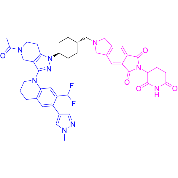
CBPD-268

Based on 1 Customer Validation
CBPD-268 is a potent and orally active CBP/p300 PROTAC degrader with an DC50 value of ≤ 0.03 nM. CBPD-268 induces CBP/p300 degradation and inhibits cell growth. CBPD-268 shows antitumor activity. CBPD-268 has the potential for the research of AR-positive prostate cancer.
(Pink: CBP/p300 ligand (HY-161483); Blue: Cereblon ligand (HY-W248665); Black: linker).
For research use only. We do not sell to patients.
- Purity : 99.23%
- CAS No.: 2832961-58-5
- Formula: C44H47F2N9O5
- Molecular Weight:819.90
-
Storage:Powder -20°C, 3 years ; In solvent -80°C, 6 months , -20°C, 1 month

To place orders, for customer services and technical support, please contact: MedChemExpress USA
Tel: 609-228-6898 E-mail: [email protected] [email protected]


-
 Biological Activity
-
Chemical Information
-
Solvent & Solubility
- Protocols
- Purity & Documentation
- References
-
 Help & FAQs


All PROTACs Isoforms
More

Biological Activity
Description
Cellular Effect
|
Cell Line

|
Type | Value | Description | References |
|---|---|---|---|---|
| CWR22R | IC50 |
3.7 nM
Compound: 33; CBPD-268
|
Antiproliferative activity against human 22Rv1 cells assessed as cell growth inhibition incubated for 4 days by celtiter-glo luminescent assay
Antiproliferative activity against human 22Rv1 cells assessed as cell growth inhibition incubated for 4 days by celtiter-glo luminescent assay
|
[PMID: 38477974] |
| LNCaP | IC50 |
10.3 nM
Compound: 33; CBPD-268
|
Antiproliferative activity against human LNCaP cells assessed as cell growth inhibition incubated for 4 days by celtiter-glo luminescent assay
Antiproliferative activity against human LNCaP cells assessed as cell growth inhibition incubated for 4 days by celtiter-glo luminescent assay
|
[PMID: 38477974] |
| VCaP | IC50 |
4.6 nM
Compound: 33; CBPD-268
|
Antiproliferative activity against human VCaP cells assessed as cell growth inhibition incubated for 4 days by celtiter-glo luminescent assay
Antiproliferative activity against human VCaP cells assessed as cell growth inhibition incubated for 4 days by celtiter-glo luminescent assay
|
[PMID: 38477974] |
In Vitro
CBPD-268 (4, 24 h) shows high degradation efficiency for CBP and p300 protein with DC50s of 0.01, 0.03 nM at 4 h in 22Rv1 cells[1].
CBPD-268 shows degradation by binding to both CBP/p300 and CRBN protein[1].
CBPD-268 (0-1000 nM; 4 days) inhibits cell growth with IC50s of 3.7, 10.3, 4.6 nM for 22Rv1, LNCaP, VCaP cells, respectively[1].
MedChemExpress (MCE) has not independently confirmed the accuracy of these methods. They are for reference only.
-
Cell Line:22Rv1, LNCaP, VCaP cells
-
Concentration:0-1000 nM
-
Incubation Time:4 days
-
Result:Inhibited cell growth with IC50s OF 3.7, 10.3, 4.6 Nm for 22Rv1, LNCaP, VCaP cells, respectively.
In Vivo
CBPD-268 (1, 3 mg/kg; p.o.; twice a week for 1 mg/kg or 3 mg/kg weekly for 4-weeks) shows antitumor activity[1].
Pharmacokinetic Parameters[1].
| Species | IV (mg/kg) | T1/2 (h) | V1/2(L/kg) | CL (mL/min/kg) | PO(mg/kg) | T1/2 (h) | Cmax (ng/ml) | AUC(h*ng/mL) | F(%) |
| Rats | 1 | 1.9 | 4.9 | 34.6 | 3 | 1.3 | 220.6 | 936.9 | 67 |
| Mice | 1 | 3.4 | 1.6 | 6.0 | 3 | 3.1 | 724.7 | 4190.4 | 60 |
MedChemExpress (MCE) has not independently confirmed the accuracy of these methods. They are for reference only.
-
Animal Model:male CB17 SCID mice (VCaP xenograft tumor)[1]
-
Dosage:0.3, 1, 3, 10, 30 mg/kg
-
Administration:P.o.; once
-
Result:Induced depletion of both CBP and p300 proteins in the VCaP tumor tissue in a dose-dependent manner.
-
Animal Model:male CB17 SCID mice (VCaP xenograft tumor model)[1]
-
Dosage:1, 3 mg/kg
-
Administration:P.o.; twice a week for 1 mg/kg or 3 mg/kg weekly for 4-weeks
-
Result:Inhibited tumor growth and shows little effect on animal body weight.
-
Animal Model:female BALB/c mice[1]
-
Dosage:3, 10, 30 mg/kg
-
Administration:P.o.; twice weekly for 5-6 weeks
-
Result:Induced no weight loss or other signs of toxicity at both 3 and 10 mg/kg dose-levels in both male and female mice.
-
Animal Model:Female Sprague–Dawley (SD) rats[1]
-
Dosage:1-10 mg/kg
-
Administration:P.o.; twice a week for 5 weeks
-
Result:Did not cause animal body weight loss during the entire experiment and did not induce any signs of toxicity during the entire experiment.


Chemical Information
-
CAS No. 2832961-58-5
-
Appearance Solid
-
Molecular Weight 819.90
-
Formula C44H47F2N9O5
-
Color White to light yellow
-
SMILES
CC(N1CC2=C(CC1)N([C@@H]3CC[C@H](CC3)CN4CC(C=C(C(N(C5CCC(NC5=O)=O)C6=O)=O)C6=C7)=C7C4)N=C2N8CCCC9=C8C=C(C(F)F)C(C%10=CN(N=C%10)C)=C9)=O
-
Shipping
Room temperature in continental US; may vary elsewhere.
-
Storage
Powder -20°C 3 years In solvent -80°C 6 months -20°C 1 month


Solvent & Solubility
In Vitro:
DMSO : 100 mg/mL (121.97 mM; Need ultrasonic; Hygroscopic DMSO has a significant impact on the solubility of product, please use newly opened DMSO)
Please refer to the solubility information to select the appropriate solvent. Once prepared, please aliquot and store the solution to prevent product inactivation from repeated freeze-thaw cycles.
Storage method and period of stock solution: -80°C, 6 months; -20°C, 1 month. When stored at -80°C, please use it within 6 months. When stored at -20°C, please use it within 1 month.
Please refer to the solubility information to select the appropriate solvent. Once prepared, please aliquot and store the solution to prevent product inactivation from repeated freeze-thaw cycles.
Storage method and period of stock solution: -80°C, 6 months; -20°C, 1 month. When stored at -80°C, please use it within 6 months. When stored at -20°C, please use it within 1 month.
Concentration (start) × Volume (start) = Concentration (final) × Volume (final)


Purity & Documentation
-
Data Sheet (289 KB)
-
SDS (252 KB)
- English - EN (252 KB)
- Français - FR (252 KB)
- Deutsch - DE (252 KB)
- Norwegian - NO (252 KB)
- Español - ES (252 KB)
- Swedish - SV (252 KB)
- Italian - IT (252 KB)
- Korean - KR (252 KB)
- Portuguese - PT (252 KB)
-
Handling Instructions (2659 KB)
References


Complete Stock Solution Preparation Table
Please refer to the solubility information to select the appropriate solvent. Once prepared, please aliquot and store the solution to prevent product inactivation from repeated freeze-thaw cycles.
Storage method and period of stock solution: -80°C, 6 months; -20°C, 1 month. When stored at -80°C, please use it within 6 months. When stored at -20°C, please use it within 1 month.
| Optional Solvent | Concentration Solvent Mass | 1 mg | 5 mg | 10 mg | 25 mg |
|---|---|---|---|---|---|
| DMSO | 1 mM | 1.2197 mL | 6.0983 mL | 12.1966 mL | 30.4915 mL |
| 5 mM | 0.2439 mL | 1.2197 mL | 2.4393 mL | 6.0983 mL | |
| 10 mM | 0.1220 mL | 0.6098 mL | 1.2197 mL | 3.0492 mL | |
| 15 mM | 0.0813 mL | 0.4066 mL | 0.8131 mL | 2.0328 mL | |
| 20 mM | 0.0610 mL | 0.3049 mL | 0.6098 mL | 1.5246 mL | |
| 25 mM | 0.0488 mL | 0.2439 mL | 0.4879 mL | 1.2197 mL | |
| 30 mM | 0.0407 mL | 0.2033 mL | 0.4066 mL | 1.0164 mL | |
| 40 mM | 0.0305 mL | 0.1525 mL | 0.3049 mL | 0.7623 mL | |
| 50 mM | 0.0244 mL | 0.1220 mL | 0.2439 mL | 0.6098 mL | |
| 60 mM | 0.0203 mL | 0.1016 mL | 0.2033 mL | 0.5082 mL | |
| 80 mM | 0.0152 mL | 0.0762 mL | 0.1525 mL | 0.3811 mL | |
| 100 mM | 0.0122 mL | 0.0610 mL | 0.1220 mL | 0.3049 mL |


CBPD-268 Related Classifications
HY-161369 Related Classifications
 Powered by Bioz