18 Results for "

plant cell staining

" in MedChemExpress (MCE) Product Catalog:
Products (18)

18 Results for "plant cell staining" in MCE Product Catalog:

17
17 Publications Verification
Cat. No.: HY-103311
CAS No.: 11103-72-3
Purity:  ≥97.0%
Synonyms: Ammoniated ruthenium oxychloride
Target:  

Calcium Channel

Research Areas:  

Cardiovascular Disease

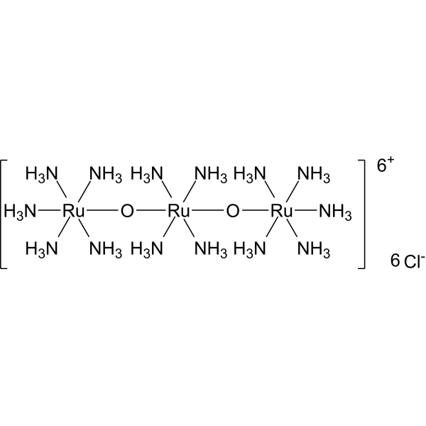
Ruthenium red (Ammoniated ruthenium oxychloride) is a polycationic dye widely used for electron microscopy (EM) of cells, tissues and vegetative bacteria. Ruthenium red strongly reacts with phospholipids and fatty acids and binds to acidic mucopolysaccharides. Ruthenium red is a L-type calcium current (ICa) blocker. When using Ruthenium Red to stain plant tissue sections affected by pathogens, the breakdown of mucilaginous substances by the pathogens prevents them from taking up the stain, whereas the cell walls of healthy tissue stain red .
loading...
    loading...
4
4 Cited Publications
Cat. No.: HY-D0220
CAS No.: 92-31-9
Synonyms: Toluidine Blue O
Target:  

Fluorescent Dye

Research Areas:  

Cancer

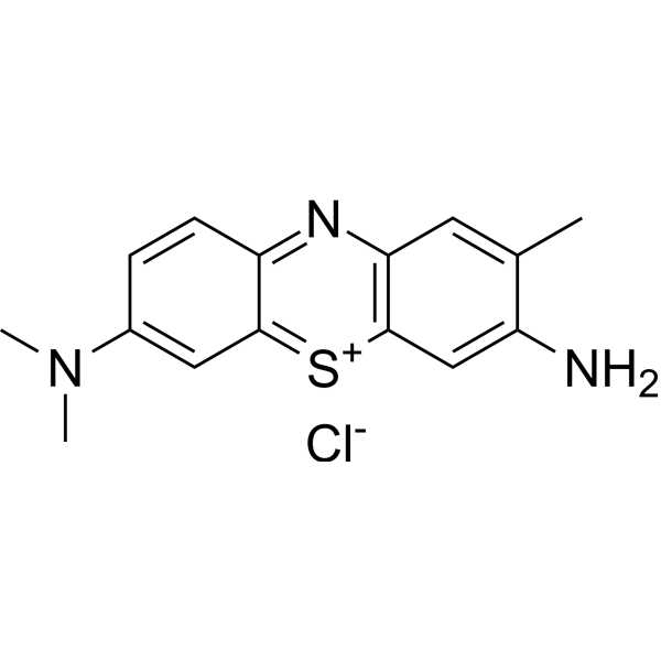
Toluidine Blue (Toluidine Blue O) is an alkaline quinonimine dye (vivo dye) with high affinity for acidic tissue components, staining nuclei blue and polysaccharides purple. Toluidine Blue shows heterostaining properties for mast cells, mucins and chondrocytes. Toluidine Blue can stain different components of plant tissues and cells in different colours. Toluidine Blue is also used as a diagnostic aid to identify malignant lesions, such as cancer .
loading...
    loading...
4
4 Cited Publications
Cat. No.: HY-D0220A
Synonyms: Toluidine Blue O (purity 36%)
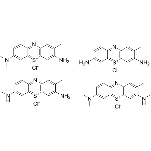
Toluidine Blue (Toluidine Blue O) purity 36% is an alkaline quinonimine dye (vivo dye) with high affinity for acidic tissue components, staining nuclei blue and polysaccharides purple. Toluidine Blue purity 36% shows heterostaining properties for mast cells, mucins and chondrocytes. Toluidine Blue purity 36% can stain different components of plant tissues and cells in different colours. Toluidine Blue purity 36% is also used as a diagnostic aid to identify malignant lesions, such as cancer .
loading...
    loading...
1
1 Cited Publications
Cat. No.: HY-NP0147
Synonyms: WGA (Fluorescein)
Wheat Germ Agglutinin (WGA) Fluorescein is a classic fluorescent label that specifically binds to sugar residues such as N-acetylglucosamine, N-acetylneuraminic acid and sialic acid. Wheat Germ Agglutinin Fluorescein performs regionally differential fluorescent staining of the ocular surface epithelial glycocalyx to assess its integrity, and causes no damage to the eye at safe concentrations. Wheat Germ Agglutinin Fluorescein is also used for staining structures including red blood cells, cultured cells, bacteria and pine wood nematodes, and facilitates the isolation of wheat-associated plant-growth-promoting rhizobacterial strains. Wheat Germ Agglutinin Fluorescein can be applied to the detection of ocular glycocalyx integrity and the research of related diseases such as pine wilt disease .
loading...
    loading...
Cat. No.: HY-D0286
CAS No.: 81-37-8
Synonyms: 2,8-Dimethylnaphtho[3,2,1-kl]xanthene
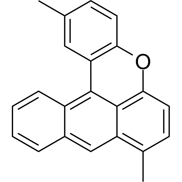
Fluorol Yellow 088 is a fluorescent dye that can be used as a biological material or an organic compound for life science research. Fluorol Yellow 088 is suitable for lipid staining in plant tissues, particularly for the fluorescence imaging of suberized cell walls and hydrophobic structures, with an excitation wavelength of 365 nm and an emission wavelength of ≥420 nm .
loading...
    loading...
Cat. No.: HY-W040198
CAS No.: 81-93-6
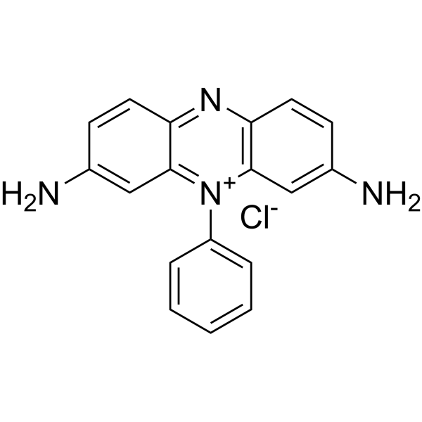
Phenosafranine is a phenazine dye. Phenosafranine exhibits higher binding affinity for triple-stranded RNA than for double-stranded RNA, and binds to both types of RNA via intercalation. Phenosafranine interacts with hemoglobin. Phenosafranine can be used for plant cell staining, as well as the detection of hemoglobin, dopamine, serotonin, etc.
loading...
    loading...
Cat. No.: HY-W540972
CAS No.: 8064-60-6
Synonyms: Primuline
Primulin is a versatile fluorescent dye and bioactive compound widely used in analytical, biological, botanical and virological studies. Primulin acts as a versatile stain that labels plant cell walls and differentiates live and dead spermatozoa via distinct fluorescence patterns. Primulin exhibits strong albumin‑binding capacity. Primulin acts as a retrograde axonal tracer in neurobiological investigations. Primulin and its derivatives inhibit HCV NS3, block dengue virus NS3-mediated ATP hydrolysis, and disrupt HCV replicase assembly .
loading...
    loading...
Cat. No.: HY-DY2043
Target:  

Fluorescent Dye

Research Areas:  

Others

Plant Cell Acidity Indicator Solution (Methyl Red Method) is a synthetic acid-base indicator and vital staining solution containing Methyl red (HY-Y0699), which can be used to indicate the pH value in plant tissues or cells. Plant Cell Acidity Indicator Solution (Methyl Red Method) exhibits a yellow-to-red color change in acid-base titrations, with a linear relationship .
loading...
    loading...
Cat. No.: HY-DY2040
Target:  

Fluorescent Dye

Research Areas:  

Others

Mucilage Staining Solution (Ruthenium Red Method) is a staining solution used to localize mucopolysaccharides in plant tissues. In the staining of plant disease and pest tissue sections, pathogens decompose mucilage substances, which thus cannot be stained, while the cell walls of healthy tissues stain red .
loading...
    loading...
Cat. No.: HY-DY2020
Target:  

Fluorescent Dye

Research Areas:  

Others

Neutral Red Staining Solution (for Live Cells) is a vital stain used for vital staining of peripheral blood cells, and it can also be used for staining other living cells or tissues. Neutral Red Staining Solution (for Live Cells) accumulates in cytoplasmic vacuoles, lysosomes of living cells, as well as plant/fungal vacuoles. It binds to anionic sites, suberin, lignin, cell walls, DNA and phenolic substances, and can intercalate into DNA. Neutral Red Staining Solution (for Live Cells) stains the cytoplasm of specific phytoplankton and estuarine zooplankton, while dead cells are not stained, enabling viability determination of such organisms .
loading...
    loading...
Cat. No.: HY-DY2036
Target:  

Fluorescent Dye

Research Areas:  

Others

Cellulose Staining Solution (Zinc Chloride-Iodine Method) is a cellulose-targeted staining agent. Cellulose Staining Solution (Zinc Chloride-Iodine Method) enables cellulose cell walls to turn blue-purple to blue by forming iodine-cellulose complexes between iodine and cellulose. Cellulose Staining Solution (Zinc Chloride-Iodine Method) can be used for the identification of plant phloem fibers, cellulose layers of algal cell walls, residual mineralized plant fibers from archaeological samples, and cellulose deposited on PET surfaces .
loading...
    loading...
Cat. No.: HY-DY2041
Target:  

Fluorescent Dye

Research Areas:  

Infection

Callose Staining Solution (Aniline Blue, Bright-Field Method) is a fluorescent dye used for detecting callose in plant tissues, green algal cell walls, plant cells, and protoplasts. Callose Staining Solution (Aniline Blue, Bright-Field Method) specifically binds to callose, a β-(1,3)-D-glucan polymer, and exhibits blue-green fluorescence under alkaline conditions with reduced background staining and non-toxicity to living cells. Callose Staining Solution (Aniline Blue, Bright-Field Method) is suitable for observation under UV or epifluorescence microscopy (Ex/Em = 365-370/397-509 nm). Callose Staining Solution (Aniline Blue, Bright-Field Method) can be used in studies related to the visualization of callose deposition in plants .
loading...
    loading...
Cat. No.: HY-DY2019
Target:  

Fluorescent Dye

Research Areas:  

Cancer

Toluidine Blue Staining Solution (1%, Phosphate Buffer) is an alkaline quinonimine dye (vivo dye) with high affinity for acidic tissue components, staining nuclei blue and polysaccharides purple. Toluidine Blue Staining Solution (1%, Phosphate Buffer) shows heterostaining properties for mast cells, mucins and chondrocytes. Toluidine Blue Staining Solution (1%, Phosphate Buffer) can stain different components of plant tissues and cells in different colours. Toluidine Blue Staining Solution (1%, Phosphate Buffer) is also used as a diagnostic aid to identify malignant lesions, such as cancer .
loading...
    loading...
Cat. No.: HY-DY2037
Target:  

Fluorescent Dye

Research Areas:  

Others

Lignin Staining Solution (Safranin Method) is a histochemical staining solution for lignin with Safranin O as the active component. Lignin Staining Solution (Safranin Method) enables lignified cell walls to appear red to deep red (pinkish in regions with low lignification) via the high affinity of the dye for lignin. Lignin Staining Solution (Safranin Method) can be used to visualize lignin distribution in ex vivo thick sections of annual shoots from woody plants and vibratome sections of stems from herbaceous plants (such as Arabidopsis and flax), as well as for in situ quantification of cell wall substructural regions (cell corners, intercellular layers, secondary walls) based on Safranin O ratiometric fluorescence .
loading...
    loading...
Cat. No.: HY-DY2039
Target:  

Fluorescent Dye

Research Areas:  

Others

Starch Staining Solution (Iodine Method), also known as Iodine Starch Staining Solution, stains starch in plants blue or dark blue-purple within a few minutes of staining with iodine solution. Glycogen accumulated in fungal cells turns red after staining with iodine solution. Starch appears blue or purplish-red when hydrolyzed into dextrins, and turns red when further decomposed into smaller-molecular-weight dextrins .
loading...
    loading...
Cat. No.: HY-DY2042
Target:  

Fluorescent Dye

Research Areas:  

Others

Plant Cell Acidity Indicator Solution (Neutral Red Method) is a basic vital stain solution using neutral red, used to indicate plant cell acidity. Plant Cell Acidity Indicator Solution (Neutral Red Method) accumulates in vacuoles via the ion trap mechanism in empty cell saps and exhibits distinct color changes based on cell sap type and pH .
loading...
    loading...
Cat. No.: HY-DY2034
Target:  

Calcium Channel

Research Areas:  

Cardiovascular Disease

Ruthenium Red Staining Solution (0.01%) is a polycationic dye widely used for electron microscopy (EM) of cells, tissues and vegetative bacteria. Ruthenium Red Staining Solution (0.01%) strongly reacts with phospholipids and fatty acids and binds to acidic mucopolysaccharides. Ruthenium Red Staining Solution (0.01%) is a L-type calcium current (ICa) blocker. When using Ruthenium Red Staining Solution (0.01%) to stain plant tissue sections affected by pathogens, the breakdown of mucilaginous substances by the pathogens prevents them from taking up the stain, whereas the cell walls of healthy tissue stain red .
loading...
    loading...
Cat. No.: HY-DY2038
Target:  

Fluorescent Dye

Research Areas:  

Infection

Lignin Staining Solution (Pianeze Method) is a multichromatic staining solution for plant histology composed of malachite green, Martius yellow and acid fuchsin. Lignin Staining Solution (Pianeze Method) enables lignified secondary walls to appear bright green, non-lignified secondary walls to appear red, and tracheid pit membranes to appear pale red after long-time immersion. Lignin Staining Solution (Pianeze Method) can be used to distinguish lignified cell walls from non-lignified structures and observe the morphology of pit membranes in plant tissue sections such as fern rhizomes. Lignin Staining Solution (Pianeze Method) can be applied to relevant studies on flax wilt and root rot .
loading...
    loading...
  • 1

