



Trametinib-d4
Based on 1 Customer Validation
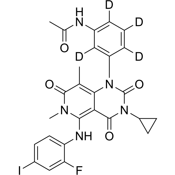
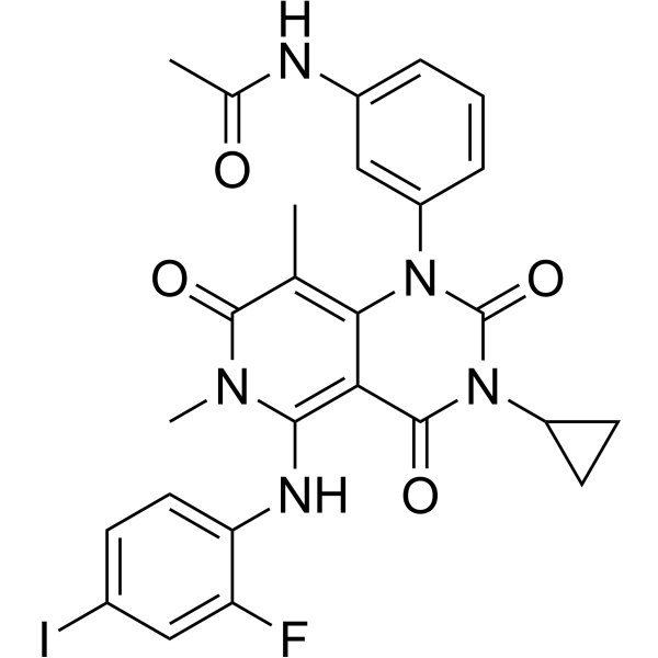
Trametinib-d4 is the deuterium labeled Trametinib. Trametinib (GSK1120212; JTP-74057) is an orally active MEK inhibitor that inhibits MEK1 and MEK2 with IC50s of about 2 nM. Trametinib activates autophagy and induces apoptosis.
Nos produits utilisent uniquement pour la recherche. Nous ne vendons pas aux patients.
- Pureté : 99.87%
- Formule: C26H19D4FIN5O4
- Masse moléculaire:619.42
-
Stockage:Powder -20°C, 3 years , 4°C, 2 years ; In solvent -80°C, 6 months , -20°C, 1 month

To place orders, for customer services and technical support, please contact: MedChemExpress USA
Tel: 609-228-6898 E-mail: [email protected] [email protected]


-
 Activité biologique
-
Chemical Information
-
Solvant et solubilité
- Protocole
- Pureté et documentation
- Références
-
 Help & FAQs


Voir tous les produits spécifiques à Isoform MEK
More

Activité biologique
Description
In Vitro
Stable heavy isotopes of hydrogen, carbon, and other elements have been incorporated into drug molecules, largely as tracers for quantitation during the drug development process. Deuteration has gained attention because of its potential to affect the pharmacokinetic and metabolic profiles of drugs[1].
MedChemExpress (MCE) has not independently confirmed the accuracy of these methods. They are for reference only.
Application
1. This compound can be used as a tracer
2. This compound can be used as an internal standard for quantitative analysis by NMR, GC-MS, or LC-MS.


Chemical Information
-
Unlabeled CAS 871700-17-3
-
Appearance Solid
-
Masse moléculaire 619.42
-
Formule C26H19D4FIN5O4
-
Color White to off-white
-
SMILES
CC(NC1=C(C([2H])=C(C(N(C(C(C(N2C3CC3)=O)=C(NC4=CC=C(I)C=C4F)N5C)=C(C)C5=O)C2=O)=C1[2H])[2H])[2H])=O
-
Livraison
Room temperature in continental US; may vary elsewhere.
-
Stockage
Powder -20°C 3 years 4°C 2 years In solvent -80°C 6 months -20°C 1 month


Solvant et solubilité
In Vitro:
DMSO : 50 mg/mL (80.72 mM; Need ultrasonic; Hygroscopic DMSO has a significant impact on the solubility of product, please use newly opened DMSO)
Please refer to the solubility information to select the appropriate solvent. Once prepared, please aliquot and store the solution to prevent product inactivation from repeated freeze-thaw cycles.
Storage method and period of stock solution: -80°C, 6 months; -20°C, 1 month. When stored at -80°C, please use it within 6 months. When stored at -20°C, please use it within 1 month.
Please refer to the solubility information to select the appropriate solvent. Once prepared, please aliquot and store the solution to prevent product inactivation from repeated freeze-thaw cycles.
Storage method and period of stock solution: -80°C, 6 months; -20°C, 1 month. When stored at -80°C, please use it within 6 months. When stored at -20°C, please use it within 1 month.
Concentration (start) × Volume (start) = Concentration (final) × Volume (final)


Pureté et documentation
-
Fiche technique (279 KB)
-
SDS (251 KB)
- English - EN (251 KB)
- Français - FR (251 KB)
- Deutsch - DE (251 KB)
- Norwegian - NO (251 KB)
- Español - ES (251 KB)
- Swedish - SV (251 KB)
- Italian - IT (251 KB)
- Korean - KR (251 KB)
- Portuguese - PT (251 KB)
-
Instruction de manipulation (2659 KB)
Références
[1]. Russak EM, et al. Impact of Deuterium Substitution on the Pharmacokinetics of Pharmaceuticals. Ann Pharmacother. 2019;53(2):211-216. [Content Brief]
[2]. Yamaguchi T, et al. Suppressive effect of an orally active MEK1/2 inhibitor in two different animal models for rheumatoid arthritis: a comparison with HWA486. Inflamm Res, 2012, 61(5), 445-454. [Content Brief]
[3]. Yamaguchi T, et al. Antitumor activities of JTP-74057 (GSK1120212), a novel MEK1/2 inhibitor, on colorectal cancer cell lines in vitro and in vivo. Int J Oncol, 2011, 39(1), 23-31. [Content Brief]
[4]. Abe H, et al. Discovery of a Highly Potent and Selective MEK Inhibitor: GSK1120212 (JTP-74057 DMSO Solvate). ACS Med Chem Lett. 2011 Feb 28;2(4):320-4. [Content Brief]
[5]. Liu H, et al. Identifying and Targeting Sporadic Oncogenic Genetic Aberrations in Mouse Models of Triple Negative Breast Cancer. Cancer Discov. 2018 Mar;8(3):354-369. [Content Brief]
[6]. Lai J, et al. Elimination of melanoma by sortase A-generated TCR-like antibody-drug conjugates (TL-ADCs) targeting intracellular melanoma antigen MART-1. Biomaterials. 2018 Sep;178:158-169. [Content Brief]


Complete Stock Solution Preparation Table
Please refer to the solubility information to select the appropriate solvent. Once prepared, please aliquot and store the solution to prevent product inactivation from repeated freeze-thaw cycles.
Storage method and period of stock solution: -80°C, 6 months; -20°C, 1 month. When stored at -80°C, please use it within 6 months. When stored at -20°C, please use it within 1 month.
| Optional Solvent | Concentration Solvent Mass | 1 mg | 5 mg | 10 mg | 25 mg |
|---|---|---|---|---|---|
| DMSO | 1 mM | 1.6144 mL | 8.0721 mL | 16.1441 mL | 40.3603 mL |
| 5 mM | 0.3229 mL | 1.6144 mL | 3.2288 mL | 8.0721 mL | |
| 10 mM | 0.1614 mL | 0.8072 mL | 1.6144 mL | 4.0360 mL | |
| 15 mM | 0.1076 mL | 0.5381 mL | 1.0763 mL | 2.6907 mL | |
| 20 mM | 0.0807 mL | 0.4036 mL | 0.8072 mL | 2.0180 mL | |
| 25 mM | 0.0646 mL | 0.3229 mL | 0.6458 mL | 1.6144 mL | |
| 30 mM | 0.0538 mL | 0.2691 mL | 0.5381 mL | 1.3453 mL | |
| 40 mM | 0.0404 mL | 0.2018 mL | 0.4036 mL | 1.0090 mL | |
| 50 mM | 0.0323 mL | 0.1614 mL | 0.3229 mL | 0.8072 mL | |
| 60 mM | 0.0269 mL | 0.1345 mL | 0.2691 mL | 0.6727 mL | |
| 80 mM | 0.0202 mL | 0.1009 mL | 0.2018 mL | 0.5045 mL |


Trametinib-d4 Related Classifications
HY-10999S Related Classifications
 Powered by Bioz