



Tulathromycin B
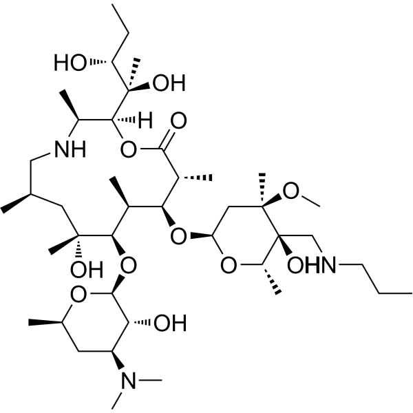
Tulathromycin B (CP 547272) is an isomer of Tulathromycin. Tulathromycin is a semi-synthetic macrolide triamilide that inhibits protein synthesis by binding to the 50S subunit of bacterial ribosomes and exhibits antibacterial activity. Tulathromycin B can be used in studies such as the analysis of Tulathromycin isomers, physicochemical properties, and veterinary drug residues.
For research use only. We do not sell to patients.
- CAS No.: 280755-12-6
- Formula: C41H79N3O12
- Molecular Weight:806.08
-
Storage:
-20°C, stored under nitrogen
* In solvent : -80°C, 6 months; -20°C, 1 month (stored under nitrogen)

To place orders, for customer services and technical support, please contact: MedChemExpress USA
Tel: 609-228-6898 E-mail: [email protected] [email protected]


-
 Biological Activity
-
Chemical Information
- Protocols
- Purity & Documentation
- References
-
 Help & FAQs


All Antibiotic Isoforms
More

Biological Activity
Description
In Vitro
MedChemExpress (MCE) has not independently confirmed the accuracy of these methods. They are for reference only.


Chemical Information
-
CAS No. 280755-12-6
-
Appearance Solid
-
Molecular Weight 806.08
-
Formula C41H79N3O12
-
Color White to off-white
-
SMILES
CO[C@@](C)([C@@](O)([C@@H](O1)C)CNCCC)C[C@@H]1O[C@H]2[C@@H]([C@H]([C@](O)(C[C@H](CN[C@H]([C@@](OC([C@@H]2C)=O)([H])[C@](C)(O)[C@H](O)CC)C)C)C)O[C@H]3[C@@H]([C@H](C[C@H](O3)C)N(C)C)O)C
-
Synonyms
CP 547272
-
Shipping
Room temperature in continental US; may vary elsewhere.
-
Storage
-20°C, stored under nitrogen
* In solvent : -80°C, 6 months; -20°C, 1 month (stored under nitrogen)


Purity & Documentation
-
Data Sheet (274 KB)
-
SDS (252 KB)
- Français - FR (252 KB)
- Deutsch - DE (252 KB)
- Norwegian - NO (252 KB)
- Español - ES (252 KB)
- Swedish - SV (252 KB)
- Italian - IT (252 KB)
- Korean - KR (252 KB)
- Portuguese - PT (252 KB)
-
Handling Instructions (2659 KB)
References

Calculators
Concentration (start) × Volume (start) = Concentration (final) × Volume (final)


Tulathromycin B Related Classifications
HY-107213 Related Classifications
 Powered by Bioz