



Voreloxin
Based on 3 publication(s) in Google Scholar
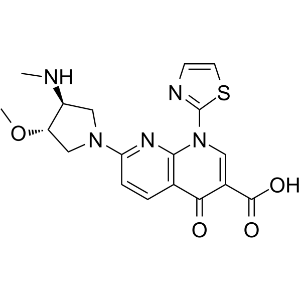
Voreloxin (SNS-595; Vosaroxin; AG 7352) is a first-in-class topoisomerase II inhibitor that intercalates DNA and induces site-selective DNA DSB, G2 arrest, and apoptosis.
For research use only. We do not sell to patients.
- Purity : 99.92%
- CAS No.: 175414-77-4
- Formula: C18H19N5O4S
- Molecular Weight:401.44
-
Storage:
4°C, protect from light, stored under nitrogen
* In solvent : -80°C, 6 months; -20°C, 1 month (protect from light, stored under nitrogen)

To place orders, for customer services and technical support, please contact: MedChemExpress USA
Tel: 609-228-6898 E-mail: [email protected] [email protected]


-
 Biological Activity
-
Chemical Information
- Protocols
- Purity & Documentation
- References
-
 Help & FAQs


-
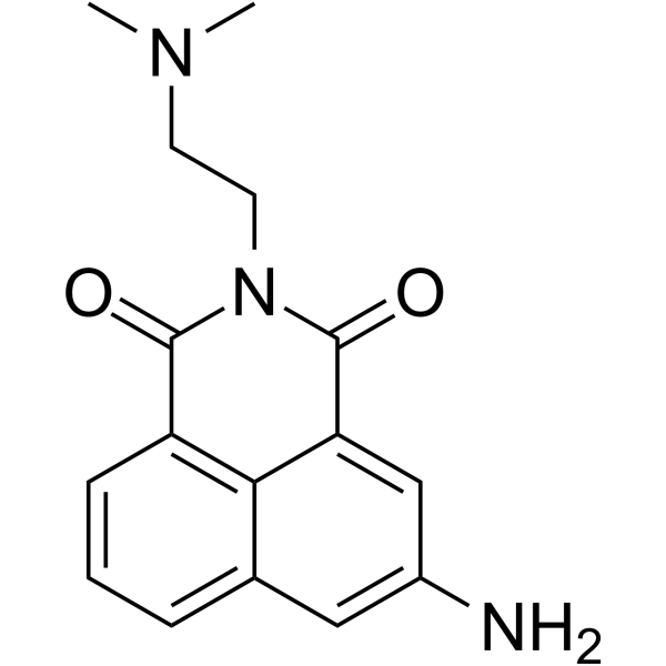
HY-10982
AmonafideTopoisomerase Inhibitor -
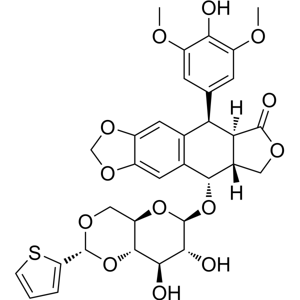
HY-13761
TeniposideTopoisomerase Inhibitor -
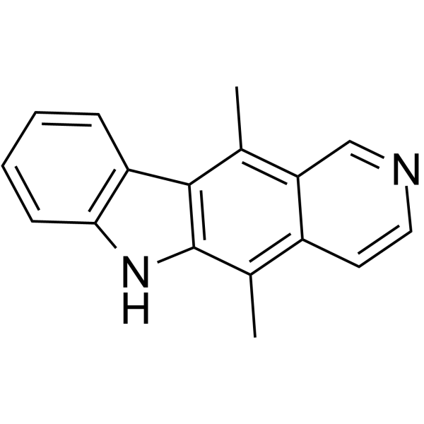
HY-15753
EllipticineTopoisomerase Inhibitor -
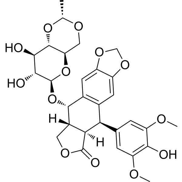
HY-13629
EtoposideTopoisomerase II Inhibitor -
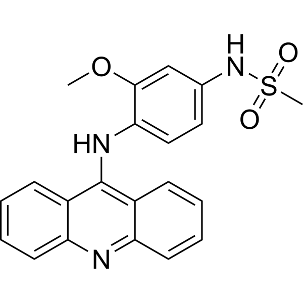
HY-13551
AmsacrineAutophagy Inducer -
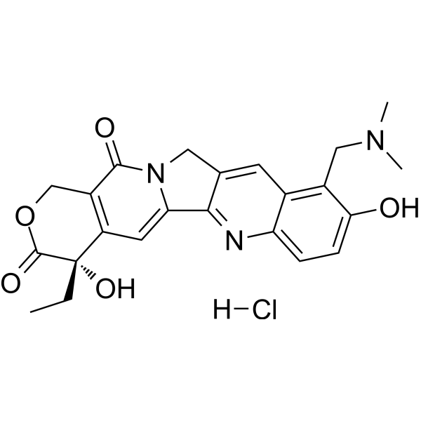
HY-13768A
Topotecan hydrochlorideTopoisomerase I Inhibitor -
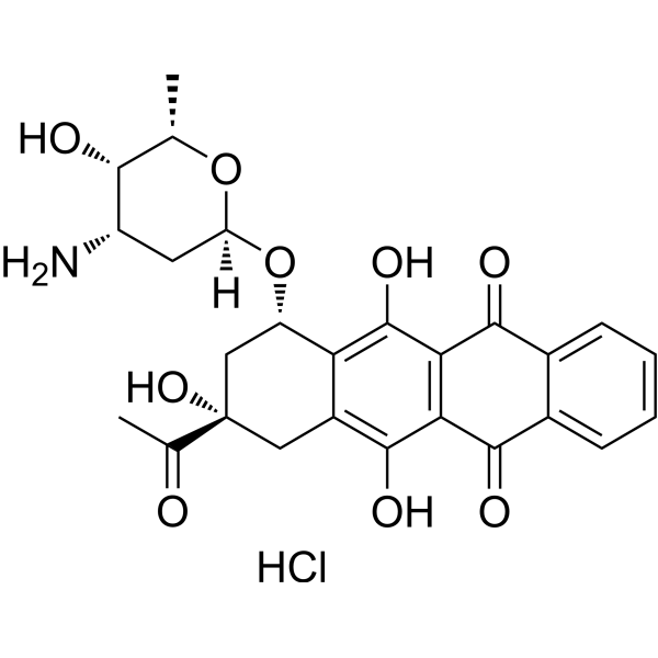
HY-17381
Idarubicin hydrochlorideTopo II Inhibitor -
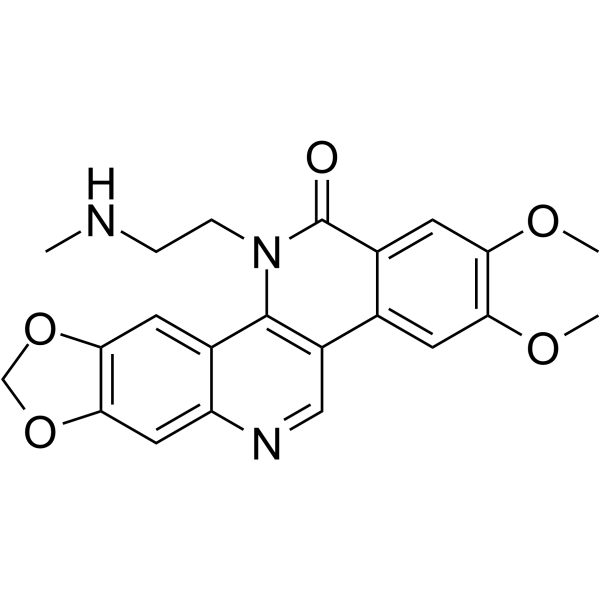
HY-16228
Genz-644282 -
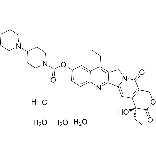
HY-16568
Irinotecan hydrochloride trihydrateTopoisomerase I Inhibitor -
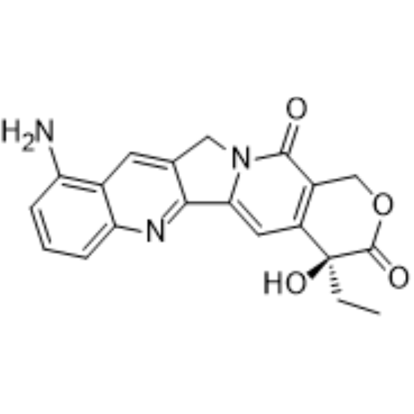
HY-100309
9-AminocamptothecinTopoisomerase I Inhibitor -

HY-10534R
Voreloxin (Standard)Reference Standard -
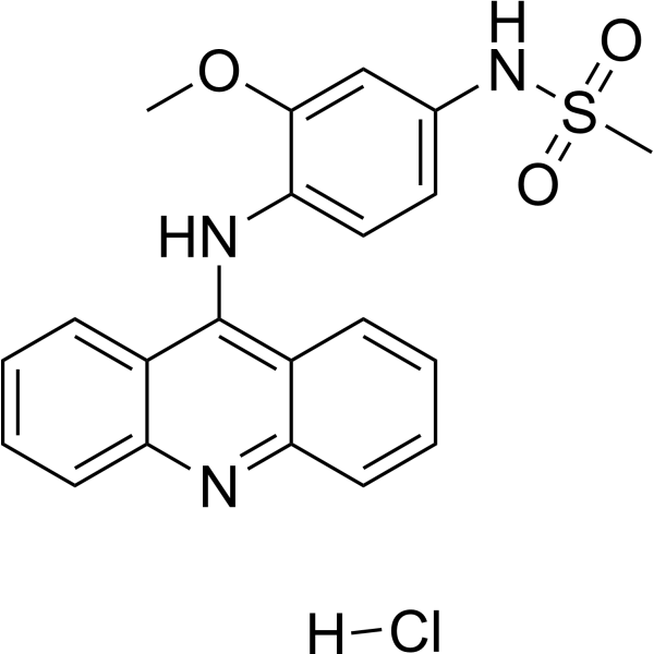
HY-13551A
Amsacrine hydrochlorideAutophagy Inducer
Publications Citing Use of MedChemExpress (MCE) Voreloxin
MoreAll Topoisomerase Isoforms
More

Biological Activity
Description
IC50 & Target
[1]|
Topoisomerase II |
Cellular Effect
|
Cell Line

|
Type | Value | Description | References |
|---|---|---|---|---|
| HL-60 | IC50 |
0.0498 μg/mL
Compound: 3
|
Cytotoxic activity against human tumor HL-60 leukemia cells
Cytotoxic activity against human tumor HL-60 leukemia cells
|
[PMID: 15056007] |
| Hs746T | IC50 |
0.194 μg/mL
Compound: 3
|
Cytotoxic activity against human tumor Hs 746.T stomach cells
Cytotoxic activity against human tumor Hs 746.T stomach cells
|
[PMID: 15056007] |
| PANC-1 | IC50 |
0.388 μg/mL
Compound: 3
|
Cytotoxic activity against human tumor PANC-1 pancreas cells
Cytotoxic activity against human tumor PANC-1 pancreas cells
|
[PMID: 15056007] |
| SBC-3 | IC50 |
0.0319 μg/mL
Compound: 3
|
Cytotoxic activity against human tumor SBC-3 lung cells
Cytotoxic activity against human tumor SBC-3 lung cells
|
[PMID: 15056007] |
| SCaBER | IC50 |
0.121 μg/mL
Compound: 3
|
Cytotoxic activity against human tumor SCaBER bladder cells
Cytotoxic activity against human tumor SCaBER bladder cells
|
[PMID: 15056007] |
| SK-OV-3 | IC50 |
0.266 μg/mL
Compound: 3
|
Cytotoxic activity against human tumor SK-OV-3 ovary cells
Cytotoxic activity against human tumor SK-OV-3 ovary cells
|
[PMID: 15056007] |
In Vitro
Voreloxin is a first-in-class topoisomerase II poison and inhibitor that intercalates DNA and induces site-selective DNA DSB, G2 arrest, and apoptosis. Voreloxin (0.1-20 μM) inhibits topoisomerase II activity and induces site-selective DNA DSB in CCRF-CEM cells. Voreloxin (0.11, 0.33, 1, 3 μM) induces G2 arrest partially through topoisomerase II in A549 lung cancer cell line. Voreloxin cytotoxic activity requires DNA intercalation. However, Voreloxin (1-9 μM) does not generate significant levels of ROS[1]. Voreloxin has potent cytotoxic activity in AML cell lines MV4-11 and HL-60, with IC50s of 95 ± 8 nM and 884 ± 114 nM, respectively. Voreloxin in combination with cytarabine shows additive or synergistic activity in acute leukemia cell lines[2]. Voreloxin is active on the primary acute myeloid leukemia (AML) with a mean LD50 of 2.3 μM. The LD50 for voreloxin in myeloid cell lines NB4 and HL-60 is 0.59 μM ± 0.25 μM. Voreloxin causes accumulation of cells in the S and G2 phases of the cell cycle and acts on topoisomerase II[3].
MedChemExpress (MCE) has not independently confirmed the accuracy of these methods. They are for reference only. Further protocols information, click here.
In Vivo
MedChemExpress (MCE) has not independently confirmed the accuracy of these methods. They are for reference only.
Clinical Trial
| NCT Number | Sponsor | Condition | Start Date |
Phase
|
|---|---|---|---|---|
| NCT01329991 | Plexxikon| | 2011-05 | PHASE1 |


Chemical Information
-
CAS No. 175414-77-4
-
Appearance Solid
-
Molecular Weight 401.44
-
Formula C18H19N5O4S
-
Color White to light yellow
-
SMILES
O=C(C1=CN(C2=NC=CS2)C3=C(C=CC(N4C[C@H](OC)[C@@H](NC)C4)=N3)C1=O)O
-
Synonyms
SNS-595; Vosaroxin; AG 7352
-
Shipping
Room temperature in continental US; may vary elsewhere.
-
Storage
4°C, protect from light, stored under nitrogen
* In solvent : -80°C, 6 months; -20°C, 1 month (protect from light, stored under nitrogen)


Publications (3)
-
Journal Impact Factor
-
Most Recent
-
Drug Des Devel Ther
Advances in the drug therapies of acute myeloid leukemia (except acute wpromyelocytic leukemia). [Abstract]2018 Apr 30:12:1009-1017. PMID: 29750014 -
-
Oncotarget
A novel, dual pan-PIM/FLT3 inhibitor SEL24 exhibits broad therapeutic potential in acute myeloid leukemia. [Abstract]2018 Mar 30;9(24):16917-16931. PMID: 29682194

Protocols
-
Apoptosis
Apoptosis, also called programmed cell death, is generally characterized by distinct morphological characteristics.
-
TUNEL staining for apoptotic DNA fragmentation
TUNEL staining detects DNA strand breaks by using terminal deoxynucleotidyl transferase to add labeled nucleotides to exposed 3′-OH DNA termini, generating either microscopic staining in fixed cells or tissue sections, or fluorescence/cytometric signal in cell suspensions. TUNEL positivity reflects DNA fragmentation but should not be interpreted alone as definitive apoptosis, because TUNEL can also label necrotic, autolytic, mechanically damaged, or DNA-repair-associated DNA breaks.
-
Annexin V plus membrane-impermeant dye apoptosis staining
Annexin V-based apoptosis assays rely on the detection of phosphatidylserine (PS) externalization from the inner leaflet of the plasma membrane to the outer leaflet, an early biochemical hallmark of apoptosis. Fluorescently labeled Annexin V binds PS in a calcium-dependent manner, enabling identification of early apoptotic cells by flow cytometry or fluorescence microscopy. When combined with a membrane-impermeant DNA-binding dye (e. g. , propidium iodide), this approach allows discrimination between viable (Annexin V−/dye−), early apoptotic (Annexin V+/dye−), and late apoptotic or necrotic (Annexin V+/dye+) cell populations by assessing membrane integrity and PS exposure.
-
Apoptosis Solutions
Apoptosis is a regulated, generally non-lytic cell-death pathway that removes unwanted, damaged, infected, or abnormal cells through coordinated morphological changes, caspase activation, DNA fragmentation, and membrane remodeling. The intrinsic apoptosis pathway is controlled mainly by mitochondrial outer membrane permeabilization, BCL-2 family proteins, cytochrome c release, apoptosome formation, caspase-9 activation, and downstream executioner caspase-3/7 activation. The extrinsic apoptosis pathway is initiated by death receptors such as Fas, TNFR, and TRAIL receptors, which recruit adaptor proteins and activate caspase-8 before engaging executioner caspases or mitochondrial amplification through BID cleavage. Apoptosis is linked to many phenotypes, including cancer cell killing, tissue homeostasis, immune regulation, neurodegeneration, infection response, and treatment-induced cytotoxicity; unresolved questions include how apoptosis interacts with necroptosis, pyroptosis, ferroptos


Purity & Documentation
-
Data Sheet (287 KB)
-
SDS (252 KB)
- English - EN (252 KB)
- Français - FR (252 KB)
- Deutsch - DE (252 KB)
- Norwegian - NO (252 KB)
- Español - ES (252 KB)
- Swedish - SV (252 KB)
- Italian - IT (252 KB)
- Korean - KR (252 KB)
- Portuguese - PT (252 KB)
-
Handling Instructions (2659 KB)
References
[1]. Hotinski AK, et al. Vosaroxin is a novel topoisomerase-II inhibitor with efficacy in relapsed and refractory acute myeloid leukaemia. Expert Opin Pharmacother. 2015 Jun;16(9):1395-402. [Content Brief]
[2]. Scatena CD, et al. Voreloxin, a first-in-class anticancer quinolone derivative, acts synergistically with cytarabine in vitro and induces bone marrow aplasia in vivo. Cancer Chemother Pharmacol. 2010 Oct;66(5):881-8. [Content Brief]
[3]. Walsby EJ, et al. The topoisomerase II inhibitor voreloxin causes cell cycle arrest and apoptosis in myeloid leukemia cells and acts in synergy with cytarabine. Haematologica. 2011 Mar;96(3):393-9. [Content Brief]

Calculators
Concentration (start) × Volume (start) = Concentration (final) × Volume (final)


Voreloxin Related Classifications
HY-10534 Related Classifications
 Powered by Bioz