



10Panx
Based on 1 publication(s) in Google Scholar
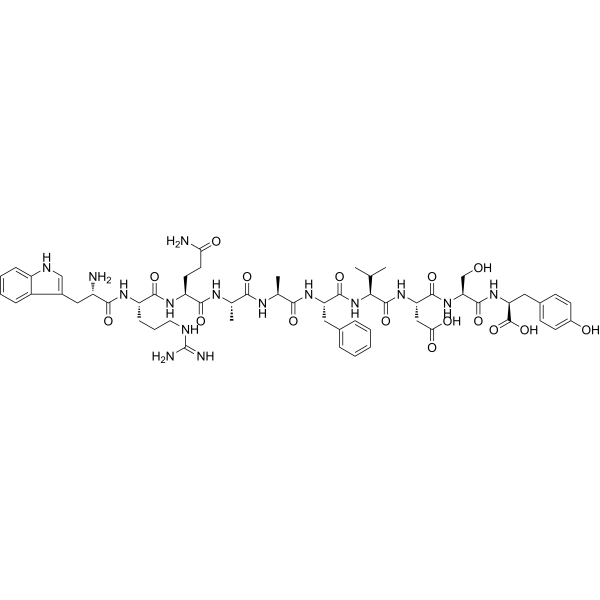
10Panx is a competitive inhibitor of selective Pannexin 1 (PANX1) channels. 10Panx blocks the opening of PANX1 channels, inhibits ATP release and downstream P2X7 receptor-mediated signaling pathways, thereby reducing cell death and inflammatory responses. 10Panx can be used in the study of diseases such as neuropathic pain, inflammatory bowel disease, and Clostridioides difficile infection. 10Panx can effectively reduce mechanical hyperalgesia and enhanced C-reflexes, and inhibit the expression of pro-inflammatory factors such as IL-6[1][2][3].
For research use only. We do not sell to patients.
- Purity : 99.34%
- CAS No.: 955091-53-9
- Formula: C58H79N15O16
- Molecular Weight:1242.34
-
Storage:
Sealed storage, away from moisture and light.
Powder -80°C, 2 years , -20°C, 1 year* In solvent : -80°C, 6 months; -20°C, 1 month (sealed storage, away from moisture and light)

To place orders, for customer services and technical support, please contact: MedChemExpress USA
Tel: 609-228-6898 E-mail: [email protected] [email protected]


-
 Biological Activity
-
Chemical Information
-
Solvent & Solubility
- Protocols
- Purity & Documentation
- References
-
 Help & FAQs


-
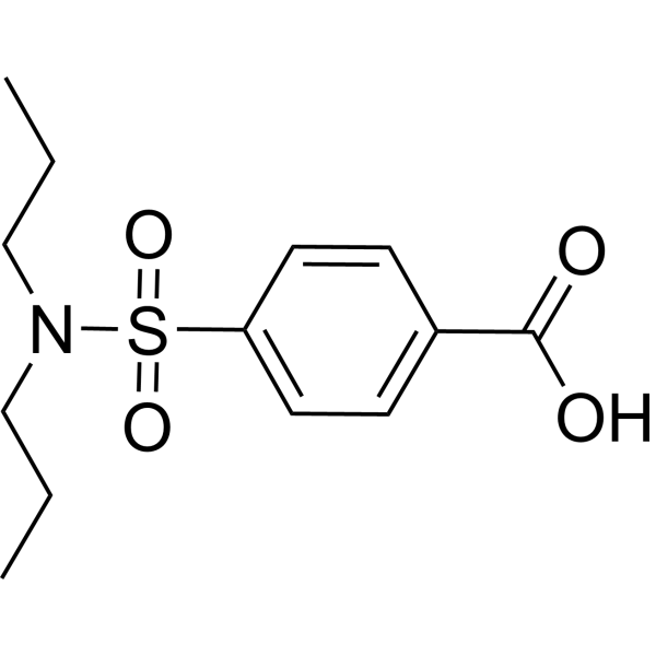
HY-B0545
ProbenecidBacterial Inhibitor -
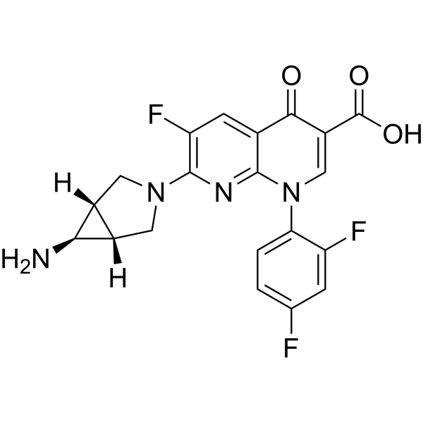
HY-A0170
TrovafloxacinAntibiotic Agent -
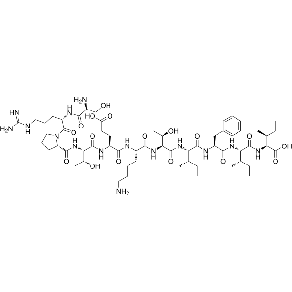
HY-P0139
Gap 27Gap Junction Inhibitor -
HY-100545
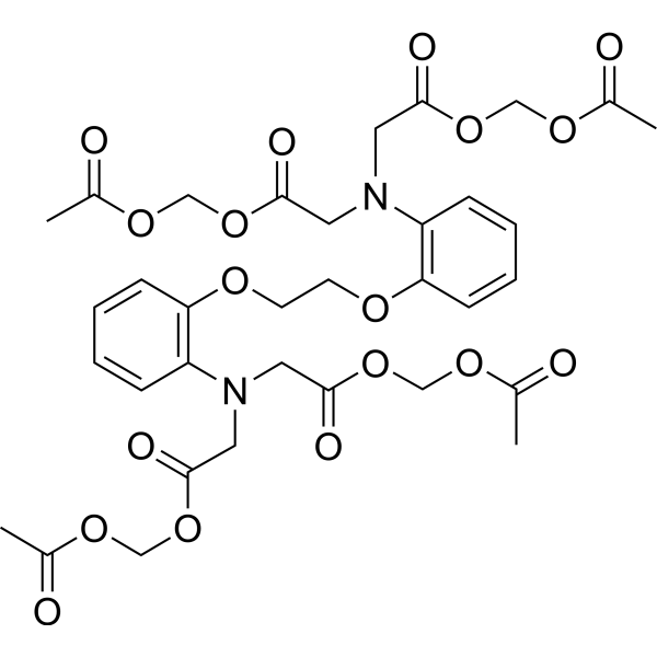
BAPTA-AMCa2+ Chelator -

HY-P1082
Gap 26Gap Junction Inhibitor -
HY-136254
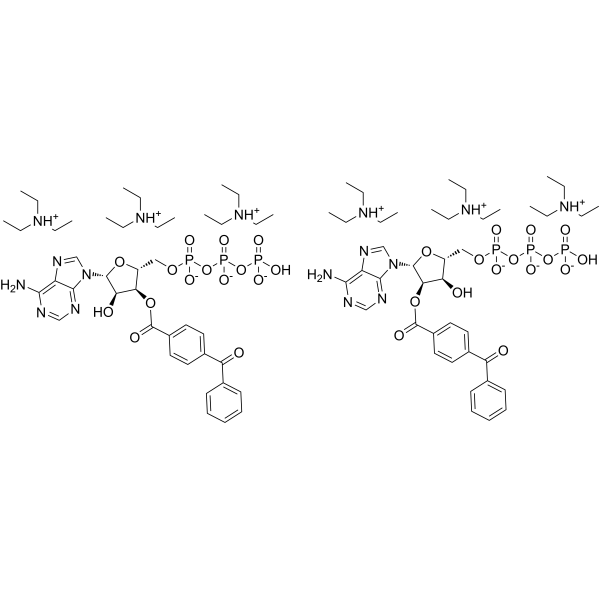
BzATP triethylammonium saltP2X Agonist -
HY-50697
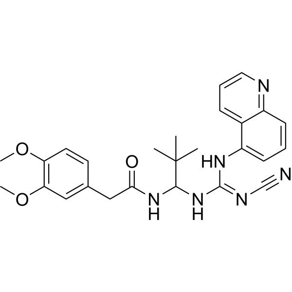
A-740003P2X Receptor Antagonist -
HY-15204
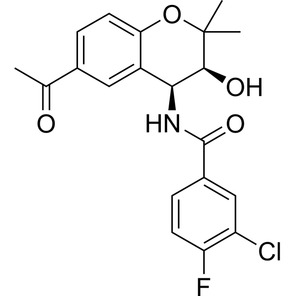
TonabersatGap Junction Modulator -

HY-P1138
Scrambled 10PanxInactive Scrambled Control Peptide -
HY-10913A
Danegaptide HydrochlorideGap Junction Modifier -
HY-13948
Angiotensin II humanVasoconstrictor -
HY-P1136
Gap19Cx43 Hemichannel Blocker
Publications Citing Use of MedChemExpress (MCE) 10Panx
More

Biological Activity
Description
In Vitro
10Panx (50 μM; 1 h pretreatment followed by 18 h co-treatment) significantly inhibited TcdA- and TcdB-induced enteric glial cell apoptosis (reduced caspase-3/7 activity and annexin V binding) but did not affect IL-6 gene expression[3].
MedChemExpress (MCE) has not independently confirmed the accuracy of these methods. They are for reference only.
In Vivo
MedChemExpress (MCE) has not independently confirmed the accuracy of these methods. They are for reference only.


Chemical Information
-
CAS No. 955091-53-9
-
Appearance Solid
-
Molecular Weight 1242.34
-
Formula C58H79N15O16
-
Color White to off-white
-
Sequence
Trp-Arg-Gln-Ala-Ala-Phe-Val-Asp-Ser-Tyr
-
Sequence Shortening
WRQAAFVDSY
-
Shipping
Room temperature in continental US; may vary elsewhere.
-
Storage
Sealed storage, away from moisture and light
Powder -80°C 2 years -20°C 1 year * In solvent : -80°C, 6 months; -20°C, 1 month (sealed storage, away from moisture and light)


Publications (1)
-
Journal Impact Factor
-
Most Recent


Solvent & Solubility
In Vitro:
H2O : 11.11 mg/mL (8.94 mM; adjust pH to 12 with 1 M NaOH)
DMSO : 6.67 mg/mL (5.37 mM; Need ultrasonic; Hygroscopic DMSO has a significant impact on the solubility of product, please use newly opened DMSO)
Please refer to the solubility information to select the appropriate solvent. Once prepared, please aliquot and store the solution to prevent product inactivation from repeated freeze-thaw cycles.
Storage method and period of stock solution: -80°C, 6 months; -20°C, 1 month (sealed storage, away from moisture and light). When stored at -80°C, please use it within 6 months. When stored at -20°C, please use it within 1 month.
* Note: If you choose water as the stock solution, please dilute it to the working solution, then filter and sterilize it with a 0.22 μm filter before use.
Please refer to the solubility information to select the appropriate solvent. Once prepared, please aliquot and store the solution to prevent product inactivation from repeated freeze-thaw cycles.
Storage method and period of stock solution: -80°C, 6 months; -20°C, 1 month (sealed storage, away from moisture and light). When stored at -80°C, please use it within 6 months. When stored at -20°C, please use it within 1 month.
* Note: If you choose water as the stock solution, please dilute it to the working solution, then filter and sterilize it with a 0.22 μm filter before use.
Concentration (start) × Volume (start) = Concentration (final) × Volume (final)
In Vivo:
Select the appropriate dissolution method based on your experimental animal and administration route.
- For the following dissolution methods, please ensure to first prepare a clear stock solution using an In Vitro approach and then sequentially add co-solvents:
- To ensure reliable experimental results, the clarified stock solution can be appropriately stored based on storage conditions. As for the working solution for In Vivo experiments, it is recommended to prepare freshly and use it on the same day.
- The percentages shown for the solvents indicate their volumetric ratio in the final prepared solution. If precipitation or phase separation occurs during preparation, heat and/or sonication can be used to aid dissolution.
Add each solvent one by one: 10% DMSO 40% PEG300 5% Tween-80 45% Saline
Solubility: ≥ 0.67 mg/mL (0.54 mM); Clear solution
This protocol yields a clear solution of ≥ 0.67 mg/mL (saturation unknown).
Taking 1 mL working solution as an example, add 100 μL DMSO stock solution (6.7 mg/mL) to 400 μL PEG300, and mix evenly; then add 50 μL Tween-80 and mix evenly; then add 450 μL Saline to adjust the volume to 1 mL.
Preparation of Saline: Dissolve 0.9 g sodium chloride in ddH₂O and dilute to 100 mL to obtain a clear Saline solution.
Add each solvent one by one: 10% DMSO 90% (20% SBE-β-CD in Saline)
Solubility: ≥ 0.67 mg/mL (0.54 mM); Clear solution
This protocol yields a clear solution of ≥ 0.67 mg/mL (saturation unknown).
Taking 1 mL working solution as an example, add 100 μL DMSO stock solution (6.7 mg/mL) to 900 μL 20% SBE-β-CD in Saline, and mix evenly.
Preparation of 20% SBE-β-CD in Saline (4°C, storage for one week): 2 g SBE-β-CD powder is dissolved in 10 mL Saline, completely dissolve until clear.
In Vivo Dissolution Calculator
Please enter the basic information of animal experiments:
-
-
-
-
Recommended: Prepare an additional quantity of animals to account for potential losses during experiments.
Please enter your animal formula composition:
-
%DMSO +
Recommended: Keep the proportion of DMSO in working solution below 2% if your animal is weak.
-
%+
-
+%Tween-80 + +
-
%Saline +
The co-solvents required include: DMSO, . All of co-solvents are available by MedChemExpress (MCE). , Tween 80. All of co-solvents are available by MedChemExpress (MCE).
Working solution concentration: 0.22 mg/mL
Method for preparing stock solution: mg drug dissolved in μL DMSO. Stock solution concentration: mg/mL. * In solvent : -80°C, 6 months; -20°C, 1 month (sealed storage, away from moisture and light)
1. Take μL DMSO stock solution;
2. Add μL .
μL , mix evenly;
3. Then add μL Tween 80, mix evenly;
4. Then add μL
Please ensure that the stock solution in the first step is dissolved to a clear state, and add co-solvents in sequence. You can use ultrasonic heating (ultrasonic cleaner, recommended frequency 20-40 kHz), vortexing, etc. to assist dissolution.


Purity & Documentation
-
Data Sheet (273 KB)
-
SDS (252 KB)
- English - EN (252 KB)
- Français - FR (252 KB)
- Deutsch - DE (252 KB)
- Norwegian - NO (252 KB)
- Español - ES (252 KB)
- Swedish - SV (252 KB)
- Italian - IT (252 KB)
- Korean - KR (252 KB)
- Portuguese - PT (252 KB)
-
Handling Instructions (2659 KB)
References
[1]. Aresta Branco MSL, et al. The Pannexin 1 Channel and the P2X7 Receptor Are in Complex Interplay to Regulate the Release of Soluble Ectonucleotidases in the Murine Bladder Lamina Propria. Int J Mol Sci. 2023 Jun 9;24(12):9964. [Content Brief]
[2]. Loureiro AV, et al. Role of Pannexin-1-P2X7R signaling on cell death and pro-inflammatory mediator expression induced by Clostridioides difficile toxins in enteric glia. Front Immunol. 2022 Aug 22;13:956340. [Content Brief]
[3]. Bravo D, et al. NMDA and P2X7 Receptors Require Pannexin 1 Activation to Initiate and Maintain Nociceptive Signaling in the Spinal Cord of Neuropathic Rats. Int J Mol Sci. 2022 Jun 16;23(12):6705. [Content Brief]


Complete Stock Solution Preparation Table
Please refer to the solubility information to select the appropriate solvent. Once prepared, please aliquot and store the solution to prevent product inactivation from repeated freeze-thaw cycles.
Storage method and period of stock solution: -80°C, 6 months; -20°C, 1 month (sealed storage, away from moisture and light). When stored at -80°C, please use it within 6 months. When stored at -20°C, please use it within 1 month.
| Optional Solvent | Concentration Solvent Mass | 1 mg | 5 mg | 10 mg | 25 mg |
|---|---|---|---|---|---|
| DMSO / H2O | 1 mM | 0.8049 mL | 4.0247 mL | 8.0493 mL | 20.1233 mL |
| 5 mM | 0.1610 mL | 0.8049 mL | 1.6099 mL | 4.0247 mL |
* Note: If you choose water as the stock solution, please dilute it to the working solution, then filter and sterilize it with a 0.22 μm filter before use.


10Panx Related Classifications
HY-P1137 Related Classifications
 Powered by Bioz