



20-Deoxyingenol
Based on 5 publication(s) in Google Scholar
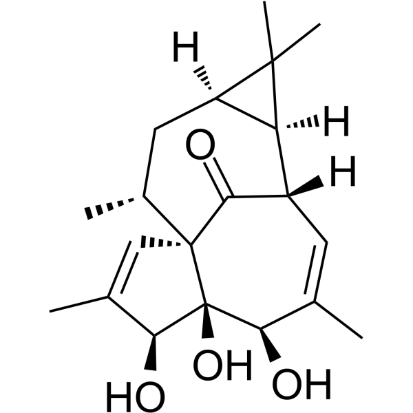
20-Deoxyingenol, a diterpene, is isolated from the roots of Euphorbia kansui. 20-Deoxyingenol can promote autophagy and lysosomal biogenesis by promoting the nuclear translocation of transcription factor EB (TFEB) in vitro. 20-Deoxyingenol can be used for the research of osteoarthritis (OA).
For research use only. We do not sell to patients.
- Purity : 98.74%
- CAS No.: 54706-99-9
- Formula: C20H28O4
- Molecular Weight:332.43
-
Storage:Powder -20°C, 3 years , 4°C, 2 years ; In solvent -80°C, 2 years , -20°C, 1 year

To place orders, for customer services and technical support, please contact: MedChemExpress USA
Tel: 609-228-6898 E-mail: [email protected] [email protected]


-
 Biological Activity
-
Chemical Information
-
Solvent & Solubility
- Protocols
- Purity & Documentation
- References
-
 Help & FAQs


Publications Citing Use of MedChemExpress (MCE) 20-Deoxyingenol
More

Biological Activity
Description
Cellular Effect
|
Cell Line

|
Type | Value | Description | References |
|---|---|---|---|---|
| MT4 | CC50 |
>12 μM
Compound: 9
|
Cytotoxicity against human MT4 cells after 3 days by CytoTox-Glo assay
Cytotoxicity against human MT4 cells after 3 days by CytoTox-Glo assay
|
[PMID: 30031972] |
In Vitro
20-Deoxyingenol (2.5-10 mM; 24 h) protects chondrocytes against Tert-butyl hydroperoxide solution (TBHP; 100 μM)-induced cell death[2].
20-Deoxyingenol (5-10 mM) decreases the TBHP-induced upregulation of apoptosis protein cleaved-caspase3 and the senescence protein p16INK4a in chondrocytes[2].
20-Deoxyingenol (2.5-40 mM; 24 h) has no cytotoxic effect on chondrocytes at the concentration less than 10 mM[2].
20-Deoxyingenol (10 mM; 24 h) restores autophagy flux in TBHP treated chondrocytes[2].
20-Deoxyingenol (4-10 mM; 24 h) promotes the nuclear level of TFEB in TBHP treated chondrocytes[2].
MedChemExpress (MCE) has not independently confirmed the accuracy of these methods. They are for reference only. Further protocols information, click here.
In Vivo
MedChemExpress (MCE) has not independently confirmed the accuracy of these methods. They are for reference only.
-
Animal Model:10-week-old C57BL/6 male wild-type (WT) mice with destabilization of the medial meniscus (DMM)[2]
-
Dosage:20 mg/kg
-
Administration:I.p. one time per day, for eight consecutive weeks
-
Result:Had a slightly wider joint space and reduced bone density and calcification compared with the DMM group.
Inhibited the decrease in the thickness of hyaline cartilage (HC), and alleviated the disorder and hypertrophy of chondrocytes in the joint tissues of mice after DMM surgery.
Had less erosion on the surface of the articular cartilage and more proteoglycan content.
Had more positive staining points of LAMP1 and LC3 II, and less cleaved-caspase3 and P16INK4a.
Increased the nuclear level of TFEB.


Chemical Information
-
CAS No. 54706-99-9
-
Appearance Solid
-
Molecular Weight 332.43
-
Formula C20H28O4
-
Color White to off-white
-
SMILES
O=C1[C@@]2(C=C(C)[C@@H]3O)[C@]3(O)[C@H](O)C(C)=C[C@@]1([H])[C@@]4([H])[C@@](C4(C)C)([H])C[C@H]2C
-
Structure Classification
-
Initial Source
-
Shipping
Room temperature in continental US; may vary elsewhere.
-
Storage
Powder -20°C 3 years 4°C 2 years In solvent -80°C 2 years -20°C 1 year


Publications (5)
-
Journal Impact Factor
-
Most Recent
-
Pharmacol Res
2021 Mar:165:105361. PMID: 33460793 -
Phytomedicine
20-Deoxyingenol attenuated doxorubicin-induced cardiotoxicity by promoting autolysosome degradation through the UCHL3-TFEB pathway. [Abstract]2025 Nov:147:157220. PMID: 40916238 -
Phytomedicine
Palmatine activation of TFEB enhances autophagy and alleviates endoplasmic reticulum stress in intervertebral disc degeneration. [Abstract]2025 Apr:139:156431. PMID: 39933468 -
Mol Neurobiol
20-Deoxyingenol Activates Mitophagy Through TFEB and Promotes Functional Recovery After Spinal Cord Injury. [Abstract]2025 Jan;62(1):445-460. PMID: 38865079 -
Naunyn Schmiedebergs Arch Pharmacol
Effect of oleuropein on morphine-induced hippocampus neurotoxicity and memory impairments in rats. [Abstract]2019 Nov;392(11):1383-1391. PMID: 31236657


Solvent & Solubility
In Vitro:
DMSO : 50 mg/mL (150.41 mM; Need ultrasonic; Hygroscopic DMSO has a significant impact on the solubility of product, please use newly opened DMSO)
Please refer to the solubility information to select the appropriate solvent. Once prepared, please aliquot and store the solution to prevent product inactivation from repeated freeze-thaw cycles.
Storage method and period of stock solution: -80°C, 2 years; -20°C, 1 year. When stored at -80°C, please use it within 2 years. When stored at -20°C, please use it within 1 year.
Please refer to the solubility information to select the appropriate solvent. Once prepared, please aliquot and store the solution to prevent product inactivation from repeated freeze-thaw cycles.
Storage method and period of stock solution: -80°C, 2 years; -20°C, 1 year. When stored at -80°C, please use it within 2 years. When stored at -20°C, please use it within 1 year.
Concentration (start) × Volume (start) = Concentration (final) × Volume (final)
In Vivo:
Select the appropriate dissolution method based on your experimental animal and administration route.
- For the following dissolution methods, please ensure to first prepare a clear stock solution using an In Vitro approach and then sequentially add co-solvents:
- To ensure reliable experimental results, the clarified stock solution can be appropriately stored based on storage conditions. As for the working solution for In Vivo experiments, it is recommended to prepare freshly and use it on the same day.
- The percentages shown for the solvents indicate their volumetric ratio in the final prepared solution. If precipitation or phase separation occurs during preparation, heat and/or sonication can be used to aid dissolution.
Add each solvent one by one: 10% DMSO 40% PEG300 5% Tween-80 45% Saline
Solubility: 2.5 mg/mL (7.52 mM); Suspended solution; Need ultrasonic
This protocol yields a suspended solution of 2.5 mg/mL. Suspended solution can be used for oral and intraperitoneal injection.
Taking 1 mL working solution as an example, add 100 μL DMSO stock solution (25.0 mg/mL) to 400 μL PEG300, and mix evenly; then add 50 μL Tween-80 and mix evenly; then add 450 μL Saline to adjust the volume to 1 mL.
Preparation of Saline: Dissolve 0.9 g sodium chloride in ddH₂O and dilute to 100 mL to obtain a clear Saline solution.
Add each solvent one by one: 10% DMSO 90% (20% SBE-β-CD in Saline)
Solubility: 2.5 mg/mL (7.52 mM); Suspended solution; Need ultrasonic
This protocol yields a suspended solution of 2.5 mg/mL. Suspended solution can be used for oral and intraperitoneal injection.
Taking 1 mL working solution as an example, add 100 μL DMSO stock solution (25.0 mg/mL) to 900 μL 20% SBE-β-CD in Saline, and mix evenly.
Preparation of 20% SBE-β-CD in Saline (4°C, storage for one week): 2 g SBE-β-CD powder is dissolved in 10 mL Saline, completely dissolve until clear.
In Vivo Dissolution Calculator
Please enter the basic information of animal experiments:
-
-
-
-
Recommended: Prepare an additional quantity of animals to account for potential losses during experiments.
Please enter your animal formula composition:
-
%DMSO +
Recommended: Keep the proportion of DMSO in working solution below 2% if your animal is weak.
-
%+
-
+%Tween-80 + +
-
%Saline +
The co-solvents required include: DMSO, . All of co-solvents are available by MedChemExpress (MCE). , Tween 80. All of co-solvents are available by MedChemExpress (MCE).
Working solution concentration: 0.22 mg/mL
Method for preparing stock solution: mg drug dissolved in μL DMSO. Stock solution concentration: mg/mL.
1. Take μL DMSO stock solution;
2. Add μL .
μL , mix evenly;
3. Then add μL Tween 80, mix evenly;
4. Then add μL
Please ensure that the stock solution in the first step is dissolved to a clear state, and add co-solvents in sequence. You can use ultrasonic heating (ultrasonic cleaner, recommended frequency 20-40 kHz), vortexing, etc. to assist dissolution.

Protocols
-
Nuclear Protein Extraction (High-Salt/Hypotonic Fractionation)
The high-salt/hypotonic fractionation method for nuclear protein extraction is based on the differential solubility of cellular components. Cytoplasmic proteins are extracted first using a hypotonic buffer that causes cell swelling and membrane rupture, followed by centrifugation to separate the cytoplasmic supernatant from the nuclear pellet. The nuclear pellet is then subjected to high-salt extraction (e. g. , 0. 4 M (NH4)2SO4 or 1 M NaCl) to solubilize tightly bound nuclear matrix proteins, including transcription factors, histones, and structural proteins associated with chromatin and the nuclear scaffold. This approach allows for the isolation of both soluble cytoplasmic proteins and salt-resistant nuclear proteins while minimizing cross-contamination.
-
Cytoplasmic-Nuclear Fractionated Protein Extraction
Cytoplasmic-nuclear fractionated protein extraction separates soluble cytoplasmic proteins from nuclear-enriched proteins by mild plasma-membrane permeabilization, differential centrifugation, washing of nuclei, and extraction of nuclear proteins for downstream immunoblotting or related molecular analysis. The readout is the relative abundance of a protein in cytoplasmic and nuclear fractions, commonly assessed by western blotting together with compartment markers such as tubulin or pyruvate kinase for cytoplasm and lamin, nucleoporin, hnRNP, H2AX, or Lamin B for nuclear fractions.
-
Autophagy
Autophagy is a process in which eukaryotic cells use lysosomes to degrade their own cytoplasmic proteins and damaged organelles under the regulation of autophagy related gene (Atg). Microtubule-associated proteins light chain 3 (LC3) is recognized as autophagy marker, which transfers from cytoplasmic LC3 (LC3-I) to membrane type (LC3-II). LC3-II/I ratio could be detected by Western Blot and fluorescence microscopy.
-
Lysosome and acidic-vesicle live-cell staining
Lysosome and acidic-vesicle live-cell staining detects acidic intracellular compartments by using membrane-permeant acidotropic probes that accumulate in low-pH vesicles, including lysosomes, late endosomes, autolysosomes, and acidic phagosomes. LysoTracker staining is commonly used as an intensity-based readout of acidic lysosomal compartment abundance or enlargement, while acridine orange produces green fluorescence in less concentrated compartments and red fluorescence after concentration-dependent accumulation in acidic vesicular organelles. Loss or reduction of acridine-orange red signal can be used as a readout of lysosomal membrane permeabilization or reduced acidic-vesicle integrity. This protocol is designed for live cultured cells and can be adapted for fluorescence microscopy, high-content imaging, plate-reader readout, or flow cytometry when the selected literature supports the readout. Because these dyes report acidotropic accumulation rather than lysosome identity alone,
-
Macroautophagy Solutions
Macroautophagy is a conserved lysosome-dependent degradation pathway in which cytoplasmic material is sequestered into double-membrane autophagosomes and delivered to lysosomes for degradation and recycling. The pathway supports cellular homeostasis during nutrient limitation, organelle stress, protein-aggregate accumulation, infection, differentiation, and tissue remodeling by coupling cargo sequestration, autophagosome maturation, lysosomal fusion, and degradation of cargo-derived macromolecules. The core molecular sequence includes initiation by nutrient- and stress-regulated autophagy machinery, autophagosome nucleation, LC3/ATG8-family conjugation to autophagosomal membranes, cargo selection through receptors such as SQSTM1/p62, autophagosome-lysosome fusion, and lysosomal degradation. LC3 was identified as a mammalian homolog of yeast Atg8 that localizes to autophagosomal membranes after processing, and p62/SQSTM1 was shown to connect ubiquitinated cargo with autophagic degradati


Purity & Documentation
-
Data Sheet (287 KB)
-
SDS (251 KB)
- English - EN (251 KB)
- Français - FR (251 KB)
- Deutsch - DE (251 KB)
- Norwegian - NO (251 KB)
- Español - ES (251 KB)
- Swedish - SV (251 KB)
- Italian - IT (251 KB)
- Korean - KR (251 KB)
- Portuguese - PT (251 KB)
-
Handling Instructions (2659 KB)
References


Complete Stock Solution Preparation Table
Please refer to the solubility information to select the appropriate solvent. Once prepared, please aliquot and store the solution to prevent product inactivation from repeated freeze-thaw cycles.
Storage method and period of stock solution: -80°C, 2 years; -20°C, 1 year. When stored at -80°C, please use it within 2 years. When stored at -20°C, please use it within 1 year.
| Optional Solvent | Concentration Solvent Mass | 1 mg | 5 mg | 10 mg | 25 mg |
|---|---|---|---|---|---|
| DMSO | 1 mM | 3.0082 mL | 15.0408 mL | 30.0815 mL | 75.2038 mL |
| 5 mM | 0.6016 mL | 3.0082 mL | 6.0163 mL | 15.0408 mL | |
| 10 mM | 0.3008 mL | 1.5041 mL | 3.0082 mL | 7.5204 mL | |
| 15 mM | 0.2005 mL | 1.0027 mL | 2.0054 mL | 5.0136 mL | |
| 20 mM | 0.1504 mL | 0.7520 mL | 1.5041 mL | 3.7602 mL | |
| 25 mM | 0.1203 mL | 0.6016 mL | 1.2033 mL | 3.0082 mL | |
| 30 mM | 0.1003 mL | 0.5014 mL | 1.0027 mL | 2.5068 mL | |
| 40 mM | 0.0752 mL | 0.3760 mL | 0.7520 mL | 1.8801 mL | |
| 50 mM | 0.0602 mL | 0.3008 mL | 0.6016 mL | 1.5041 mL | |
| 60 mM | 0.0501 mL | 0.2507 mL | 0.5014 mL | 1.2534 mL | |
| 80 mM | 0.0376 mL | 0.1880 mL | 0.3760 mL | 0.9400 mL | |
| 100 mM | 0.0301 mL | 0.1504 mL | 0.3008 mL | 0.7520 mL |


20-Deoxyingenol Related Classifications
HY-N0866 Related Classifications
 Powered by Bioz