



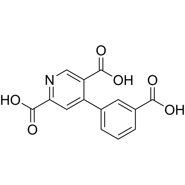
4-CPPC

Based on 1 Customer Validation
4-CPPC is a selective MIF-2 inhibitor with a Ki of 33 µM. 4-CPPC inhibits the COX2/PGE2 pathway, reduces the levels of COX2 and mPGES-1, and decreases the production of PGE2. 4-CPPC attenuates the formation of early atherosclerotic lesions in hyperlipidemic mice, and lowers the levels of circulating inflammatory cytokines and chemokines. 4-CPPC inhibits MIF-2-triggered cell migration. 4-CPPC improves hindlimb motor function in rats with contusion-induced spinal cord injury. 4-CPPC can be used in studies related to atherosclerosis and spinal cord injury.
Nur für Forschungszwecke. Wir verkaufen nicht an Patienten.
- Reinheit : 98.20%
- CAS. Nr.: 29553-70-6
- Formel: C14H9NO6
- Molecular Weight:287.22
-
Speicherung:Powder -20°C, 3 years , 4°C, 2 years ; In solvent -80°C, 6 months , -20°C, 1 month

To place orders, for customer services and technical support, please contact: MedChemExpress USA
Tel: 609-228-6898 E-mail: [email protected] [email protected]


-
 Biologische Aktivität
-
Chemical Information
-
Lösungsmittel & Löslichkeit
- Protokoll
- Reinheit & Dokumentation
- Verweise
-
 Help & FAQs


-
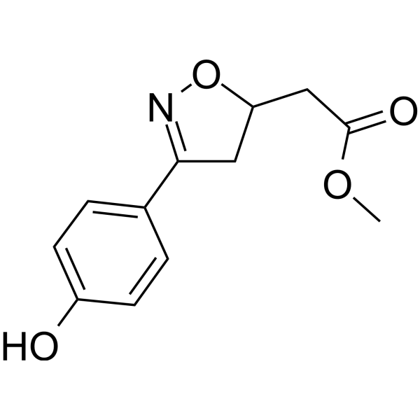
HY-16692
ISO-1MIF Antagonist -
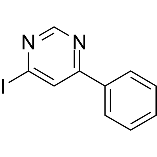
HY-110063
4-IPPMIF Inhibitor -
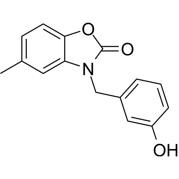
HY-147390
MIF098MIF Antagonist -
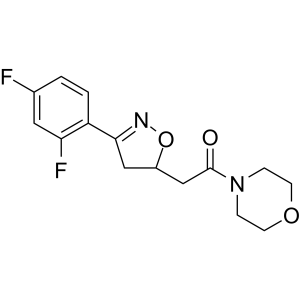
HY-110095
(±)-CPSI-1306MIF Antagonist -
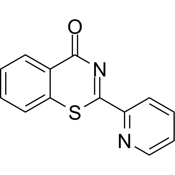
HY-110084
BTZO-1MIF Binder -
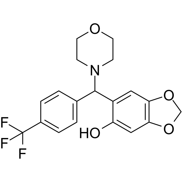
HY-W164560
RDR 03785MIF Inhibitor -
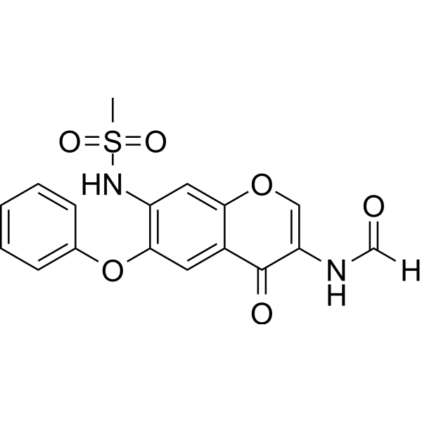
HY-17009
IguratimodAnti-rheumatic Agent -

HY-16692R
ISO-1 (Standard)MIF Antagonist -

HY-17009R
Iguratimod (Standard)Anti-rheumatic Agent


Biologische Aktivität
Beschreibung
|
MIF-2 33 μM (Ki) |
MIF-1 431 μM (Ki) |
COX-2 |
In Vitro
4-CPPC (4-160 nM; 1 h) dose-dependently inhibits MIF-2-induced chemotactic migration of primary peripheral blood mononuclear cells[1].
4-CPPC (100 μM; 24 h) inhibits D-DT-induced activation of the COX2/PGE2 pathway in primary astrocytes, reduces the protein levels of COX2 and mPGES-1, and decreases PGE2 production[2].
4-CPPC competitively inhibits the tautomerase activity of MIF-2 with a Ki value of 33 μM, and exhibits 13-fold selectivity over MIF-1[3].
MedChemExpress (MCE) has not independently confirmed the accuracy of these methods. They are for reference only. Further protocols information, click here.
In Vivo
MedChemExpress (MCE) has not independently confirmed the accuracy of these methods. They are for reference only.
-
Animal Model:C57BL/6 background (male, 8 weeks old, atherosclerotic model via cholesterol-rich high-fat diet)[1]
-
Dosage:5 mg/kg
-
Administration:i.p.; three times per week; 4.5 weeks
-
Result:Markedly reduced aortic root atherosclerotic lesion area.
Reduced lesional CD68-positive macrophage area.
Reduced plasma levels of IL-1α, IL-6, IL-15, and IFN-γ.


Chemical Information
-
CAS. Nr. 29553-70-6
-
Appearance Solid
-
Molecular Weight 287.22
-
Formel C14H9NO6
-
Color White to off-white
-
SMILES
O=C(C1=NC=C(C(O)=O)C(C2=CC=CC(C(O)=O)=C2)=C1)O
-
Versand
Room temperature in continental US; may vary elsewhere.
-
Speicherung
Powder -20°C 3 years 4°C 2 years In solvent -80°C 6 months -20°C 1 month


Lösungsmittel & Löslichkeit
In Vitro:
DMSO : 100 mg/mL (348.17 mM; Need ultrasonic; Hygroscopic DMSO has a significant impact on the solubility of product, please use newly opened DMSO)
Please refer to the solubility information to select the appropriate solvent. Once prepared, please aliquot and store the solution to prevent product inactivation from repeated freeze-thaw cycles.
Storage method and period of stock solution: -80°C, 6 months; -20°C, 1 month. When stored at -80°C, please use it within 6 months. When stored at -20°C, please use it within 1 month.
Please refer to the solubility information to select the appropriate solvent. Once prepared, please aliquot and store the solution to prevent product inactivation from repeated freeze-thaw cycles.
Storage method and period of stock solution: -80°C, 6 months; -20°C, 1 month. When stored at -80°C, please use it within 6 months. When stored at -20°C, please use it within 1 month.
Konzentration (Stammlösung) × Volumen (Stammlösung) = Konzentration (Ziellösung) × Volumen (Ziellösung)

Protokoll
-
Multiplex immunofluorescence IHC
Multiplex immunofluorescence IHC detects multiple protein biomarkers in one tissue section by sequential antibody staining, HRP-mediated tyramide fluorophore deposition, heat-mediated antibody stripping, nuclear counterstaining, multispectral imaging, spectral unmixing, and digital cell phenotyping; TSA deposits fluorophore near the antigen so the fluorescence signal remains after primary and secondary antibodies are removed, enabling repeated staining cycles, including with antibodies from the same host species. Classic FFPE tumor immune-profiling applications use panels such as CD3, CD8, CD68/CD163, FOXP3, PD-1, PD-L1, pancytokeratin, Ki67, and DAPI to identify tumor cells, immune-cell subsets, checkpoint-marker expression, co-expression phenotypes, cell density, and spatial relationships in the tumor microenvironment.
-
Scratch/Wound-Healing Migration Assay
The scratch/wound-healing migration assay measures collective migration of adherent cells into an experimentally created cell-free gap in a confluent monolayer. The readout is generated by imaging the gap immediately after scratching and at later time points, then quantifying reduction in wound area, wound width, or percentage closure as cells move into the denuded region. Gap closure reflects cell migration but may also include cell proliferation, so interpretation should distinguish migration-focused conditions from proliferation-driven closure when possible, such as by using short assay windows, serum-controlled conditions, cell counting, or proliferation controls reported in published protocols.
-
Cell migration
Cell migration is a method that plays an important role in wound healing, cell differentiation, embryonic development, etc.
-
Neural Crest/Neuronal Cell Migration Explant Assay
Neural crest (NC) and neuronal cell migration explant assays are in vitro systems in which neural tube-derived tissues are cultured to allow premigratory or newly emigrated neural crest cells to undergo epithelial-to-mesenchymal transition (EMT), migrate away from the explant, and form a measurable radial outgrowth that reflects migratory capacity and environmental responsiveness. These assays typically quantify migration by measuring the expansion of cell outgrowth from neural tube or neural plate border explants over time, often comparing early and later timepoints to derive a migration index such as a radius ratio, which reflects net cell dispersal from the explant core. Neural tube explant cultures preserve key aspects of neural crest behavior, including EMT, migration, and early differentiation, making them suitable for assessing intrinsic migratory ability and extrinsic cue dependence. However, studies emphasize that migratory outgrowth from neural tube explants may include non-n
-
Multiplex immunohistochemistry
Multiplex immunohistochemistry (mIHC), also known as tyramide dignal amplification (TSA), is an enzymatic detection method that uses horseradish peroxidase (HRP) to perform high-density in-situ labeling of target proteins or nucleic acids.
-
Research Protocol for Cardiovascular Diseases
Cardiovascular disease can be modeled as maladaptive cardiac remodeling, where ischemic injury or pressure overload activates inflammatory signaling, fibroblast activation, extracellular-matrix deposition, cardiomyocyte hypertrophy, vascular remodeling, and progressive ventricular dysfunction. The TGF-β/SMAD axis is a central profibrotic pathway after myocardial injury and pressure overload, while innate immune and cytokine pathways regulate leukocyte recruitment, scar formation, and adverse remodeling. Key unresolved questions include which inflammatory signals are reparative versus harmful, when fibrosis is protective versus maladaptive, and whether pathway inhibition improves function without weakening necessary infarct healing or compensatory remodeling.
-
LPS-Induced Endotoxemia/Systemic Inflammation
Lipopolysaccharide (LPS)-induced endotoxemia is a widely used in vivo model of acute systemic inflammation in which LPS, a Gram-negative bacterial endotoxin, activates innate immune signaling primarily through TLR4, leading to rapid and transient induction of pro-inflammatory cytokines such as TNF-α, IL-6, and IL-1β in circulation and tissues. This cytokine surge is commonly used as a measurable readout of systemic inflammatory activation and immune dysregulation, and is typically assessed within hours after intraperitoneal LPS administration in mouse models of endotoxemia. The model captures key features of systemic inflammatory response syndrome, including cytokine release, immune cell activation, and downstream tissue responses, and has been used to evaluate anti-inflammatory interventions such as cytokine modulation, lipid mediators, and immune cell-targeting therapies.
-
Chemotaxis Gradient Chamber Assay 1
The chemotaxis gradient chamber assay is based on the principle of observing directional cell migration in response to a stable, linear or quasi-linear concentration gradient of a chemoattractant formed between two fluid reservoirs separated by a narrow observation chamber. Cells placed within the chamber respond to the gradient by polarized movement toward higher chemoattractant concentrations, allowing quantification of chemotactic behavior in real time under microscopy. The classic Zigmond chamber design enables simultaneous visualization of gradient formation and individual cell trajectories, making it suitable for studying leukocyte chemotaxis and other motile cell types in vitro.
-
Under-Agarose Cell Migration Assay
The under-agarose cell migration assay is a classical in vitro chemotaxis method designed to evaluate directed cell movement through a semi-solid agarose matrix toward soluble chemoattractant gradients, enabling visualization and quantification of leukocyte or motile cell migration in a confined 2D-like environment. In this system, cells and chemoattractants are placed in separate wells cut into an agarose gel, allowing diffusion-driven gradient formation that guides directional migration, which is typically assessed by measuring migration distance, cell morphology changes, and accumulation toward the chemoattractant source. This assay has been widely used to study neutrophil and leukocyte chemotaxis as a simple alternative to filter-based migration systems and allows direct microscopic observation of migrating cells under near-physiological confinement conditions.
-
Transwell/Boyden Chamber Migration Assay
The Transwell/Boyden chamber migration assay measures cell movement through a porous membrane separating an upper and lower chamber, usually after a chemoattractant gradient is established by placing cells in the upper chamber and chemoattractant-containing medium in the lower chamber. The readout is generated by quantifying cells that traverse the membrane and appear on the lower membrane surface or in the lower chamber, depending on whether the cell type is adherent or non-adherent. This assay reflects chemotactic or haptotactic migration rather than matrix invasion unless an extracellular-matrix barrier is added to the membrane.
-
3D Collagen/Hydrogel Matrix Migration Assay
The 3D collagen/hydrogel matrix migration assay is based on embedding cells within or on top of a fibrillar collagen type I-rich three-dimensional matrix to model in vivo-like extracellular matrix (ECM) architecture, enabling analysis of cell migration through a physically and biochemically relevant scaffold. In contrast to 2D migration systems, cells in 3D matrices interact with fibrillar collagen networks, requiring coordinated adhesion remodeling and proteolytic or non-proteolytic deformation mechanisms to move through confined spaces, thereby providing a more physiologically relevant readout of invasive and migratory behavior in tissue-like environments. Cell movement in 3D collagen matrices is typically quantified by tracking single-cell trajectories, invasion depth, or matrix penetration over time, reflecting combined effects of cytoskeletal dynamics, cell-ECM adhesion turnover, and ECM remodeling. These systems are widely used to study tumor cell invasion and stromal cell motili
-
Cell-Exclusion Zone Migration Assay
The Cell-Exclusion Zone (CEZ) migration assay is an in vitro 2D cell migration method in which a defined cell-free area is created using removable physical barriers such as silicone stoppers, allowing cells to be seeded around the barrier and subsequently migrate into the cleared zone after barrier removal. This approach enables quantification of collective cell migration by monitoring repopulation of the initially cell-free region over time using microscopy-based imaging. Compared with scratch-based wound healing assays, barrier-based exclusion methods are designed to avoid mechanical damage to the extracellular matrix and reduce injury-induced effects on boundary cells, thereby improving interpretability of migration behavior in vitro. The assay readout is typically the progressive reduction in the cell-free area or the number of cells invading the exclusion zone, reflecting coordinated cell motility relevant to physiological processes such as wound healing, epithelial repair, and ca
-
Chemotaxis Gradient Chamber Assay 2
Chemotaxis gradient chamber assays measure directional cell migration in response to a soluble chemical gradient by imaging cells as they move across a defined observation region; the readout is generated from time-lapse cell trajectories, displacement toward the gradient, forward migration index, trajectory plots, rose/polar plots, and statistical tests of non-random directionality. The Dunn chamber is a direct-viewing glass chamber in which cells migrate across a bridge between control and chemoattractant wells, allowing observation of cells in a linear concentration gradient; related direct-viewing formats include the Insall chamber, which supports defined unidirectional gradients and high numerical-aperture microscopy, and the μ-Slide Chemotaxis chamber, which supports long-term live-cell imaging and gradient characterization with fluorescent dye.


Reinheit & Dokumentation
-
Data Sheet (283 KB)
-
SDS (251 KB)
- English - EN (251 KB)
- Français - FR (251 KB)
- Deutsch - DE (251 KB)
- Norwegian - NO (251 KB)
- Español - ES (251 KB)
- Swedish - SV (251 KB)
- Italian - IT (251 KB)
- Korean - KR (251 KB)
- Portuguese - PT (251 KB)
-
Handling Instructions (2659 KB)
Verweise
[1]. El Bounkari O, et al. An atypical atherogenic chemokine that promotes advanced atherosclerosis and hepatic lipogenesis. Nature communications. 2025 Mar 07;16(1):2297. [Content Brief]
[3]. Pantouris G, et al. Structural Plasticity in the C-Terminal Region of Macrophage Migration Inhibitory Factor-2 Is Associated with an Induced Fit Mechanism for a Selective Inhibitor. Biochemistry. 2018 Jul 03;57(26):3599-3605. [Content Brief]


Complete Stock Solution Preparation Table
Please refer to the solubility information to select the appropriate solvent. Once prepared, please aliquot and store the solution to prevent product inactivation from repeated freeze-thaw cycles.
Storage method and period of stock solution: -80°C, 6 months; -20°C, 1 month. When stored at -80°C, please use it within 6 months. When stored at -20°C, please use it within 1 month.
| Optional Solvent | Concentration Solvent Mass | 1 mg | 5 mg | 10 mg | 25 mg |
|---|---|---|---|---|---|
| DMSO | 1 mM | 3.4817 mL | 17.4083 mL | 34.8165 mL | 87.0413 mL |
| 5 mM | 0.6963 mL | 3.4817 mL | 6.9633 mL | 17.4083 mL | |
| 10 mM | 0.3482 mL | 1.7408 mL | 3.4817 mL | 8.7041 mL | |
| 15 mM | 0.2321 mL | 1.1606 mL | 2.3211 mL | 5.8028 mL | |
| 20 mM | 0.1741 mL | 0.8704 mL | 1.7408 mL | 4.3521 mL | |
| 25 mM | 0.1393 mL | 0.6963 mL | 1.3927 mL | 3.4817 mL | |
| 30 mM | 0.1161 mL | 0.5803 mL | 1.1606 mL | 2.9014 mL | |
| 40 mM | 0.0870 mL | 0.4352 mL | 0.8704 mL | 2.1760 mL | |
| 50 mM | 0.0696 mL | 0.3482 mL | 0.6963 mL | 1.7408 mL | |
| 60 mM | 0.0580 mL | 0.2901 mL | 0.5803 mL | 1.4507 mL | |
| 80 mM | 0.0435 mL | 0.2176 mL | 0.4352 mL | 1.0880 mL | |
| 100 mM | 0.0348 mL | 0.1741 mL | 0.3482 mL | 0.8704 mL |


4-CPPC Related Classifications
HY-123814 Related Classifications
 Powered by Bioz