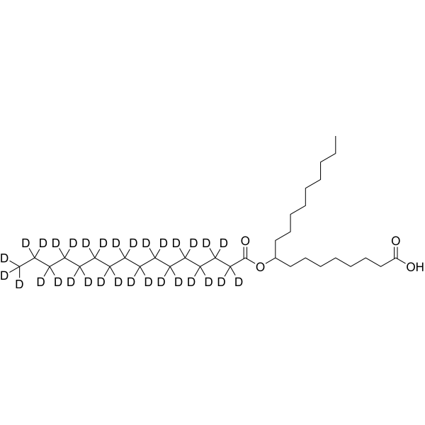
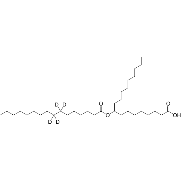
9-PAHSA-d4
9-PAHSA-d4 is the deuterium labeled 9-PAHSA. 9-PAHSA is an orally active endogenous GPR120 agonist (EC50=18 μM). 9-PAHSA significantly inhibits LPS-induced inflammatory responses by blocking the NF-κB pathway. 9-PAHSA induces adipocyte browning, enhances glucose uptake and reduces lipid accumulation, while improving mitochondrial function and the survival rate of steatotic hepatocytes. In terms of neuroprotection, 9-PAHSA regulates the expression of REST and BDNF in the prefrontal cortex of diabetic mice, and effectively prevents spatial working memory deficits and abnormal social behaviors. 9-PAHSA does not directly regulate insulin secretion or improve systemic insulin sensitivity, and possesses specific anti-inflammatory, metabolic regulatory and neuroprotective properties. 9-PAHSA can be used in the research of diabetes-related cognitive impairment, obesity and non-alcoholic fatty liver disease.
For research use only. We do not sell to patients.
- CAS No.: 2704278-85-1
- Formula: C34H62D4O4
- Molecular Weight:542.91
-
Storage:
Please store the product under the recommended conditions in the Certificate of Analysis.
Biological Activity
Description
In Vitro
Stable heavy isotopes of hydrogen, carbon, and other elements have been incorporated into drug molecules, largely as tracers for quantitation during the drug development process. Deuteration has gained attention because of its potential to affect the pharmacokinetic and metabolic profiles of drugs[1].
MedChemExpress (MCE) has not independently confirmed the accuracy of these methods. They are for reference only. Further protocols information, click here.
Application
1. This compound can be used as a tracer
2. This compound can be used as an internal standard for quantitative analysis by NMR, GC-MS, or LC-MS.
Chemical Information
-
CAS No. 2704278-85-1
-
Unlabeled CAS 1481636-31-0
-
Molecular Weight 542.91
-
Formula C34H62D4O4
-
SMILES
OC(CCCCCCCC(CCCCCCCCC)OC(CCCCCC([2H])(C([2H])(CCCCCCCC)[2H])[2H])=O)=O
-
Shipping
Room temperature in continental US; may vary elsewhere.
-
Storage
Please store the product under the recommended conditions in the Certificate of Analysis.
Protocols
-
3T3-L1 preadipocyte-to-adipocyte differentiation
3T3-L1 preadipocytes are induced to differentiate after growth arrest using adipogenic media containing insulin, dexamethasone, and IBMX; differentiation is assessed by lipid-droplet accumulation, triglyceride increase, Oil Red O staining, and adipocyte-marker induction such as PPARγ and C/EBPα.
-
RNA extraction experimental
By lysing cells, releasing RNA, and removing impurities such as proteins and DNA, high-purity RNA products are finally obtained. The commonly used traditional method is the guanidine isothiocyanate/phenol/chloroform method (Trizol), which is suitable for a variety of animal materials including animal tissues, microorganisms, cultured cells, etc., and most plant materials.
-
Mitochondrial membrane-potential fluorescent assay
Mitochondrial membrane potential fluorescent assays estimate ΔΨm in living cells using lipophilic cationic dyes such as TMRM, TMRE, rhodamine 123, and JC-1, which accumulate in mitochondria according to membrane polarization; loss of signal after FCCP or CCCP treatment is interpreted as mitochondrial depolarization. TMRM/TMRE and rhodamine 123 are commonly used for semi-quantitative live-cell microscopy or flow cytometry, while JC-1 can report a shift from red aggregate fluorescence to green monomer fluorescence during depolarization; interpretation requires controls because dye concentration, quenching mode, cell type, dye efflux, and mitochondrial mass can affect fluorescence independently of ΔΨm.
-
Large-size fat particle sorting
Large-size fat particle sorting is widely used to isolate cells up to 200 μm in diameter. Single-cell flow sorting will allow greater insight into adipocyte heterogeneity by identifying gene expression, protein composition, and metabolic signatures at the single-cell level.
-
Research Protocol for Endocrine Diseases
Endocrine diseases often arise from disrupted hormone production, hormone signaling, or target-tissue responsiveness; for diabetes-focused endocrine disease models, insulin signaling regulates glucose uptake, hepatic glucose output, lipid metabolism, and β-cell compensation. Type 2 diabetes develops through interacting defects in insulin resistance, β-cell dysfunction, adipose inflammation, hepatic glucose overproduction, altered incretin signaling, and ectopic lipid metabolism. A major unresolved question is whether endocrine dysfunction is driven primarily by target-tissue insulin resistance, intrinsic β-cell failure, immune/inflammatory stress, or combined multi-organ failure that differs by disease stage.
-
Mitochondrial membrane-potential and mitochondrial mass staining
Mitochondrial membrane potential staining measures the electrochemical polarization across the mitochondrial inner membrane in live cells using lipophilic cationic fluorescent probes; early rhodamine-based work showed that selective mitochondrial dye accumulation is lost when the mitochondrial transmembrane potential is dissipated. JC-1 reports mitochondrial polarization by shifting from green monomer fluorescence to red J-aggregate fluorescence as dye concentration increases within energized mitochondria; therefore, the red/green fluorescence ratio is used as a relative readout of mitochondrial membrane potential. TMRE or TMRM staining provides a single-channel relative readout because these cationic rhodamine esters accumulate in polarized mitochondria, and lower fluorescence indicates reduced mitochondrial polarization when acquisition and dye-loading conditions are controlled. Mitochondrial mass staining is commonly performed with MitoTracker Green FM or related MitoTracker dyes as
-
Human Islet Cell Culture
The method of preserving islets in vitro, with purified reduced immunogenicity. The steps are islet isolation, islet cell purification, in vitro determination of islet function and islet cell culture.
-
Research Protocol for Inflammation-related Diseases
The NLRP3 inflammasome is a cytosolic innate immune signaling platform that integrates priming signals and danger-signal activation to promote caspase-1 activation, maturation of IL-1β and IL-18, and gasdermin D-mediated pyroptotic cell death. The core experimental logic is to determine whether inflammatory disease phenotypes are driven by increased NLRP3 expression, ASC-containing inflammasome assembly, caspase-1 cleavage, GSDMD cleavage, and extracellular release of IL-1β/IL-18 rather than by nonspecific cell injury alone. The pathway is strongly linked to inflammation-related disease phenotypes because monosodium urate crystals activate NALP3/NLRP3 inflammasome signaling in gout-like crystal inflammation, cholesterol crystals activate NLRP3 inflammasomes in atherogenesis models, and DSS-induced intestinal inflammation has been reported to involve NLRP3 inflammasome activity. However, experimental colitis studies also show context-dependent protective effects of NLRP3 inflammasome co
-
Research Protocol for Metabolic Diseases
AMP-activated protein kinase, AMPK, is a conserved cellular energy sensor that responds to reduced cellular energy status and coordinates metabolism by increasing ATP-generating catabolic pathways while suppressing ATP-consuming anabolic processes. In metabolic disease research, the AMPK pathway is experimentally relevant because it regulates hepatic lipid synthesis, fatty acid oxidation, glucose production, skeletal-muscle glucose disposal, mTORC1-linked biosynthesis, autophagy, mitochondrial homeostasis, and whole-body energy balance. The central pathway logic is that energy stress, metformin, exercise-like stimulation, or direct AMPK activators increase AMPKα Thr172 phosphorylation and downstream substrate phosphorylation, including ACC and RAPTOR. Phosphorylation of ACC suppresses lipogenesis and supports fatty acid oxidation, whereas phosphorylation of RAPTOR suppresses mTORC1 signaling and links cellular energy status to growth and protein synthesis control. The pathway is linked
-
Lipid Droplets: Oil Red O/Sudan Dye Lipid Staining
Lipid droplets are intracellular organelles with a neutral-lipid core that stores triacylglycerols and sterol esters, and Oil Red O or Sudan dyes detect these hydrophobic lipid deposits by partitioning into retained lipids in fresh or frozen specimens. Oil Red O stains neutral triglycerides and lipids in frozen tissue sections or air-dried cytologic preparations, while Sudan Black B has also been used as a histochemical fat stain for lipid-rich tissue structures.
-
Alzheimer’s Disease Modeling
Alzheimer’s Disease (AD) is a neurodegenerative disorder characterized by a progressive decline in cognitive functions and loss of specific types of neurons and synapses. Alzheimer's symptoms can be simulated in mice by injecting drugs (such as Aβ) or genetically modified.
Purity & Documentation
References
[1]. Russak EM, et al. Impact of Deuterium Substitution on the Pharmacokinetics of Pharmaceuticals. Ann Pharmacother. 2019 Feb;53(2):211-216. [Content Brief]
[2]. Pflimlin E, et al. Acute and Repeated Treatment with 5-PAHSA or 9-PAHSA Isomers Does Not Improve Glucose Control in Mice. Cell Metab. 2018;28(2):217-227.e13. [Content Brief]
[3]. Wen XH, et al. Effect of 9 - PAHSA on cognitive dysfunction in diabetic mice and its possible mechanism. Biochem Biophys Res Commun. 2020;524(3):525-532. [Content Brief]
[5]. Schultz Moreira AR, et al. 9-PAHSA Prevents Mitochondrial Dysfunction and Increases the Viability of Steatotic Hepatocytes. Int J Mol Sci. 2020;21(21):8279. Published 2020 Nov 5. [Content Brief]
Calculators
Concentration (start) × Volume (start) = Concentration (final) × Volume (final)