



Butaprost
Based on 2 publication(s) in Google Scholar
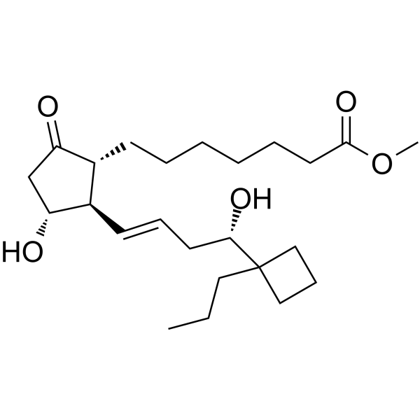
Butaprost is a selective prostaglandin E receptor (EP2) agonist with an EC50 of 33 nM and a Ki of 2.4 μM for murine EP2 receptor. Butaprost is less activity against murine EP1, EP3 and EP4 receptors. Butaprost attenuates fibrosis by hampering TGF-β/Smad2 signalling.
For research use only. We do not sell to patients.
- Purity : 99.85%
- CAS No.: 69685-22-9
- Formula: C24H40O5
- Molecular Weight:408.57
-
Storage:
Solution, -20°C, 2 years

To place orders, for customer services and technical support, please contact: MedChemExpress USA
Tel: 609-228-6898 E-mail: [email protected] [email protected]


-
 Biological Activity
-
Chemical Information
- Protocols
- Purity & Documentation
- References
-
 Help & FAQs


Publications Citing Use of MedChemExpress (MCE) Butaprost
More

Biological Activity
Description
IC50 & Target
[3]|
2400 nM (Ki) |
33 nM (EC50) |
In Vitro
Butaprost (1-100 nM; 0.5-24 hours) induces about a five-fold upregulation of Nur77 mRNA expression in hEP2-HEK293/EBNA cells in a dose- and time-dependent. Butaprost upregulated Nur77 gene expression through the PKC pathway[1].
Butaprost (50 μM; 24 hours) reduces TGF-β-induced fibronectin (FN) expression, Smad2 phosphorylation and epithelial-mesenchymal transition in Madin-Darby Canine Kidney (MDCK) cells[2].
MedChemExpress (MCE) has not independently confirmed the accuracy of these methods. They are for reference only. Further protocols information, click here.
-
Cell Line:HEK 293/EBNA cells stably expressing the human EP2 receptor (hEP2-HEK 293/EBNA cells)
-
Concentration:1 nM, 10 nM, 100 nM
-
Incubation Time:0.5 hours, 1 hours, 6 hours, 24 hours
-
Result:Induced about a five-fold upregulation of Nur77 mRNA expression in hEP2-HEK293/EBNA cells.
In Vivo
MedChemExpress (MCE) has not independently confirmed the accuracy of these methods. They are for reference only.
-
Animal Model:Male C57BL/6 mice (8 weeks of age; 21 g) bearing unilateral ureteral obstruction surgery[2]
-
Dosage:1 mg/kg, 2 mg/kg, 4 mg/kg
-
Administration:Intraperitoneal injection; twice daily; for 7 days
-
Result:Attenuated the development of fibrosis in mice that underwent unilateral ureteral obstruction surgery.


Chemical Information
-
CAS No. 69685-22-9
-
Appearance Liquid
-
Molecular Weight 408.57
-
Formula C24H40O5
-
Color Colorless to light yellow
-
SMILES
COC(CCCCCC[C@@H](C(C[C@H]1O)=O)[C@H]1/C=C/C[C@@H](C2(CCC2)CCC)O)=O
-
Shipping
Room temperature in continental US; may vary elsewhere.
-
Storage
Solution, -20°C, 2 years


Publications (2)
-
Journal Impact Factor
-
Most Recent
-
J Neuroinflammation
Taprenepag restores maternal-fetal interface homeostasis for the treatment of neurodevelopmental disorders. [Abstract]2024 Nov 28;21(1):307. PMID: 39609821 -

Protocols
-
Fibrosis/Collagen Morphometry
Fibrosis and collagen morphometry is based on the quantitative visualization of fibrillar collagen deposition in tissue sections using histochemical stains such as Sirius Red (Picrosirius Red) or Masson's trichrome, followed by image-based or polarization-enhanced analysis to estimate collagen proportional area as a surrogate of extracellular matrix accumulation during fibrotic remodeling. Sirius Red combined with polarized light microscopy enhances detection of collagen fibers due to birefringence properties, enabling more specific visualization of collagen type I and III fibrils compared to conventional bright-field histology, while whole-section or region-restricted digital morphometry reduces field-selection bias in fibrosis assessment. Alternative quantitative approaches include second harmonic generation (SHG) and two-photon excited fluorescence microscopy, which enable label-free detection of fibrillar collagen and have been validated against histological staining and biochemica
-
Collagen: Sirius Red Staining
Sirius Red or picrosirius red staining is a histochemical method for visualizing collagen-rich extracellular matrix in tissue sections, and collagen fibers are detected as red-stained structures under bright-field microscopy with enhanced birefringence under polarized light. Picrosirius red is useful for assessing total collagen organization, distribution, and fibrosis burden, but polarized color should not be interpreted as a definitive collagen type I versus type III readout because color is affected by fiber orientation, thickness, and packing.
-
Connective Tissue: Masson's Trichrome/Collagen Trichrome Staining
Masson’s Trichrome (collagen/trichrome staining) is a histological technique that differentially stains tissue compartments using sequential acidic dyes to distinguish collagen from muscle and cytoplasmic components based on dye affinity and tissue permeability differences, enabling visualization of fibrosis and connective tissue architecture in histological sections. The classical formulation typically uses Weigert's iron hematoxylin for nuclear staining, Biebrich scarlet-acid fuchsin for cytoplasm and muscle, and aniline blue (or light green variants) for collagen, producing a characteristic blue/green collagen signal contrasted against red cytoplasm and dark nuclei. The staining principle relies on selective displacement of smaller dye molecules by larger anionic dyes in collagen-rich regions under controlled acidified conditions, which enhances collagen-specific dye retention. This property makes the method widely used for fibrosis assessment in organs such as heart, liver, lung, a


Purity & Documentation
-
Data Sheet (287 KB)
-
SDS (394 KB)
- English - EN (394 KB)
- Français - FR (394 KB)
- Deutsch - DE (394 KB)
- Norwegian - NO (394 KB)
- Español - ES (394 KB)
- Swedish - SV (394 KB)
- Italian - IT (394 KB)
- Korean - KR (394 KB)
- Portuguese - PT (394 KB)
-
Handling Instructions (2659 KB)
References
[1]. Yanbin Liang, et al. Upregulation of orphan nuclear receptor Nur77 following PGF(2alpha), Bimatoprost, and Butaprost treatments. Essential role of a protein kinase C pathway involved in EP(2) receptor activated Nur77 gene transcription. Br J Pharmacol. 2004 Jun;142(4):737-48. [Content Brief]
[2]. Michael Schou Jensen, et al. Activation of the prostaglandin E 2 EP 2 receptor attenuates renal fibrosis in unilateral ureteral obstructed mice and human kidney slices. Acta Physiol (Oxf). 2019 Sep;227(1):e13291. [Content Brief]
[3]. K Tani, et al. Design and synthesis of a highly selective EP2-receptor agonist. Bioorg Med Chem Lett. 2001 Aug 6;11(15):2025-8. [Content Brief]

Calculators
Concentration (start) × Volume (start) = Concentration (final) × Volume (final)


Butaprost Related Classifications
HY-100448A Related Classifications
 Powered by Bioz