



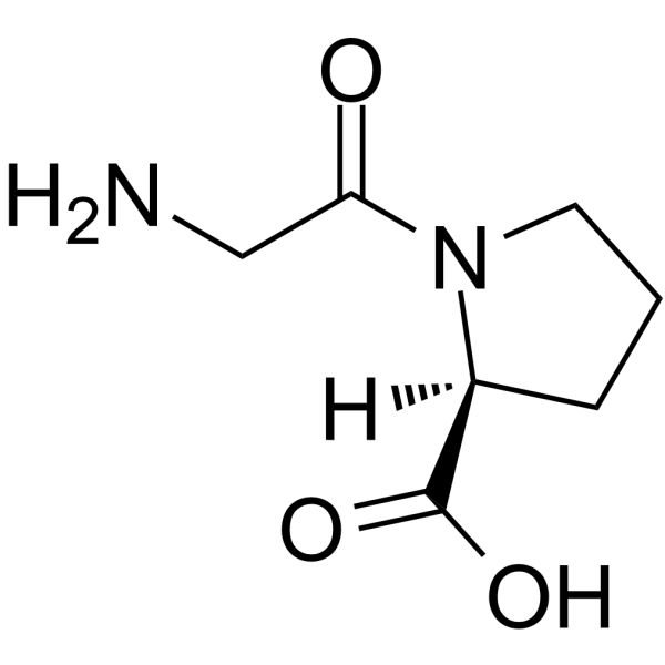
Glycyl-L-proline
Based on 1 publication(s) in Google Scholar
Glycyl-L-proline (H-Gly-Pro-OH) is a dipeptide. Glycyl-L-proline can induce MCF-7 cells apoptosis. Glycyl-L-proline can enhance the inhibitory effect of the PRODH/POX knockout on collagen and DNA biosynthesis. Glycyl-L-proline can inhibit Gly-Sar and L-proline transport. Glycyl-L-proline can be used for the researches of cancer and metabolic disease, such as breast cancer.
For research use only. We do not sell to patients.
- Purity : 99.51%
- CAS No.: 704-15-4
- Formula: C7H12N2O3
- Molecular Weight:172.18
-
Storage:
Sealed storage, away from moisture.
Powder -80°C, 2 years , -20°C, 1 year* In solvent : -80°C, 6 months; -20°C, 1 month (sealed storage, away from moisture)

To place orders, for customer services and technical support, please contact: MedChemExpress USA
Tel: 609-228-6898 E-mail: [email protected] [email protected]


-
 Biological Activity
-
Chemical Information
-
Solvent & Solubility
- Protocols
- Purity & Documentation
- References
-
 Help & FAQs


-
HY-W013494
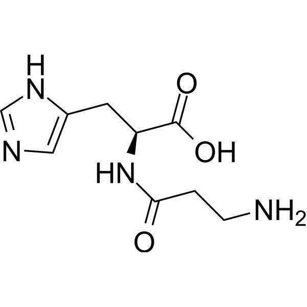
L-CarnosineFree Radical Scavenger -
HY-111174
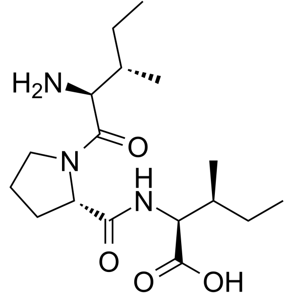
Diprotin ADipeptidyl Peptidase Inhibitor -
HY-76082
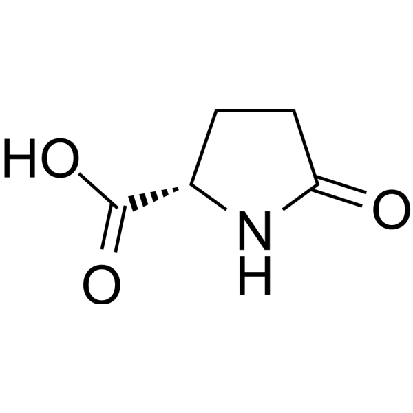
L-Pyroglutamic acidEndogenous Metabolite -
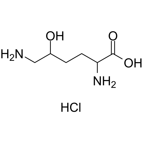
HY-W014930
DL-5-Hydroxylysine hydrochlorideAmino Acid -

HY-W016887R
Glycyl-L-proline (Standard) -

HY-W013494R
L-Carnosine (Standard) -
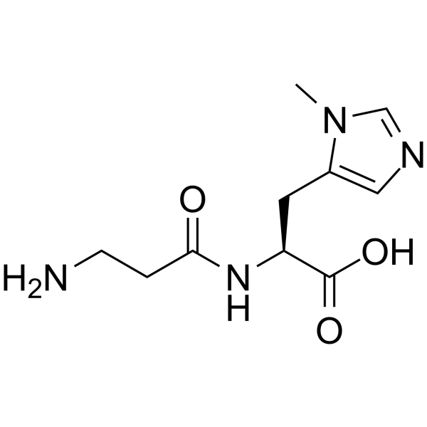
HY-113354
AnserineEndogenous Metabolite -
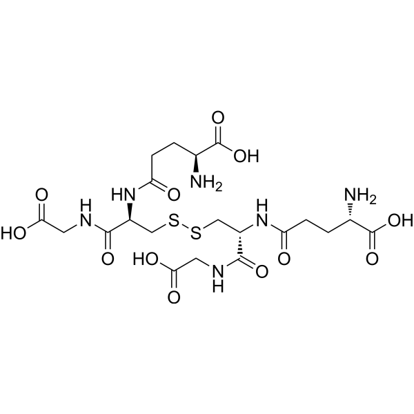
HY-D0844
Glutathione oxidizedAntioxidant -
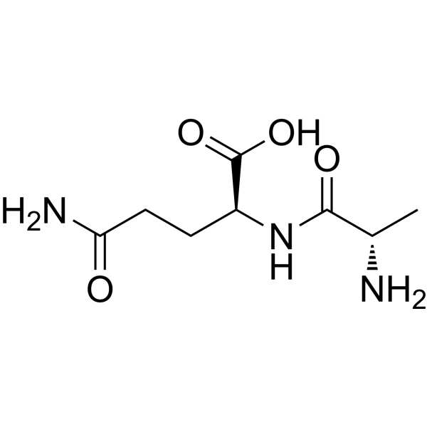
HY-W014102
L-Alanyl-L-glutamine -
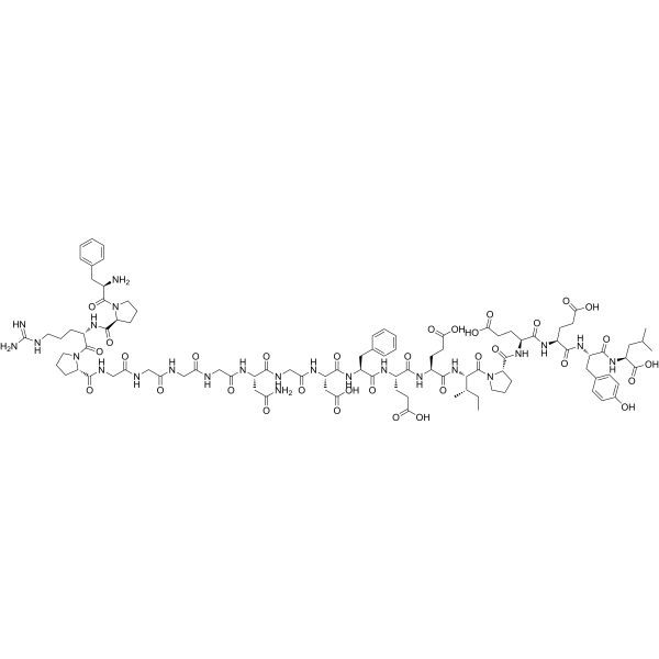
HY-P1929
BivalirudinThrombin Inhibitor -

HY-D0844R
Glutathione oxidized (Standard)Antioxidant -
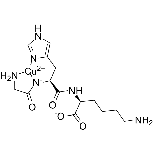
HY-P0063
Copper tripeptideTripeptide
Publications Citing Use of MedChemExpress (MCE) Glycyl-L-proline
MoreAll Endogenous Metabolite Isoforms
More

Biological Activity
Description
IC50 & Target
|
Human Endogenous Metabolite |
Cellular Effect
|
Cell Line

|
Type | Value | Description | References |
|---|---|---|---|---|
| MDCK | EC50 |
0.13 mM
Compound: 36
|
Activation of human PEPT1 expressed in MDCK cells
Activation of human PEPT1 expressed in MDCK cells
|
[PMID: 16759105] |
| MDCK | IC50 |
0.33 mM
Compound: 36
|
Binding affinity to human PEPT1 assessed as inhibition of [14C]Gly-Sar uptake in MDCK cells
Binding affinity to human PEPT1 assessed as inhibition of [14C]Gly-Sar uptake in MDCK cells
|
[PMID: 16759105] |
In Vitro
Glycyl-L-proline (17,22 μg/ml; 24 h) does not significantly affect cell viability in MCF-7 and MCF-7shPRODH/POX (knocked-down PRODH/POX) cells[1].
Glycyl-L-proline (17,22 μg/ml; 24 h) induces apoptosis in MCF-7 cells[1].
Glycyl-L-proline (17,22 μg/ml; 24 h) decreases collagen biosynthesis and DNA biosynthesis in MCF-7shPRODH/POX[1].
Glycyl-L-proline (10-50000 μM; 10 mins) decreases intracellular Gly-Sar uptake in Caco-2 cells with an IC50 of 257 µM[2].
MedChemExpress (MCE) has not independently confirmed the accuracy of these methods. They are for reference only. Further protocols information, click here.
-
Cell Line:MCF-7 and MCF-7shPRODH/POX
-
Concentration:17,22 μg/ml
-
Incubation Time:24 h
-
Result:Showed no significant difference in cell viability between MCF-7 shPRODH/POX and MCF-7 control cells.
-
Cell Line:MCF-7 and MCF-7shPRODH/POX
-
Concentration:17,22 μg/ml
-
Incubation Time:24 h
-
Result:Decreased the expression of PRODH/POX, p53, Caspase 9 in MCF-7 cells.
Increased Caspase 3, cleaved Caspase 3, cleaved Caspase 9, iNOS, NF-κB, COX-2 and AMPKβ levels in MCF-7 cells.
Increased Caspase 9 levles in MCF-7shPRODH/POX cells.


Chemical Information
-
CAS No. 704-15-4
-
Appearance Solid
-
Molecular Weight 172.18
-
Formula C7H12N2O3
-
Color White to off-white
-
Synonyms
H-Gly-Pro-OH; Gly-Pro
-
Structure Classification
-
Initial Source
-
Shipping
Room temperature in continental US; may vary elsewhere.
-
Storage
Sealed storage, away from moisture
Powder -80°C 2 years -20°C 1 year * In solvent : -80°C, 6 months; -20°C, 1 month (sealed storage, away from moisture)


Publications (1)
-
Journal Impact Factor
-
Most Recent
-
Redox Biol
ReDisulphID: A discovery platform for thiol redox sensors identifies a druggable site regulating p53 activation. [Abstract]2026 Jul:94:104196. PMID: 42097034


Solvent & Solubility
In Vitro:
H2O : 20 mg/mL (116.16 mM; ultrasonic and warming and heat to 60°C)
DMSO : 1 mg/mL (5.81 mM; ultrasonic and warming and heat to 60°C; Hygroscopic DMSO has a significant impact on the solubility of product, please use newly opened DMSO)
Please refer to the solubility information to select the appropriate solvent. Once prepared, please aliquot and store the solution to prevent product inactivation from repeated freeze-thaw cycles.
Storage method and period of stock solution: -80°C, 6 months; -20°C, 1 month (sealed storage, away from moisture). When stored at -80°C, please use it within 6 months. When stored at -20°C, please use it within 1 month.
* Note: If you choose water as the stock solution, please dilute it to the working solution, then filter and sterilize it with a 0.22 μm filter before use.
Please refer to the solubility information to select the appropriate solvent. Once prepared, please aliquot and store the solution to prevent product inactivation from repeated freeze-thaw cycles.
Storage method and period of stock solution: -80°C, 6 months; -20°C, 1 month (sealed storage, away from moisture). When stored at -80°C, please use it within 6 months. When stored at -20°C, please use it within 1 month.
* Note: If you choose water as the stock solution, please dilute it to the working solution, then filter and sterilize it with a 0.22 μm filter before use.
Concentration (start) × Volume (start) = Concentration (final) × Volume (final)

Protocols
-
Gene Editing
Gene editing modify specific sites within the genome through gene deletions, insertions or conversions to study functionally unknown genes or conduct gene therapy. It is also used to change the biological traits of organisms to establish new varieties. Gene editing techniques include zinc finger nuclease (ZFN), transcription activator-like effector nuclease (TALEN), and clustered regularly interspaced short palindromic repeats (CRISPR)/CRISPR-associated protein 9 (Cas 9) (CRISPR/Cas9).
-
Apoptosis
Apoptosis, also called programmed cell death, is generally characterized by distinct morphological characteristics.
-
TUNEL staining for apoptotic DNA fragmentation
TUNEL staining detects DNA strand breaks by using terminal deoxynucleotidyl transferase to add labeled nucleotides to exposed 3′-OH DNA termini, generating either microscopic staining in fixed cells or tissue sections, or fluorescence/cytometric signal in cell suspensions. TUNEL positivity reflects DNA fragmentation but should not be interpreted alone as definitive apoptosis, because TUNEL can also label necrotic, autolytic, mechanically damaged, or DNA-repair-associated DNA breaks.
-
Annexin V plus membrane-impermeant dye apoptosis staining
Annexin V-based apoptosis assays rely on the detection of phosphatidylserine (PS) externalization from the inner leaflet of the plasma membrane to the outer leaflet, an early biochemical hallmark of apoptosis. Fluorescently labeled Annexin V binds PS in a calcium-dependent manner, enabling identification of early apoptotic cells by flow cytometry or fluorescence microscopy. When combined with a membrane-impermeant DNA-binding dye (e. g. , propidium iodide), this approach allows discrimination between viable (Annexin V−/dye−), early apoptotic (Annexin V+/dye−), and late apoptotic or necrotic (Annexin V+/dye+) cell populations by assessing membrane integrity and PS exposure.
-
Apoptosis Solutions
Apoptosis is a regulated, generally non-lytic cell-death pathway that removes unwanted, damaged, infected, or abnormal cells through coordinated morphological changes, caspase activation, DNA fragmentation, and membrane remodeling. The intrinsic apoptosis pathway is controlled mainly by mitochondrial outer membrane permeabilization, BCL-2 family proteins, cytochrome c release, apoptosome formation, caspase-9 activation, and downstream executioner caspase-3/7 activation. The extrinsic apoptosis pathway is initiated by death receptors such as Fas, TNFR, and TRAIL receptors, which recruit adaptor proteins and activate caspase-8 before engaging executioner caspases or mitochondrial amplification through BID cleavage. Apoptosis is linked to many phenotypes, including cancer cell killing, tissue homeostasis, immune regulation, neurodegeneration, infection response, and treatment-induced cytotoxicity; unresolved questions include how apoptosis interacts with necroptosis, pyroptosis, ferroptos
-
Research Protocol for Metabolic Diseases
AMP-activated protein kinase, AMPK, is a conserved cellular energy sensor that responds to reduced cellular energy status and coordinates metabolism by increasing ATP-generating catabolic pathways while suppressing ATP-consuming anabolic processes. In metabolic disease research, the AMPK pathway is experimentally relevant because it regulates hepatic lipid synthesis, fatty acid oxidation, glucose production, skeletal-muscle glucose disposal, mTORC1-linked biosynthesis, autophagy, mitochondrial homeostasis, and whole-body energy balance. The central pathway logic is that energy stress, metformin, exercise-like stimulation, or direct AMPK activators increase AMPKα Thr172 phosphorylation and downstream substrate phosphorylation, including ACC and RAPTOR. Phosphorylation of ACC suppresses lipogenesis and supports fatty acid oxidation, whereas phosphorylation of RAPTOR suppresses mTORC1 signaling and links cellular energy status to growth and protein synthesis control. The pathway is linked
-
Breast Cancer Modeling
Breast cancer is a heterogeneous cancer, and it has been distinguished into four subtypes: luminal A, luminal B, HER2-positive and basal-like. Molecular mutations, epigenetic alterations, hormone exposure and immune microenvironment are related to the progression of breast cancer.


Purity & Documentation
-
Data Sheet (278 KB)
-
SDS (393 KB)
- English - EN (393 KB)
- Français - FR (393 KB)
- Deutsch - DE (393 KB)
- Norwegian - NO (393 KB)
- Español - ES (393 KB)
- Swedish - SV (393 KB)
- Italian - IT (393 KB)
- Korean - KR (393 KB)
- Portuguese - PT (393 KB)
-
Handling Instructions (2659 KB)
References
[1]. Zareba I, et al. Proline oxidase silencing induces proline-dependent pro-survival pathways in MCF-7 cells. Oncotarget. 2018;9(17):13748-13757. Published 2018 Feb 9. [Content Brief]
[2]. von Linde T, et al. Rapid and Sensitive Quantification of Intracellular Glycyl-Sarcosine for Semi-High-Throughput Screening for Inhibitors of PEPT-1. Pharmaceutics. 2021;13(7):1019. Published 2021 Jul 3. [Content Brief]
[3]. Boyd CA, et al. L-proline transport by brush border membrane vesicles prepared from human placenta. J Physiol. 1981;315:9-19. [Content Brief]


Complete Stock Solution Preparation Table
Please refer to the solubility information to select the appropriate solvent. Once prepared, please aliquot and store the solution to prevent product inactivation from repeated freeze-thaw cycles.
Storage method and period of stock solution: -80°C, 6 months; -20°C, 1 month (sealed storage, away from moisture). When stored at -80°C, please use it within 6 months. When stored at -20°C, please use it within 1 month.
| Optional Solvent | Concentration Solvent Mass | 1 mg | 5 mg | 10 mg | 25 mg |
|---|---|---|---|---|---|
| DMSO / H2O | 1 mM | 5.8079 mL | 29.0394 mL | 58.0788 mL | 145.1969 mL |
| 5 mM | 1.1616 mL | 5.8079 mL | 11.6158 mL | 29.0394 mL | |
| H2O | 10 mM | 0.5808 mL | 2.9039 mL | 5.8079 mL | 14.5197 mL |
| 15 mM | 0.3872 mL | 1.9360 mL | 3.8719 mL | 9.6798 mL | |
| 20 mM | 0.2904 mL | 1.4520 mL | 2.9039 mL | 7.2598 mL | |
| 25 mM | 0.2323 mL | 1.1616 mL | 2.3232 mL | 5.8079 mL | |
| 30 mM | 0.1936 mL | 0.9680 mL | 1.9360 mL | 4.8399 mL | |
| 40 mM | 0.1452 mL | 0.7260 mL | 1.4520 mL | 3.6299 mL | |
| 50 mM | 0.1162 mL | 0.5808 mL | 1.1616 mL | 2.9039 mL | |
| 60 mM | 0.0968 mL | 0.4840 mL | 0.9680 mL | 2.4199 mL | |
| 80 mM | 0.0726 mL | 0.3630 mL | 0.7260 mL | 1.8150 mL | |
| 100 mM | 0.0581 mL | 0.2904 mL | 0.5808 mL | 1.4520 mL |
* Note: If you choose water as the stock solution, please dilute it to the working solution, then filter and sterilize it with a 0.22 μm filter before use.


Glycyl-L-proline Related Classifications
HY-W016887 Related Classifications
 Powered by Bioz