



Salirasib
Based on 16 publication(s) in Google Scholar
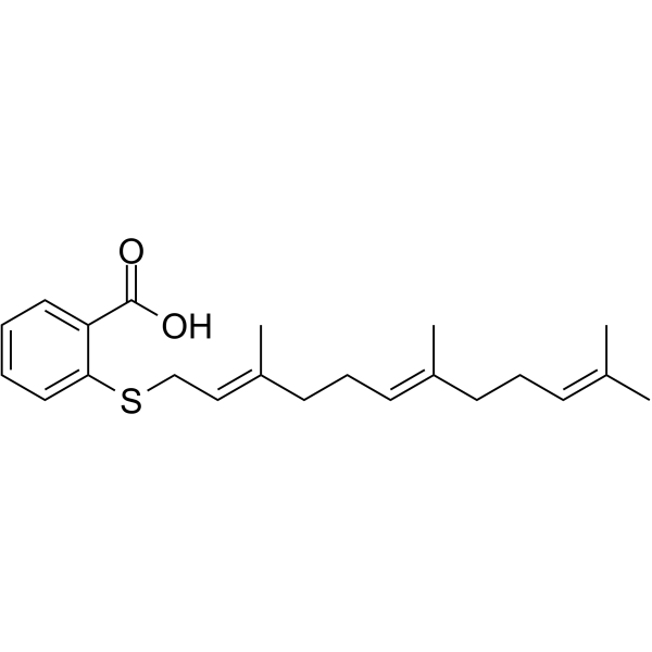
Salirasib is a Ras inhibitor that inhibits specifically both oncogenically activated Ras and growth factor receptor-mediated Ras activation, resulting in the inhibition of Ras-dependent tumor growth.
For research use only. We do not sell to patients.
- Purity : 99.04%
- CAS No.: 162520-00-5
- Formula: C22H30O2S
- Molecular Weight:358.54
-
Storage:Powder -20°C, 3 years , 4°C, 2 years ; In solvent -80°C, 2 years , -20°C, 1 year

To place orders, for customer services and technical support, please contact: MedChemExpress USA
Tel: 609-228-6898 E-mail: [email protected] [email protected]


-
 Biological Activity
-
Chemical Information
-
Solvent & Solubility
- Protocols
- Purity & Documentation
- References
-
 Help & FAQs


-
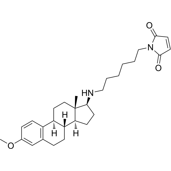
HY-13419
U-73122Phospholipase C Inhibitor -
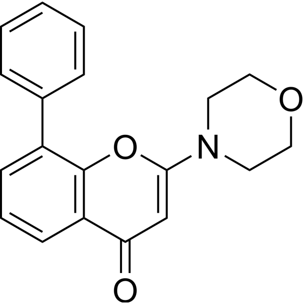
HY-10108
LY294002PI3K Inhibitor -
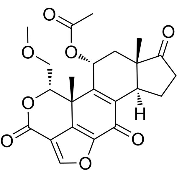
HY-10197
WortmanninPI3K Inhibitor -
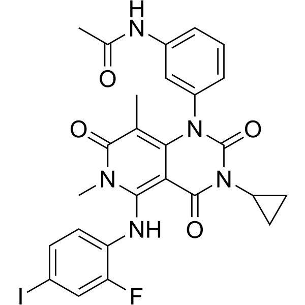
HY-10999
TrametinibMEK Inhibitor -
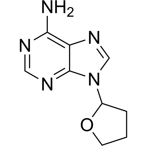
HY-100396
SQ22536Adenylate Cyclase Inhibitor -
HY-K0021
Phosphatase Inhibitor Cocktail I (100× in DMSO) -
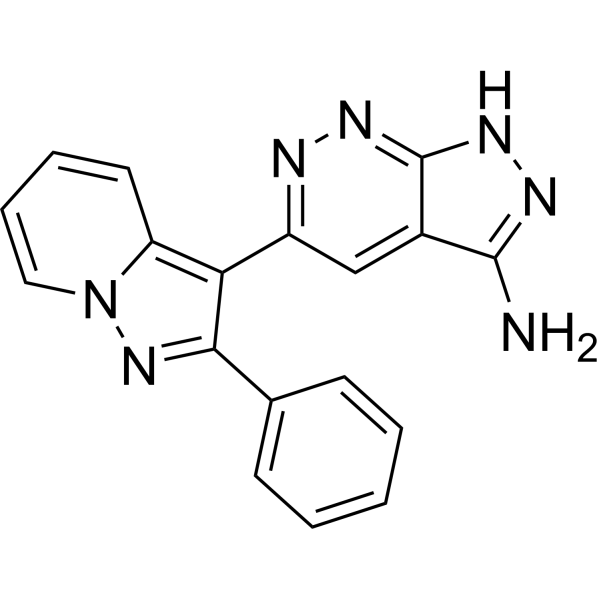
HY-12275
FR 180204ERK Inhibitor -
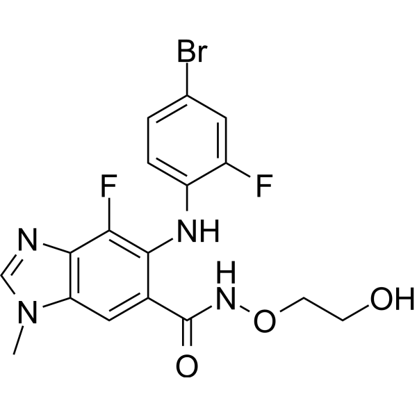
HY-15202
BinimetinibMEK1/2 Inhibitor -
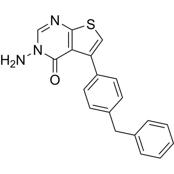
HY-119706
Barbadinβ-arrestin/β2-adaptin Interaction Inhibitor -
HY-K0010
Protease Inhibitor Cocktail (EDTA-Free, 100× in DMSO) -
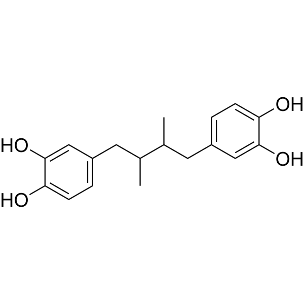
HY-N0198
Nordihydroguaiaretic acidAutophagy Inducer -
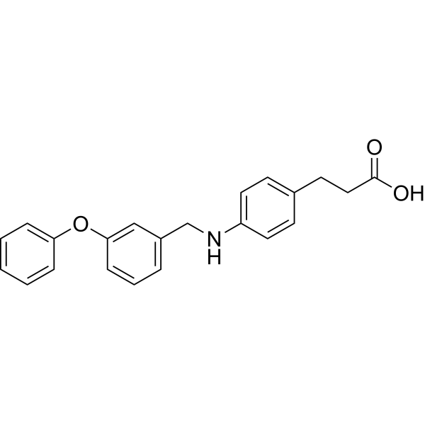
HY-15589
GW9508GPR40 Agonist
Publications Citing Use of MedChemExpress (MCE) Salirasib
More- Adv Sci (Weinh). 2021 Jan 29;8(6):2002831. [Abstract]
- Cell Death Dis. 2018 Dec 5;9(12):1170. [Abstract]
- Free Radic Biol Med. 2026 Apr:247:469-485. [Abstract]
- Clin Transl Med. 2025 Oct;15(10):e70484. [Abstract]
- Life Sci. 2024 Aug 1:350:122763. [Abstract]
- Life Sci. 2021 Jun 1:274:119332. [Abstract]
- Int J Pharm. 2019 Dec 15:572:118823. [Abstract]
- Int J Mol Sci. 2024 Dec 27;26(1):143. [Abstract]
- ACS Appl Bio Mater. 2020 Feb 17;3(2):1129-1138. [Abstract]
- Neoplasia. 2021 Jun;23(6):607-623. [Abstract]
- Lab Invest. 2022 Dec;102(12):1389-1399. [Abstract]
- Cytokine. 2026 Aug:204:157169. [Abstract]
- Discov Oncol. 2025 Jun 14;16(1):1104. [Abstract]
- Epilepsy Res. 2026 May 14:225:107820. [Abstract]
- bioRxiv. 2025 Oct 25.
- Research Square Preprint. 2024 Dec 25.
 Customer Validation & Images

-
WB

-
RT-PCR

-
RT-PCR

-
WB

-
IF


Biological Activity
Description
IC50 & Target
Ki: 2.6 μM (PPMTase)
Cellular Effect
|
Cell Line

|
Type | Value | Description | References |
|---|---|---|---|---|
| A549 | GI50 |
34 μM
Compound: 12
|
Antiproliferative activity against human A549 cells assessed as inhibition of culture growth incubated for 48 hrs by SRB assay
Antiproliferative activity against human A549 cells assessed as inhibition of culture growth incubated for 48 hrs by SRB assay
|
[PMID: 37531922] |
| Bel-7402 | IC50 |
76.13 μM
Compound: 1, FTS, S-trans,trans-FTS, Salirasib
|
Cytotoxicity against human Bel7402 cells after 48 hrs by MTT based ELISA
Cytotoxicity against human Bel7402 cells after 48 hrs by MTT based ELISA
|
[PMID: 21504204] |
| BXPC-3 | EC50 |
>20 μM
Compound: FTS, Salirasib
|
Cytotoxicity against human BxPC3 cells after 48 hrs by fluorescence assay
Cytotoxicity against human BxPC3 cells after 48 hrs by fluorescence assay
|
[PMID: 24852279] |
| CHO | EC50 |
5 μM
Compound: 10
|
Agonist activity at human TRPA1 channel expressed in CHO cells assessed as increase in intracellular calcium levels
Agonist activity at human TRPA1 channel expressed in CHO cells assessed as increase in intracellular calcium levels
|
[PMID: 20356305] |
| EJ | EC50 |
7.5 μM
Compound: 2
|
Effective concentration required for 50% inhibition of growth of human Ha-ras-transformed rat 1 (EJ)cells
Effective concentration required for 50% inhibition of growth of human Ha-ras-transformed rat 1 (EJ)cells
|
[PMID: 7731012] |
| EJ | IC50 |
47.6 μM
Compound: FTS
|
Cytotoxicity against human EJ cells after 48 hrs by MTT assay
Cytotoxicity against human EJ cells after 48 hrs by MTT assay
|
[PMID: 24300920] |
| EJ | IC50 |
48.6 μM
Compound: Farnesyl Thiosalicylic acid
|
Cytotoxicity against human EJ cells assessed as inhibition of cell growth by MTT assay
Cytotoxicity against human EJ cells assessed as inhibition of cell growth by MTT assay
|
[PMID: 32791404] |
| HBL-100 | GI50 |
22 μM
Compound: 12
|
Antiproliferative activity against human HBL-100 cells assessed as inhibition of culture growth incubated for 48 hrs by SRB assay
Antiproliferative activity against human HBL-100 cells assessed as inhibition of culture growth incubated for 48 hrs by SRB assay
|
[PMID: 37531922] |
| HEK293 | EC50 |
0.8 μM
Compound: FTS
|
Agonist activity at human TRPA1 expressed in HEK293 cells assessed as increase in calcium influx by Fluo-4-AM dye based fluorescence assay
Agonist activity at human TRPA1 expressed in HEK293 cells assessed as increase in calcium influx by Fluo-4-AM dye based fluorescence assay
|
[PMID: 30878828] |
| HEK293 | EC50 |
7 μM
Compound: FTS
|
Agonist activity at human TRPA1 expressed in HEK293 cells assessed as increase in calcium influx by Fluo-3/acetoxymethyl ester dye based FLIPR analysis
Agonist activity at human TRPA1 expressed in HEK293 cells assessed as increase in calcium influx by Fluo-3/acetoxymethyl ester dye based FLIPR analysis
|
[PMID: 30878828] |
| HeLa | GI50 |
34 μM
Compound: 12
|
Antiproliferative activity against human HeLa cells assessed as inhibition of culture growth incubated for 48 hrs by SRB assay
Antiproliferative activity against human HeLa cells assessed as inhibition of culture growth incubated for 48 hrs by SRB assay
|
[PMID: 37531922] |
| Hep 3B2 | IC50 |
63.75 μM
Compound: 1, FTS, S-trans,trans-FTS, Salirasib
|
Cytotoxicity against human Hep3B cells after 48 hrs by MTT based ELISA
Cytotoxicity against human Hep3B cells after 48 hrs by MTT based ELISA
|
[PMID: 21504204] |
| HepG2 | IC50 |
107.51 μM
Compound: 1, FTS, S-trans,trans-FTS, Salirasib
|
Cytotoxicity against human HepG2 cells after 48 hrs by MTT based ELISA
Cytotoxicity against human HepG2 cells after 48 hrs by MTT based ELISA
|
[PMID: 21504204] |
| HepG2 | IC50 |
48.6 μM
Compound: Farnesyl Thiosalicylic acid
|
Cytotoxicity against human HepG2 cells assessed as inhibition of cell growth by MTT assay
Cytotoxicity against human HepG2 cells assessed as inhibition of cell growth by MTT assay
|
[PMID: 32791404] |
| MCF7 | IC50 |
46.75 μM
Compound: FTA
|
Cytotoxicity against human MCF7 cells after 48 hrs by MTT assay
Cytotoxicity against human MCF7 cells after 48 hrs by MTT assay
|
[PMID: 20435479] |
| MCF7 | IC50 |
48.6 μM
Compound: Farnesyl Thiosalicylic acid
|
Cytotoxicity against human MCF7 cells assessed as inhibition of cell growth by MTT assay
Cytotoxicity against human MCF7 cells assessed as inhibition of cell growth by MTT assay
|
[PMID: 32791404] |
| MCF7 | IC50 |
49.1 μM
Compound: FTS
|
Cytotoxicity against human MCF7 cells after 48 hrs by MTT assay
Cytotoxicity against human MCF7 cells after 48 hrs by MTT assay
|
[PMID: 24300920] |
| MDA-MB-231 | IC50 |
51.22 μM
Compound: FTA
|
Cytotoxicity against human MDA-MB-231 cells after 48 hrs by MTT assay
Cytotoxicity against human MDA-MB-231 cells after 48 hrs by MTT assay
|
[PMID: 20435479] |
| MEF | IC50 |
14.3 μM
Compound: FTS
|
Cytotoxicity against icmt-double mutant MEF cells after 5 days by MTT assay
Cytotoxicity against icmt-double mutant MEF cells after 5 days by MTT assay
|
[PMID: 22754607] |
| MEF | IC50 |
15.3 μM
Compound: FTS
|
Cytotoxicity against icmt-positive MEF cells after 5 days by MTT assay
Cytotoxicity against icmt-positive MEF cells after 5 days by MTT assay
|
[PMID: 22754607] |
| MIA PaCa-2 | EC50 |
>20 μM
Compound: FTS, Salirasib
|
Cytotoxicity against human MIAPaCa2 cells harboring K-Ras mutant oncogene after 48 hrs by fluorescence assay
Cytotoxicity against human MIAPaCa2 cells harboring K-Ras mutant oncogene after 48 hrs by fluorescence assay
|
[PMID: 24852279] |
| NCI-H1299 | GI50 |
85 μM
Compound: 12
|
Antiproliferative activity against human H1299 cells assessed as inhibition of culture growth incubated for 48 hrs by SRB assay
Antiproliferative activity against human H1299 cells assessed as inhibition of culture growth incubated for 48 hrs by SRB assay
|
[PMID: 37531922] |
| NCI-H460 | IC50 |
48.6 μM
Compound: Farnesyl Thiosalicylic acid
|
Cytotoxicity against human NCI-H460 cells assessed as inhibition of cell growth by MTT assay
Cytotoxicity against human NCI-H460 cells assessed as inhibition of cell growth by MTT assay
|
[PMID: 32791404] |
| NCI-H460 | IC50 |
49.2 μM
Compound: FTS
|
Cytotoxicity against human H460 cells after 48 hrs by MTT assay
Cytotoxicity against human H460 cells after 48 hrs by MTT assay
|
[PMID: 24300920] |
| PANC-1 | IC50 |
35 μM
Compound: FTS
|
Cytotoxicity against human PANC1 cells after 5 to 7 days
Cytotoxicity against human PANC1 cells after 5 to 7 days
|
[PMID: 19072665] |
| PANC-1 | IC50 |
35.2 μM
Compound: Salirasib
|
Antiproliferative activity against human PANC-1 cells assessed as cell growth inhibition
Antiproliferative activity against human PANC-1 cells assessed as cell growth inhibition
|
[PMID: 33228976] |
| PANC-1 | IC50 |
53.6 μM
Compound: FTS
|
Cytotoxicity against human PANC1 cells after 48 hrs by MTT assay
Cytotoxicity against human PANC1 cells after 48 hrs by MTT assay
|
[PMID: 24300920] |
| Rat1 | EC50 |
7.5 μM
Compound: Salirasib
|
Inhibition of cell growth in rat Rat1 cells transfected with HRAS
Inhibition of cell growth in rat Rat1 cells transfected with HRAS
|
[PMID: 33228976] |
| SGC-7901 | IC50 |
37.74 μM
Compound: FTA
|
Cytotoxicity against human SGC7901 cells after 48 hrs by MTT assay
Cytotoxicity against human SGC7901 cells after 48 hrs by MTT assay
|
[PMID: 20435479] |
| SGC-7901 | IC50 |
41.3 μM
Compound: FTS
|
Cytotoxicity against human SGC7901 cells after 48 hrs by MTT assay
Cytotoxicity against human SGC7901 cells after 48 hrs by MTT assay
|
[PMID: 24300920] |
| SK-OV-3 | IC50 |
51.2 μM
Compound: FTS
|
Cytotoxicity against human SKOV3 cells after 48 hrs by MTT assay
Cytotoxicity against human SKOV3 cells after 48 hrs by MTT assay
|
[PMID: 24300920] |
| SMMC-7721 | IC50 |
48.6 μM
Compound: Farnesyl Thiosalicylic acid
|
Cytotoxicity against human SMMC-7721 cells assessed as inhibition of cell growth by MTT assay
Cytotoxicity against human SMMC-7721 cells assessed as inhibition of cell growth by MTT assay
|
[PMID: 32791404] |
| SMMC-7721 | IC50 |
6.7 μM
Compound: FTS
|
Cytotoxicity against human SMMC-7721 cells assessed as cell growth inhibition incubated for 48 hrs by MTT assay
Cytotoxicity against human SMMC-7721 cells assessed as cell growth inhibition incubated for 48 hrs by MTT assay
|
[PMID: 33143937] |
| SMMC-7721 | IC50 |
69.7 μM
Compound: FTS
|
Cytotoxicity against human SMMC7721 cells after 48 hrs by MTT assay
Cytotoxicity against human SMMC7721 cells after 48 hrs by MTT assay
|
[PMID: 24300920] |
| SMMC-7721 | IC50 |
71.22 μM
Compound: 1, FTS, S-trans,trans-FTS, Salirasib
|
Cytotoxicity against human SMMC7721 cells after 48 hrs by MTT based ELISA
Cytotoxicity against human SMMC7721 cells after 48 hrs by MTT based ELISA
|
[PMID: 21504204] |
| SW1573 | GI50 |
41 μM
Compound: 12
|
Antiproliferative activity against human SW1573 cells assessed as inhibition of culture growth incubated for 48 hrs by SRB assay
Antiproliferative activity against human SW1573 cells assessed as inhibition of culture growth incubated for 48 hrs by SRB assay
|
[PMID: 37531922] |
| T47D | GI50 |
42 μM
Compound: 12
|
Antiproliferative activity against human T47D cells assessed as inhibition of culture growth incubated for 48 hrs by SRB assay
Antiproliferative activity against human T47D cells assessed as inhibition of culture growth incubated for 48 hrs by SRB assay
|
[PMID: 37531922] |
| U-251 | IC50 |
46.91 μM
Compound: FTA
|
Cytotoxicity against human U251 cells after 48 hrs by MTT assay
Cytotoxicity against human U251 cells after 48 hrs by MTT assay
|
[PMID: 20435479] |
| U-87MG ATCC | IC50 |
50 μM
Compound: FTS
|
Cytotoxicity against human U87 cells after 5 to 7 days
Cytotoxicity against human U87 cells after 5 to 7 days
|
[PMID: 19072665] |
| U-87MG ATCC | IC50 |
53.95 μM
Compound: FTA
|
Cytotoxicity against human U87 cells after 48 hrs by MTT assay
Cytotoxicity against human U87 cells after 48 hrs by MTT assay
|
[PMID: 20435479] |
| Vero | IC50 |
210 μM
Compound: 1j
|
Cytotoxicity against African green monkey Vero cells after 72 hrs by MTT assay
Cytotoxicity against African green monkey Vero cells after 72 hrs by MTT assay
|
[PMID: 31803400] |
In Vitro
Salirasib (12.5-100 μM) inhibits the proliferation of ELT3 cells in a dose-dependent manner with an average IC50 of 58.57±4.59 μM. The effects of Salirasib on the TSC2-null cells are evidently mimicked by DN-Rheb but not by DN-Ras. Salirasib reduces Rheb in TSC2-null cells and TSC2 expression rescues the cells from the inhibitory effect of Salirasib. Salirasib reduces phosphorylation of S6K but not of ERK in the TSC2-null ELT3 cells[1]. Salirasib (50, 100, 150 μM) induces a dose- and time-dependent decrease of cell growth in HCC cells. Salirasib reduces cell proliferation through modulation of cell cycle effectors and inhibitors. Salirasib induces apoptosis in HepG2 and Hep3B cells. The growth inhibitory effect of salirasib in HCC cell lines is associated with mTOR inhibition independent of ERK or Akt activation[2].
MedChemExpress (MCE) has not independently confirmed the accuracy of these methods. They are for reference only. Further protocols information, click here.
In Vivo
MedChemExpress (MCE) has not independently confirmed the accuracy of these methods. They are for reference only.
Clinical Trial
| NCT Number | Sponsor | Condition | Start Date |
Phase
|
|---|---|---|---|---|
| NCT01329991 | Plexxikon| | 2011-05 | PHASE1 |


Chemical Information
-
CAS No. 162520-00-5
-
Appearance Solid
-
Molecular Weight 358.54
-
Formula C22H30O2S
-
Color White to light yellow
-
SMILES
O=C(O)C1=CC=CC=C1SC/C=C(C)/CC/C=C(C)/CC/C=C(C)/C
-
Synonyms
S-Farnesylthiosalicylic acid; Farnesyl Thiosalicylic Acid; FTS
-
Shipping
Room temperature in continental US; may vary elsewhere.
-
Storage
Powder -20°C 3 years 4°C 2 years In solvent -80°C 2 years -20°C 1 year


Publications (16)
-
Journal Impact Factor
-
Most Recent
-
Adv Sci (Weinh)
PADI2-Catalyzed MEK1 Citrullination Activates ERK1/2 and Promotes IGF2BP1-Mediated SOX2 mRNA Stability in Endometrial Cancer. [Abstract]2021 Jan 29;8(6):2002831. PMID: 33747724
Salirasib purchased from MedChemExpress. Usage Cited in: Adv Sci (Weinh). 2021 Jan 29;8(6):2002831. [Abstract]
Salirasib (50 μM) decreased the protein expression of p-MEK1 in HEK293 cells overexpressed with PADI2 (WT or C647S).
-
Cell Death Dis
UBIAD1 suppresses the proliferation of bladder carcinoma cells by regulating H-Ras intracellular trafficking via interaction with the C-terminal domain of H-Ras. [Abstract]2018 Dec 5;9(12):1170. PMID: 30518913
Salirasib purchased from MedChemExpress. Usage Cited in: Cell Death Dis. 2018 Dec 5;9(12):1170. [Abstract]
Salirasib (35 μM; 72 h) prevented ERK phosphorylation in the absence of UBIAD1 in HEK293T cells.
-
Free Radic Biol Med
Modulation of glutamate metabolic reprogramming via Ras-Raf-MEK/ERK signaling alleviates immune inflammation of astrocytes in glaucomatous neurodegeneration. [Abstract]2026 Apr:247:469-485. PMID: 41692317
Salirasib purchased from MedChemExpress. Usage Cited in: Free Radic Biol Med. 2026 Apr:247:469-485. [Abstract]
Salirasib (FTS; 50 μM; 72 h) increased LPS-induced c-Raf, p-ERK, p-MEK, Ras protein expression in primary astrocytes.
Salirasib purchased from MedChemExpress. Usage Cited in: Free Radic Biol Med. 2026 Apr:247:469-485. [Abstract]
Salirasib (FTS; 50 μM; 72 h) rescued the LPS-induced downregulation of EAAT1, EAAT2, and GS in primary astrocytes and C8-D1A cells.
Salirasib purchased from MedChemExpress. Usage Cited in: Free Radic Biol Med. 2026 Apr:247:469-485. [Abstract]
Salirasib (FTS; 50 μM; 72 h) rescued the LPS-induced upregulation of GFAP, IL-1β, IL-6, and TNF-α mRNA levels (RT-qPCR) in primary astrocytes.
Salirasib purchased from MedChemExpress. Usage Cited in: Free Radic Biol Med. 2026 Apr:247:469-485. [Abstract]
Salirasib (FTS; 5 mg/kg; i.p.; daily for 28 days) significantly downregulated the expression of Ras, c-Raf, MEK, ERK, and abnormally phosphorylated MEK and ERK proteins in the retinas of C57BL/6J mice with EAG model.
Salirasib purchased from MedChemExpress. Usage Cited in: Free Radic Biol Med. 2026 Apr:247:469-485. [Abstract]
Salirasib (FTS; 5 mg/kg; i.p.; daily for 28 days) prevented RGC loss by immunolabeling for Brn3a in C57BL/6J mice with EAG model.

Salirasib purchased from MedChemExpress. Usage Cited in: Free Radic Biol Med. 2026 Apr:247:469-485. [Abstract]
Salirasib (FTS; 5 mg/kg; i.p.; daily for 28 days) observed an increased number of cells in the ganglion cell layer, neatly arranged cells in the optic nerves, and preservation of myelin integrity in the optic nerves in C57BL/6J mice with EAG model.
-
Clin Transl Med
Targeting c-Myc enhances immunotherapy efficacy in combination with Ras inhibitor in triple-negative breast cancer. [Abstract]2025 Oct;15(10):e70484. PMID: 41020311 -
Life Sci
Low GPR81 in ER+ breast cancer cells drives tamoxifen resistance through inducing PPARα-mediated fatty acid oxidation. [Abstract]2024 Aug 1:350:122763. PMID: 38823505 -
Life Sci
2021 Jun 1:274:119332. PMID: 33711384 -
Int J Pharm
2019 Dec 15:572:118823. PMID: 31715346 -
Int J Mol Sci
Neutrophil Extracellular Trap Formation Model Induced by Monosodium Urate and Phorbol Myristate Acetate: Involvement in MAPK Signaling Pathways. [Abstract]2024 Dec 27;26(1):143. PMID: 39796001 -
ACS Appl Bio Mater
Mediated Imaging and Improved Targeting of Farnesylthiosalicylic Acid Delivery for Pancreatic Cancer via Conjugation with Near-Infrared Fluorescence Heptamethine Carbocyanine Dye. [Abstract]2020 Feb 17;3(2):1129-1138. PMID: 35019314 -
Neoplasia
STAMBP promotes lung adenocarcinoma metastasis by regulating the EGFR/MAPK signaling pathway. [Abstract]2021 Jun;23(6):607-623. PMID: 34102455 -
Lab Invest
Upregulation of KLHL17 promotes the proliferation and migration of non-small cell lung cancer by activating the Ras/MAPK signaling pathway. [Abstract]2022 Dec;102(12):1389-1399. PMID: 35978057 -
Cytokine
2026 Aug:204:157169. PMID: 42143915 -
Discov Oncol
Syndecan-4 promotes gastric cancer progression through activating TGF-β1 induced lipid reprogramming and contributes positive loop circuits. [Abstract]2025 Jun 14;16(1):1104. PMID: 40516011 -
Epilepsy Res
Vitamin D3 prevents epileptic seizures by modulating the ras signalling pathway via Gnb1 and Casr in acute epilepsy mouse models. [Abstract]2026 May 14:225:107820. PMID: 42150281 -
-


Solvent & Solubility
In Vitro:
DMSO : ≥ 50 mg/mL (139.45 mM; Hygroscopic DMSO has a significant impact on the solubility of product, please use newly opened DMSO)
* "≥" means soluble, but saturation unknown.
Please refer to the solubility information to select the appropriate solvent. Once prepared, please aliquot and store the solution to prevent product inactivation from repeated freeze-thaw cycles.
Storage method and period of stock solution: -80°C, 2 years; -20°C, 1 year. When stored at -80°C, please use it within 2 years. When stored at -20°C, please use it within 1 year.
Please refer to the solubility information to select the appropriate solvent. Once prepared, please aliquot and store the solution to prevent product inactivation from repeated freeze-thaw cycles.
Storage method and period of stock solution: -80°C, 2 years; -20°C, 1 year. When stored at -80°C, please use it within 2 years. When stored at -20°C, please use it within 1 year.
Concentration (start) × Volume (start) = Concentration (final) × Volume (final)
In Vivo:
Select the appropriate dissolution method based on your experimental animal and administration route.
- For the following dissolution methods, please ensure to first prepare a clear stock solution using an In Vitro approach and then sequentially add co-solvents:
- To ensure reliable experimental results, the clarified stock solution can be appropriately stored based on storage conditions. As for the working solution for In Vivo experiments, it is recommended to prepare freshly and use it on the same day.
- The percentages shown for the solvents indicate their volumetric ratio in the final prepared solution. If precipitation or phase separation occurs during preparation, heat and/or sonication can be used to aid dissolution.
Add each solvent one by one: 10% DMSO 40% PEG300 5% Tween-80 45% Saline
Solubility: ≥ 2.5 mg/mL (6.97 mM); Clear solution
This protocol yields a clear solution of ≥ 2.5 mg/mL (saturation unknown).
Taking 1 mL working solution as an example, add 100 μL DMSO stock solution (25.0 mg/mL) to 400 μL PEG300, and mix evenly; then add 50 μL Tween-80 and mix evenly; then add 450 μL Saline to adjust the volume to 1 mL.
Preparation of Saline: Dissolve 0.9 g sodium chloride in ddH₂O and dilute to 100 mL to obtain a clear Saline solution.
Add each solvent one by one: 10% DMSO 90% (20% SBE-β-CD in Saline)
Solubility: 2.5 mg/mL (6.97 mM); Suspended solution; Need ultrasonic
This protocol yields a suspended solution of 2.5 mg/mL. Suspended solution can be used for oral and intraperitoneal injection.
Taking 1 mL working solution as an example, add 100 μL DMSO stock solution (25.0 mg/mL) to 900 μL 20% SBE-β-CD in Saline, and mix evenly.
Preparation of 20% SBE-β-CD in Saline (4°C, storage for one week): 2 g SBE-β-CD powder is dissolved in 10 mL Saline, completely dissolve until clear.
In Vivo Dissolution Calculator
Please enter the basic information of animal experiments:
-
-
-
-
Recommended: Prepare an additional quantity of animals to account for potential losses during experiments.
Please enter your animal formula composition:
-
%DMSO +
Recommended: Keep the proportion of DMSO in working solution below 2% if your animal is weak.
-
%+
-
+%Tween-80 + +
-
%Saline +
The co-solvents required include: DMSO, . All of co-solvents are available by MedChemExpress (MCE). , Tween 80. All of co-solvents are available by MedChemExpress (MCE).
Working solution concentration: 0.22 mg/mL
Method for preparing stock solution: mg drug dissolved in μL DMSO. Stock solution concentration: mg/mL.
1. Take μL DMSO stock solution;
2. Add μL .
μL , mix evenly;
3. Then add μL Tween 80, mix evenly;
4. Then add μL
Please ensure that the stock solution in the first step is dissolved to a clear state, and add co-solvents in sequence. You can use ultrasonic heating (ultrasonic cleaner, recommended frequency 20-40 kHz), vortexing, etc. to assist dissolution.

Protocols
-
Patient-Derived Xenograft (PDX)
Patient-derived xenograft (PDX) models are generated by engrafting primary human tumor tissue directly into immunodeficient mice, allowing in vivo propagation of patient tumor biology without initial in vitro adaptation. These models are used to preserve key histopathological and molecular characteristics of the original tumor and enable assessment of tumor growth dynamics and therapeutic response in a living organism. The biological readout is tumor engraftment and subsequent growth in the murine host, which reflects the ability of human tumor cells to survive, vascularize, and expand in an immunocompromised microenvironment.
-
Patient-Derived Orthotopic Xenograft (PDOX)
Patient-derived orthotopic xenograft (PDOX) modeling implants fresh patient tumor tissue or patient-derived tumor cells into the anatomically corresponding organ or tissue site of immunodeficient mice, usually by surgical orthotopic implantation, to preserve patient tumor histology, local microenvironmental context, invasion, metastatic behavior, and treatment-response features better than subcutaneous implantation. PDOX readouts include tumor engraftment, orthotopic tumor growth, local invasion, metastasis, recurrence after resection, histologic similarity to the donor tumor, biomarker retention, molecular concordance, survival, and response or resistance to therapy. PDOX models are used for preclinical drug testing and individualized therapy evaluation, but engraftment success varies by tumor type and specimen quality.
-
Subcutaneous Cell-Line-Derived Xenograft
Subcutaneous cell-line-derived xenograft (CDX) models are established by implanting cultured human cancer cell lines into immunodeficient mice, where the injected cells form localized tumors that can be monitored in vivo as a measure of tumorigenic potential, growth kinetics, and treatment response. These models are widely used in oncology research because they allow reproducible tumor formation and enable comparative assessment of tumor growth between different cell lines or genetic manipulations in a controlled in vivo microenvironment. Subcutaneous implantation of cancer cells in immunodeficient mice is a standard approach for evaluating tumor growth behavior and therapeutic response across multiple cancer types, including prostate, esophageal, pancreatic, and colon cancer models.
-
Orthotopic Cell-Line Xenograft
Orthotopic cell-line xenograft models involve implantation of human cancer cell lines into the anatomically corresponding organ of immunodeficient mice to reproduce tumor growth within a native microenvironment, enabling more clinically relevant tumor behavior compared with subcutaneous models. These models are widely used because orthotopic placement better recapitulates tumor progression, including invasion and metastatic spread, which are often underrepresented in heterotopic implantation systems. Compared with conventional xenografts, orthotopic implantation is described as more technically complex but provides improved simulation of tumor-microenvironment interactions and metastatic behavior, making it particularly valuable for translational oncology research. Surgical orthotopic implantation approaches have been emphasized as enabling faithful reproduction of clinical cancer features, including metastasis and disease progression patterns that align with the tumor’s organ of origi


Purity & Documentation
-
Data Sheet (276 KB)
-
SDS (393 KB)
- English - EN (393 KB)
- Français - FR (393 KB)
- Deutsch - DE (393 KB)
- Norwegian - NO (393 KB)
- Español - ES (393 KB)
- Swedish - SV (393 KB)
- Italian - IT (393 KB)
- Korean - KR (393 KB)
- Portuguese - PT (393 KB)
-
Handling Instructions (2659 KB)
References
[1]. Makovski V, et al. Farnesylthiosalicylic acid (salirasib) inhibits Rheb in TSC2-null ELT3 cells: a potential treatment for lymphangioleiomyomatosis. Int J Cancer. 2012 Mar 15;130(6):1420-9. [Content Brief]
[2]. Nevo Y, et al. Chapman J. The Ras antagonist, farnesylthiosalicylic acid (FTS), decreases fibrosis and improves muscle strength in dy/dy mouse model of muscular dystrophy. PLoS One. 2011 Mar 22;6(3):e18049. [Content Brief]
[3]. Charette N, et al. Salirasib inhibits the growth of hepatocarcinoma cell lines in vitro and tumor growth in vivo through ras and mTOR inhibition. Mol Cancer. 2010 Sep 22;9:256. [Content Brief]


Complete Stock Solution Preparation Table
Please refer to the solubility information to select the appropriate solvent. Once prepared, please aliquot and store the solution to prevent product inactivation from repeated freeze-thaw cycles.
Storage method and period of stock solution: -80°C, 2 years; -20°C, 1 year. When stored at -80°C, please use it within 2 years. When stored at -20°C, please use it within 1 year.
| Optional Solvent | Concentration Solvent Mass | 1 mg | 5 mg | 10 mg | 25 mg |
|---|---|---|---|---|---|
| DMSO | 1 mM | 2.7891 mL | 13.9454 mL | 27.8909 mL | 69.7272 mL |
| 5 mM | 0.5578 mL | 2.7891 mL | 5.5782 mL | 13.9454 mL | |
| 10 mM | 0.2789 mL | 1.3945 mL | 2.7891 mL | 6.9727 mL | |
| 15 mM | 0.1859 mL | 0.9297 mL | 1.8594 mL | 4.6485 mL | |
| 20 mM | 0.1395 mL | 0.6973 mL | 1.3945 mL | 3.4864 mL | |
| 25 mM | 0.1116 mL | 0.5578 mL | 1.1156 mL | 2.7891 mL | |
| 30 mM | 0.0930 mL | 0.4648 mL | 0.9297 mL | 2.3242 mL | |
| 40 mM | 0.0697 mL | 0.3486 mL | 0.6973 mL | 1.7432 mL | |
| 50 mM | 0.0558 mL | 0.2789 mL | 0.5578 mL | 1.3945 mL | |
| 60 mM | 0.0465 mL | 0.2324 mL | 0.4648 mL | 1.1621 mL | |
| 80 mM | 0.0349 mL | 0.1743 mL | 0.3486 mL | 0.8716 mL | |
| 100 mM | 0.0279 mL | 0.1395 mL | 0.2789 mL | 0.6973 mL |


Salirasib Related Classifications
HY-14754 Related Classifications
 Powered by Bioz