



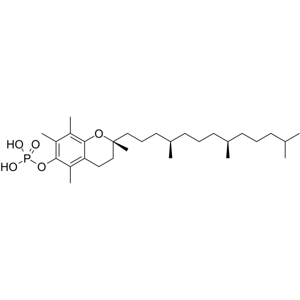
α-Tocopherol phosphate
Based on 8 publication(s) in Google Scholar
α-Tocopherol phosphate is an antioxidant that protects against long-wave UVA1 induced cell death and scavenge UVA1 induced ROS in a skin cell model. α-Tocopherol phosphate exhibits angiogenesis-promoting activity.
For research use only. We do not sell to patients.
- Purity : 98.0%
- CAS No.: 38976-17-9
- Formula: C29H51O5P
- Molecular Weight:510.69
-
Storage:Powder -20°C, 3 years , 4°C, 2 years ; In solvent -80°C, 2 years , -20°C, 1 year

To place orders, for customer services and technical support, please contact: MedChemExpress USA
Tel: 609-228-6898 E-mail: [email protected] [email protected]


-
 Biological Activity
-
Chemical Information
-
Solvent & Solubility
- Protocols
- Purity & Documentation
- References
-
 Help & FAQs


-
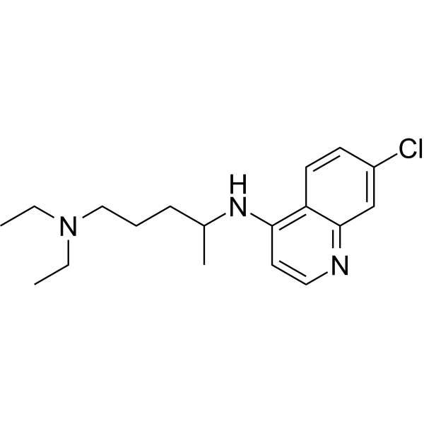
HY-17589A
ChloroquineAntimalarial Agent -

HY-B0215
AcetylcysteineMucolytic Agent -
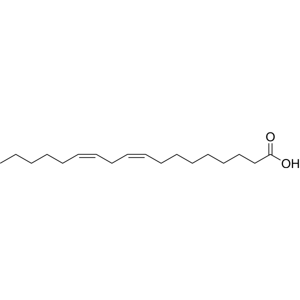
HY-N0729
Linoleic acidHuman Endogenous Metabolite -

HY-10182
LaduviglusibGSK-3 Inhibitor -
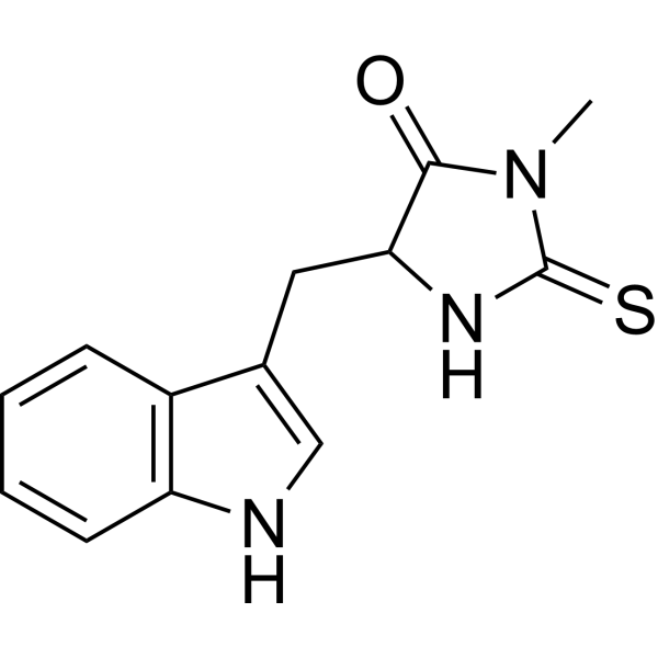
HY-15760
Necrostatin-1RIPK1 Inhibitor -
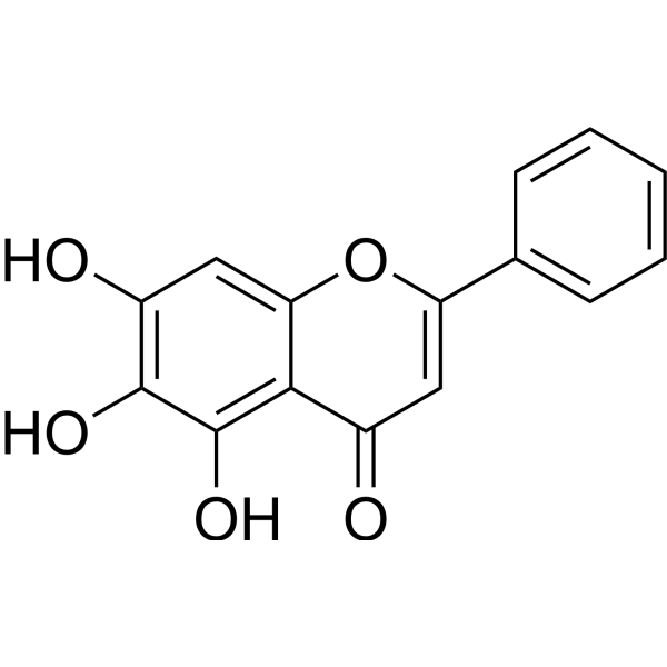
HY-N0196
BaicaleinXanthine Oxidase Inhibitor -
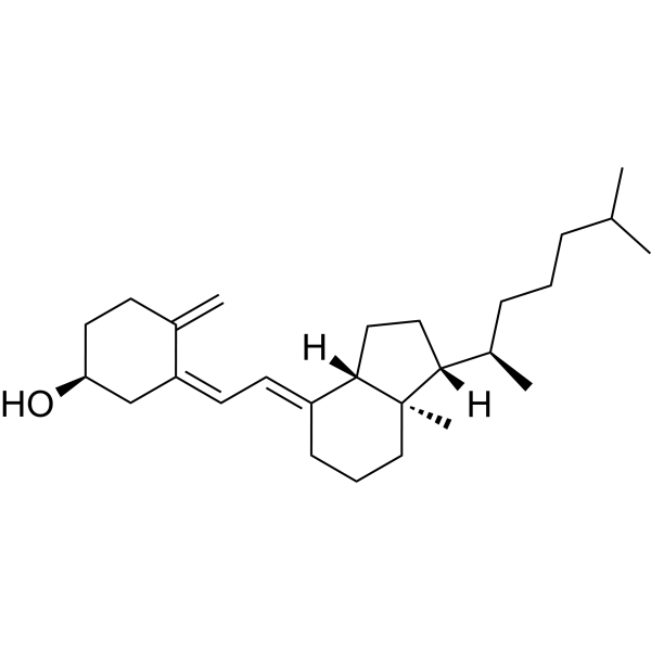
HY-15398
Vitamin D3Vitamin -

HY-100579
Ferrostatin-1Ferroptosis Inhibitor -
HY-13750
EbselenVDCC Inhibitor -
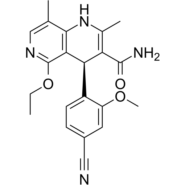
HY-111372
FinerenoneMineralocorticoid Receptor Antagonist
Publications Citing Use of MedChemExpress (MCE) α-Tocopherol phosphate
More- Cell Death Dis. 2022 Jan 21;13(1):73. [Abstract]
- Cell Death Discov. 2025 Aug 22;11(1):398. [Abstract]
- Cancer Cell Int. 2025 Dec 10. [Abstract]
- Appl Mater Today. 2021, 101066.
- Pharmacol Res Perspect. 2026 Feb;14(1):e70213. [Abstract]
- bioRxiv. 2025 Sep 9:2025.09.04.674327. [Abstract]
- Research Square Preprint. 2024 Dec 29.
- Nigerian Journal of Scientific Research. 18 (3): 2019.


Biological Activity
Description
In Vitro
α-Tocopherol phosphate (100 μM, 24 h) inhibits high Glucose (HY-B0389)/ischemic-induced apoptosis and migration in endothelial progenitor cells, promotes capillary-like tube formation through regulation of Bcl-2, Akt, NF-κB, p38MAPK and JNK expressions[2].
MedChemExpress (MCE) has not independently confirmed the accuracy of these methods. They are for reference only.
-
Cell Line:Endothelial progenitor cells derived from rat bone marrow
-
Concentration:100 μM
-
Incubation Time:24 h
-
Result:Upregulated the expression of Bcl-2 and Akt.
Downregulated the expression of Tie2, Notch1, p38MAPK and JNK.
-
Cell Line:Endothelial progenitor cells derived from rat bone marrow
-
Concentration:100 μM
-
Incubation Time:24 h
-
Result:Promoted the cell migration.
In Vivo
MedChemExpress (MCE) has not independently confirmed the accuracy of these methods. They are for reference only.
-
Animal Model:Rats type 2 diabetes and ischemia models[2]
-
Dosage:100 μM α-Tocopherol phosphate incubated with EPCs
-
Administration:24 incubation, intramuscular injection of EPC into rats hind limbs
-
Result:Increased the capillary density of the ischemic hindlimbs.
- Human pluripotent stem cell endothelial-cell differentiation
- Cell Cytotoxicity Assay
- Mammalian live/dead viability and cytotoxicity staining
- Vascular/Branching Fractal Analysis
- ROS/oxidative-stress fluorescent staining
- Endothelial Tube Formation Assay
- Apoptosis Solutions
- Endothelial Cell Migration/Angiogenic Sprouting Assay


Chemical Information
-
CAS No. 38976-17-9
-
Appearance Solid
-
Molecular Weight 510.69
-
Formula C29H51O5P
-
Color White to off-white
-
SMILES
CC1=C(O[C@@](CCC[C@H](C)CCC[C@H](C)CCCC(C)C)(C)CC2)C2=C(C)C(OP(O)(O)=O)=C1C
-
Synonyms
alpha-Tocopherol phosphate; TocP; vitamin E phosphate
-
Shipping
Room temperature in continental US; may vary elsewhere.
-
Storage
Powder -20°C 3 years 4°C 2 years In solvent -80°C 2 years -20°C 1 year


Publications (8)
-
Journal Impact Factor
-
Most Recent
-
Cell Death Dis
RNA binding protein NKAP protects glioblastoma cells from ferroptosis by promoting SLC7A11 mRNA splicing in an m6A-dependent manner. [Abstract]2022 Jan 21;13(1):73. PMID: 35064112 -
Cell Death Discov
ERBB3 influences the ferroptosis pathway via modulation of lipid peroxidation and GSH synthesis in gastric cancer. [Abstract]2025 Aug 22;11(1):398. PMID: 40846695 -
Cancer Cell Int
Polysaccharides from Huaier induce autophagy-dependent ferroptosis to inhibit breast cancer stem cells in triple-negative breast cancer. [Abstract]2025 Dec 10. PMID: 41372887 -
-
Pharmacol Res Perspect
2026 Feb;14(1):e70213. PMID: 41445272 -
bioRxiv
Unravelling the anti-cancer mechanisms elicited by non-covalent thioredoxin reductase inhibitors for triple negative breast cancer therapy. [Abstract]2025 Sep 9:2025.09.04.674327. PMID: 40964317 -
-


Solvent & Solubility
In Vitro:
DMSO : ≥ 100 mg/mL (195.81 mM; Hygroscopic DMSO has a significant impact on the solubility of product, please use newly opened DMSO)
H2O : < 0.1 mg/mL (insoluble)
* "≥" means soluble, but saturation unknown.
Please refer to the solubility information to select the appropriate solvent. Once prepared, please aliquot and store the solution to prevent product inactivation from repeated freeze-thaw cycles.
Storage method and period of stock solution: -80°C, 2 years; -20°C, 1 year. When stored at -80°C, please use it within 2 years. When stored at -20°C, please use it within 1 year.
Please refer to the solubility information to select the appropriate solvent. Once prepared, please aliquot and store the solution to prevent product inactivation from repeated freeze-thaw cycles.
Storage method and period of stock solution: -80°C, 2 years; -20°C, 1 year. When stored at -80°C, please use it within 2 years. When stored at -20°C, please use it within 1 year.
Concentration (start) × Volume (start) = Concentration (final) × Volume (final)
In Vivo:
Select the appropriate dissolution method based on your experimental animal and administration route.
- For the following dissolution methods, please ensure to first prepare a clear stock solution using an In Vitro approach and then sequentially add co-solvents:
- To ensure reliable experimental results, the clarified stock solution can be appropriately stored based on storage conditions. As for the working solution for In Vivo experiments, it is recommended to prepare freshly and use it on the same day.
- The percentages shown for the solvents indicate their volumetric ratio in the final prepared solution. If precipitation or phase separation occurs during preparation, heat and/or sonication can be used to aid dissolution.
Add each solvent one by one: 10% DMSO 40% PEG300 5% Tween-80 45% Saline
Solubility: ≥ 2.5 mg/mL (4.90 mM); Clear solution
This protocol yields a clear solution of ≥ 2.5 mg/mL (saturation unknown).
Taking 1 mL working solution as an example, add 100 μL DMSO stock solution (25.0 mg/mL) to 400 μL PEG300, and mix evenly; then add 50 μL Tween-80 and mix evenly; then add 450 μL Saline to adjust the volume to 1 mL.
Preparation of Saline: Dissolve 0.9 g sodium chloride in ddH₂O and dilute to 100 mL to obtain a clear Saline solution.
In Vivo Dissolution Calculator
Please enter the basic information of animal experiments:
-
-
-
-
Recommended: Prepare an additional quantity of animals to account for potential losses during experiments.
Please enter your animal formula composition:
-
%DMSO +
Recommended: Keep the proportion of DMSO in working solution below 2% if your animal is weak.
-
%+
-
+%Tween-80 + +
-
%Saline +
The co-solvents required include: DMSO, . All of co-solvents are available by MedChemExpress (MCE). , Tween 80. All of co-solvents are available by MedChemExpress (MCE).
Working solution concentration: 0.22 mg/mL
Method for preparing stock solution: mg drug dissolved in μL DMSO. Stock solution concentration: mg/mL.
1. Take μL DMSO stock solution;
2. Add μL .
μL , mix evenly;
3. Then add μL Tween 80, mix evenly;
4. Then add μL
Please ensure that the stock solution in the first step is dissolved to a clear state, and add co-solvents in sequence. You can use ultrasonic heating (ultrasonic cleaner, recommended frequency 20-40 kHz), vortexing, etc. to assist dissolution.


Purity & Documentation
-
Data Sheet (274 KB)
-
SDS (393 KB)
- English - EN (393 KB)
- Français - FR (393 KB)
- Deutsch - DE (393 KB)
- Norwegian - NO (393 KB)
- Español - ES (393 KB)
- Swedish - SV (393 KB)
- Italian - IT (393 KB)
- Korean - KR (393 KB)
- Portuguese - PT (393 KB)
-
Handling Instructions (2659 KB)
References
[1]. Saleh MM, et al. The photoprotective properties of α-tocopherol phosphate against long-wave UVA1 (385 nm) radiation in keratinocytes in vitro. Sci Rep. 2021 Nov 17;11(1):22400. [Content Brief]
[2]. Wu Z, Zheng X, Meng L, et al. α-Tocopherol, especially α-tocopherol phosphate, exerts antiapoptotic and angiogenic effects on rat bone marrow-derived endothelial progenitor cells under high-glucose and hypoxia conditions. J Vasc Surg. 2018;67(4):1263-1273 [Content Brief]


Complete Stock Solution Preparation Table
Please refer to the solubility information to select the appropriate solvent. Once prepared, please aliquot and store the solution to prevent product inactivation from repeated freeze-thaw cycles.
Storage method and period of stock solution: -80°C, 2 years; -20°C, 1 year. When stored at -80°C, please use it within 2 years. When stored at -20°C, please use it within 1 year.
| Optional Solvent | Concentration Solvent Mass | 1 mg | 5 mg | 10 mg | 25 mg |
|---|---|---|---|---|---|
| DMSO | 1 mM | 1.9581 mL | 9.7907 mL | 19.5814 mL | 48.9534 mL |
| 5 mM | 0.3916 mL | 1.9581 mL | 3.9163 mL | 9.7907 mL | |
| 10 mM | 0.1958 mL | 0.9791 mL | 1.9581 mL | 4.8953 mL | |
| 15 mM | 0.1305 mL | 0.6527 mL | 1.3054 mL | 3.2636 mL | |
| 20 mM | 0.0979 mL | 0.4895 mL | 0.9791 mL | 2.4477 mL | |
| 25 mM | 0.0783 mL | 0.3916 mL | 0.7833 mL | 1.9581 mL | |
| 30 mM | 0.0653 mL | 0.3264 mL | 0.6527 mL | 1.6318 mL | |
| 40 mM | 0.0490 mL | 0.2448 mL | 0.4895 mL | 1.2238 mL | |
| 50 mM | 0.0392 mL | 0.1958 mL | 0.3916 mL | 0.9791 mL | |
| 60 mM | 0.0326 mL | 0.1632 mL | 0.3264 mL | 0.8159 mL | |
| 80 mM | 0.0245 mL | 0.1224 mL | 0.2448 mL | 0.6119 mL | |
| 100 mM | 0.0196 mL | 0.0979 mL | 0.1958 mL | 0.4895 mL |


α-Tocopherol phosphate Related Classifications
HY-16686 Related Classifications
 Powered by Bioz